Basics of Light Microscopy Imaging - AOMF
Basics of Light Microscopy Imaging - AOMF
Basics of Light Microscopy Imaging - AOMF

Create successful ePaper yourself
Turn your PDF publications into a flip-book with our unique Google optimized e-Paper software.
light microscopy. Note here that resolution<br />
is NOT directly dependent on the<br />
magnification. Furthermore the end<br />
magnification should not be higher than<br />
1000x the NA <strong>of</strong> the objective, because<br />
then the image will be only enlarged but<br />
no further resolution will be visible. This<br />
is called empty magnification.<br />
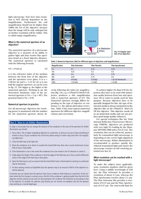
What is the numerical aperture <strong>of</strong> an<br />
objective<br />
The numerical aperture <strong>of</strong> a microscope<br />
objective is a measure <strong>of</strong> its ability to<br />
gather light and thus resolve fine specimen<br />
detail at a fixed objective distance.<br />
The numerical aperture is calculated<br />
with the following formula:<br />
NA = n*(sin µ) (3)<br />
n is the refractive index <strong>of</strong> the medium<br />
between the front lens <strong>of</strong> the objective<br />
and the specimen cover glass. It is n =<br />
1.00 for air and n = 1.51 for oil. µ is one<br />
half the angular aperture, as can be seen<br />
in fig. 13. The bigger µ, the higher is the<br />
numerical aperture. Working in air, the<br />
theoretical maximum value <strong>of</strong> the numerical<br />
aperture is NA = 1 (µ = 90°). The<br />
practical limit is NA = 0.95.<br />
Numerical aperture in practice<br />
For all microscope objectives the resolving<br />
power is mentioned with the number<br />
for the numerical aperture shown di-<br />
Table 2: Numerical Apertures (NA) for different types <strong>of</strong> objectives and magnifications<br />
Fig. 13: Angular aperture<br />
<strong>of</strong> an objective.<br />
Magnification Plan Achromat Plan Fluorite Plan Apochromat<br />
4 x 0.1 0.13 0.16<br />
10x 0.25 0.3 0.4<br />
20x 0.4 0.5 0.75<br />
40x 0.65 0.75 0.9<br />
60x 0.8 (dry) 0.9 (dry) 1.42 (oil immersion)<br />
100x 1.25 (oil) 1.3 (oil) 1.4 (oil)<br />
rectly following the index for magnification<br />
(fig. 14), e.g. a UPlanFLN 60x/0.9 objective<br />
produces a 60x magnification<br />
with a numerical aperture <strong>of</strong> 0.9. The<br />
Numerical Aperture strongly differs depending<br />
on the type <strong>of</strong> objective or condenser,<br />
i.e. the optical aberration correction.<br />
Table 2 lists some typical numerical<br />
apertures for different objective magnifications<br />
and corrections.<br />
Box 2: How to set Köhler illumination<br />
To align a microscope that can change the distance <strong>of</strong> the condenser to the area <strong>of</strong> the specimen for Köhler<br />
illumination you should:<br />
1. Focus with a 10x or higher magnifying objective on a specimen, so that you can see at least something <strong>of</strong><br />
interest in focus. (If your condenser has a front lens please swing it in when using more then 10x magnifying<br />
objectives.)<br />
2. Close the field stop at the light exit.<br />
3. Move the condenser up or down to visualise the closed field stop. Now only a round central part <strong>of</strong> your<br />
field <strong>of</strong> view is illuminated.<br />
4. If the illumination is not in the centre the condenser has to be moved in the XY direction to centre it.<br />
5. Finely adjust the height <strong>of</strong> the condenser so that the edges <strong>of</strong> the field stop are in focus and the diffraction<br />
colour at this edge <strong>of</strong> the field stop is blue green.<br />
6. Open the field stop to such an amount that the total field <strong>of</strong> view is illuminated (re-centring in xy direction<br />
may be necessary).<br />
7. For best viewing contrast at brightfield close the aperture stop to an amount <strong>of</strong> 80 % <strong>of</strong> the objective numerical<br />
aperture.<br />
In practice, you can slowly close the aperture stop <strong>of</strong> your condenser while looking at a specimen. At the moment<br />
when the first change in contrast occurs, the NA <strong>of</strong> the condenser is getting smaller then the NA <strong>of</strong> the<br />
objective and this setting can be used. For documentation the condenser NA should be set according to that<br />
<strong>of</strong> the objective. To visualise the aperture stop directly you can remove one eyepiece and see the aperture<br />
stop working as a normal diaphragm.<br />
To achieve higher NA than 0.95 for objectives<br />
they have to be used with immersion<br />
media between front lens and specimen.<br />
Oil or water is mostly used for that<br />
purpose. Because objectives have to be<br />
specially designed for this, the type <strong>of</strong> immersion<br />
media is always mentioned on the<br />
objective like on the UPlanFLN 60x/1.25<br />
Oil Iris objective. This objective needs oil<br />
as an immersion media and can not produce<br />
good image quality without it.<br />
For special techniques like the Total<br />
Internal Reflection Fluorescence <strong>Microscopy</strong><br />
(TIRFM), objectives are produced<br />
with very high NA leading by the Olympus<br />
APO100x OHR with a NA <strong>of</strong> 1.65. The<br />
resolution that can be achieved, particularly<br />
for transmitted light microscopy, is<br />
depending on the correct light alignment<br />
<strong>of</strong> the microscope. Köhler illumination is<br />
recommended to produce equally distributed<br />
transmitted light and ensure the<br />
microscope reaches its full resolving potential<br />
(see box 2).<br />
What resolution can be reached with a<br />
light microscope<br />
To make the subject more applicable,<br />
some resolution numbers shall be given<br />
here. Using a middle wavelength <strong>of</strong> 550<br />
nm, the Plan Achromat 4x provides a<br />
resolution <strong>of</strong> about 3.3 µm, whereas the<br />
Plan Apochromat reaches about 2.1 µm.<br />
The Plan Achromat 40x provides a resolution<br />
<strong>of</strong> 0.51 µm and the Plan Apochromat<br />
<strong>of</strong> 0.37 µm. The real-world limit for<br />
14 • <strong>Basics</strong> <strong>of</strong> light <strong>Microscopy</strong> & <strong>Imaging</strong> the Resolving Power