
You also want an ePaper? Increase the reach of your titles
YUMPU automatically turns print PDFs into web optimized ePapers that Google loves.
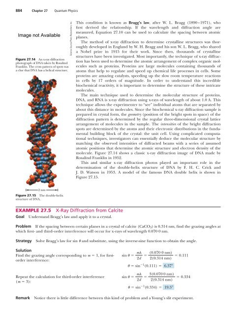
884 Chapter 27 <strong>Quantum</strong> <strong>Physics</strong>Figure 27.14 An x-ray diffractionphotograph of DNA taken by RosalindFranklin. The cross pattern of spots wasa clue that DNA has a helical structure.Science Source/Photo Researchers, Inc.This condition is known as Bragg’s law, after W. L. Bragg (1890–1971), whofirst derived the relationship. If the wavelength and diffraction angle aremeasured, Equation 27.10 can be used to calculate the spacing between atomicplanes.The method of x-ray diffraction to determine crystalline structures was thoroughlydeveloped in England by W. H. Bragg and his son W. L. Bragg, who shareda Nobel prize in 1915 for their work. Since then, thousands of crystallinestructures have been investigated. Most importantly, the technique of x-ray diffractionhas been used to determine the atomic arrangement of complex organic moleculessuch as proteins. Proteins are large molecules containing thousands ofatoms that help to regulate and speed up chemical life processes in cells. Someproteins are amazing catalysts, speeding up the slow room temperature reactionsin cells by 17 orders of magnitude. In order to understand this incrediblebiochemical reactivity, it is important to determine the structure of these intricatemolecules.The main technique used to determine the molecular structure of proteins,DNA, and RNA is x-ray diffraction using x-rays of wavelength of about 1.0 A. Thistechnique allows the experimenter to “see” individual atoms that are separated byabout this distance in molecules. Since the biochemical x-ray diffraction sample isprepared in crystal form, the geometry (position of the bright spots in space) of thediffraction pattern is determined by the regular three-dimensional crystal latticearrangement of molecules in the sample. The intensities of the bright diffractionspots are determined by the atoms and their electronic distributions in the fundamentalbuilding block of the crystal: the unit cell. Using complicated computationaltechniques, investigators can essentially deduce the molecular structure bymatching the observed intensities of diffracted beams with a series of assumedatomic positions that determine the atomic structure and electron density of themolecule. Figure 27.14 shows a classic x-ray diffraction image of DNA made byRosalind Franklin in 1952.This and similar x-ray diffraction photos played an important role in thedetermination of the double-helix structure of DNA by F. H. C. Crick andJ. D. Watson in 1953. A model of the famous DNA double helix is shown inFigure 27.15.2 nmFigure 27.15 The double-helixstructure of DNA.EXAMPLE 27.5 X-Ray Diffraction from CalciteGoal Understand Bragg’s law and apply it to a crystal.Problem If the spacing between certain planes in a crystal of calcite (CaCO 3 ) is 0.314 nm, find the grazing angles atwhich first- and third-order interference will occur for x-rays of wavelength 0.070 0 nm.StrategySolve Bragg’s law for sin and substitute, using the inverse-sine function to obtain the angle.SolutionFind the grazing angle corresponding to m 1, for firstorderinterference:sin m (0.070 0 nm)2d 2(0.314 nm) 0.111 sin 1 (0.111) 6.37Repeat the calculation for third-order interference(m 3):sin m 3(0.070 0 nm)2d 2(0.314 nm) sin 1 (0.334) 19.5 0.334RemarkNotice there is little difference between this kind of problem and a Young’s slit experiment.