29.7 Medical Applications of Radiation 961Second, a conventional x-ray absorption picture is indicative of the averageamount of absorption along a particular direction in the body, leading to somewhatobscured pictures. To overcome these problems, an instrument called a CATscanner was developed in England in 1973; the device is capable of producing picturesof much greater clarity and detail than previously possible.The operation of a CAT scanner can be understood by considering the followinghypothetical experiment: suppose a box consists of four compartments,labeled A, B, C, and D, as in Figure 29.13a. Each compartment has a differentamount of absorbing material from any other compartment. What set of experimentalprocedures will enable us to determine the relative amounts of material ineach compartment? The following steps outline one method that will provide thisinformation: first, a beam of x-rays is passed through compartments A and C, as inFigure 29.13b. The intensity of the exiting radiation is reduced by absorption bysome number that we assign as 8. (The number 8 could mean, for example, thatthe intensity of the exiting beam is reduced by eight-tenths of 1% from its initialvalue.) Because we don’t know which of the compartments, A or C, was responsiblefor this reduction in intensity, half the loss is assigned to each compartment, asin Figure 29.13c. Next, a beam of x-rays is passed through compartments B and D,as in Figure 29.13b. The reduction in intensity for this beam is 10, and again we assignhalf the loss to each compartment. We now redirect the x-ray source so that itsends one beam through compartments A and B and another through compartmentsC and D, as in Figure 29.13d. Once more, we measure the absorption. Supposethe absorption through compartments A and B in this experiment is measuredto be 7 units. On the basis of our first experiment, we would have guessed itwould be 9 units: 4 by compartment A and 5 by compartment B. Thus, we have reducedthe guessed absorption for each compartment by 1 unit, so that the sum is 7rather than 9, giving the numbers shown in Figure 29.13e. Likewise, when thebeam is passed through compartments C and D, as in Figure 29.13d, we may findthe total absorption to be 11 as compared to our first experiment of 9. In this case,we add 1 unit of absorption to each compartment to give a sum of 11, as in Figure29.13e. This somewhat crude procedure could be improved by measuring the absorptionalong other paths. However, these simple measurements are sufficient toenable us to conclude that compartment D contains the most absorbing materialand A the least. A visual representation of these results can be obtained by assigning,to each compartment, a shade of gray corresponding to the particular numberAPPLICATIONCAT ScansExitbeam810ABABCD(a)CD(b)Incidentbeam4 5A B4 5C D(c)IncidentbeamACBD(d)711Exitbeam3 4A B5 6C D(e)Figure 29.13 An experimental procedure fordetermining the relative amounts of x-ray absorptionby four different compartments in a box.
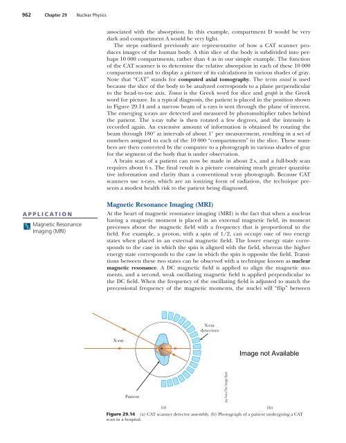
962 Chapter 29 Nuclear <strong>Physics</strong>associated with the absorption. In this example, compartment D would be verydark and compartment A would be very light.The steps outlined previously are representative of how a CAT scanner producesimages of the human body. A thin slice of the body is subdivided into perhaps10 000 compartments, rather than 4 as in our simple example. The functionof the CAT scanner is to determine the relative absorption in each of these 10 000compartments and to display a picture of its calculations in various shades of gray.Note that “CAT” stands for computed axial tomography. The term axial is usedbecause the slice of the body to be analyzed corresponds to a plane perpendicularto the head-to-toe axis. Tomos is the Greek word for slice and graph is the Greekword for picture. In a typical diagnosis, the patient is placed in the position shownin Figure 29.14 and a narrow beam of x-rays is sent through the plane of interest.The emerging x-rays are detected and measured by photomultiplier tubes behindthe patient. The x-ray tube is then rotated a few degrees, and the intensity isrecorded again. An extensive amount of information is obtained by rotating thebeam through 180° at intervals of about 1° per measurement, resulting in a set ofnumbers assigned to each of the 10 000 “compartments” in the slice. These numbersare then converted by the computer to a photograph in various shades of grayfor the segment of the body that is under observation.A brain scan of a patient can now be made in about 2 s, and a full-body scanrequires about 6 s. The final result is a picture containing much greater quantitativeinformation and clarity than a conventional x-ray photograph. Because CATscanners use x-rays, which are an ionizing form of radiation, the technique presentsa modest health risk to the patient being diagnosed.APPLICATIONMagnetic ResonanceImaging (MRI)Magnetic Resonance Imaging (MRI)At the heart of magnetic resonance imaging (MRI) is the fact that when a nucleushaving a magnetic moment is placed in an external magnetic field, its momentprecesses about the magnetic field with a frequency that is proportional to thefield. For example, a proton, with a spin of 1/2, can occupy one of two energystates when placed in an external magnetic field. The lower energy state correspondsto the case in which the spin is aligned with the field, whereas the higherenergy state corresponds to the case in which the spin is opposite the field. Transitionsbetween these two states can be observed with a technique known as nuclearmagnetic resonance. A DC magnetic field is applied to align the magnetic moments,and a second, weak oscillating magnetic field is applied perpendicular tothe DC field. When the frequency of the oscillating field is adjusted to match theprecessional frequency of the magnetic moments, the nuclei will “flip” betweenX-raydetectorsX-rayPatient(a)Figure 29.14 (a) CAT scanner detector assembly. (b) Photograph of a patient undergoing a CATscan in a hospital.Jay Freis/The Image Bank(b)
- Page 1 and 2:
Color-enhanced scanning electronmic
- Page 3:
876 Chapter 27 Quantum PhysicsSolve
- Page 6 and 7:
27.2 The Photoelectric Effect and t
- Page 8 and 9:
27.3 X-Rays 881even when black card
- Page 10 and 11:
27.4 Diffraction of X-Rays by Cryst
- Page 12 and 13:
27.5 The Compton Effect 885Exercise
- Page 14 and 15:
27.6 The Dual Nature of Light and M
- Page 16 and 17:
27.6 The Dual Nature of Light and M
- Page 18 and 19:
27.8 The Uncertainty Principle 891w
- Page 20 and 21:
27.8 The Uncertainty Principle 893E
- Page 22 and 23:
27.9 The Scanning Tunneling Microsc
- Page 24 and 25:
Problems 897The probability per uni
- Page 26 and 27:
Problems 89917. When light of wavel
- Page 28 and 29:
Problems 90151.time of 5.00 ms. Fin
- Page 30 and 31:
“Neon lights,” commonly used in
- Page 32 and 33:
28.2 Atomic Spectra 905l(nm) 400 50
- Page 34 and 35:
28.3 The Bohr Theory of Hydrogen 90
- Page 36 and 37:
28.3 Th Bohr Theory of Hydrogen 909
- Page 38 and 39: 28.4 Modification of the Bohr Theor
- Page 40 and 41: 28.6 Quantum Mechanics and the Hydr
- Page 42 and 43: 28.7 The Spin Magnetic Quantum Numb
- Page 44 and 45: 28.9 The Exclusion Principle and th
- Page 46 and 47: 28.9 The Exclusion Principle and th
- Page 48 and 49: 28.11 Atomic Transitions 921electro
- Page 50 and 51: 28.12 Lasers and Holography 923is u
- Page 52 and 53: 28.13 Energy Bands in Solids 925Ene
- Page 54 and 55: 28.13 Energy Bands in Solids 927Ene
- Page 56 and 57: 28.14 Semiconductor Devices 929I (m
- Page 58 and 59: Summary 931(a)Figure 28.32 (a) Jack
- Page 60 and 61: Problems 9335. Is it possible for a
- Page 62 and 63: Problems 935tum number n. (e) Shoul
- Page 64 and 65: Problems 93748. A dimensionless num
- Page 66 and 67: Aerial view of a nuclear power plan
- Page 68 and 69: 29.1 Some Properties of Nuclei 941T
- Page 70 and 71: 29.2 Binding Energy 943130120110100
- Page 72 and 73: 29.3 Radioactivity 94529.3 RADIOACT
- Page 74 and 75: 29.3 Radioactivity 947INTERACTIVE E
- Page 76 and 77: 29.4 The Decay Processes 949Alpha D
- Page 78 and 79: 29.4 The Decay Processes 951Strateg
- Page 80 and 81: 29.4 The Decay Processes 953they we
- Page 82 and 83: 29.6 Nuclear Reactions 955wounds on
- Page 84 and 85: 29.6 Nuclear Reactions 957EXAMPLE 2
- Page 86 and 87: 29.7 Medical Applications of Radiat
- Page 90 and 91: 29.8 Radiation Detectors 963Figure
- Page 92 and 93: Summary 965Photo Researchers, Inc./
- Page 94 and 95: Problems 967CONCEPTUAL QUESTIONS1.
- Page 96 and 97: Problems 96924. A building has beco
- Page 98 and 99: Problems 97157. A by-product of som
- Page 100 and 101: This photo shows scientist MelissaD
- Page 102 and 103: 30.1 Nuclear Fission 975Applying Ph
- Page 104 and 105: 30.2 Nuclear Reactors 977Courtesy o
- Page 106 and 107: 30.2 Nuclear Reactors 979events in
- Page 108 and 109: 30.3 Nuclear Fusion 981followed by
- Page 110 and 111: 30.3 Nuclear Fusion 983VacuumCurren
- Page 112 and 113: 30.6 Positrons and Other Antipartic
- Page 114 and 115: 30.7 Mesons and the Beginning of Pa
- Page 116 and 117: 30.9 Conservation Laws 989LeptonsLe
- Page 118 and 119: 30.10 Strange Particles and Strange
- Page 120 and 121: 30.12 Quarks 993n pΣ _ Σ 0 Σ + S
- Page 122 and 123: 30.12 Quarks 995charm C 1, its anti
- Page 124 and 125: 30.14 Electroweak Theory and the St
- Page 126 and 127: 30.15 The Cosmic Connection 999prot
- Page 128 and 129: 30.16 Problems and Perspectives 100
- Page 130 and 131: Problems 100330.12 Quarks &30.13 Co
- Page 132 and 133: Problems 1005particles fuse to prod
- Page 134 and 135: Problems 100740. Assume binding ene
- Page 136 and 137: A.1 MATHEMATICAL NOTATIONMany mathe
- Page 138 and 139:
A.3 Algebra A.3by 8, we have8x8 32
- Page 140 and 141:
A.3 Algebra A.5EXERCISESSolve the f
- Page 142 and 143:
A.5 Trigonometry A.7When natural lo
- Page 144 and 145:
APPENDIX BAn Abbreviated Table of I
- Page 146 and 147:
An Abbreviated Table of Isotopes A.
- Page 148 and 149:
An Abbreviated Table of Isotopes A.
- Page 150 and 151:
Some Useful Tables A.15TABLE C.3The
- Page 152 and 153:
Answers to Quick Quizzes,Odd-Number
- Page 154 and 155:
Answers to Quick Quizzes, Odd-Numbe
- Page 156 and 157:
Answers to Quick Quizzes, Odd-Numbe
- Page 158 and 159:
Answers to Quick Quizzes, Odd-Numbe
- Page 160 and 161:
Answers to Quick Quizzes, Odd-Numbe
- Page 162 and 163:
Answers to Quick Quizzes, Odd-Numbe
- Page 164 and 165:
Answers to Quick Quizzes, Odd-Numbe
- Page 166 and 167:
Answers to Quick Quizzes, Odd-Numbe
- Page 168 and 169:
IndexPage numbers followed by “f
- Page 170 and 171:
Current, 568-573, 586direction of,
- Page 172 and 173:
Index I.5Fissionnuclear, 973-976, 9
- Page 174 and 175:
Index I.7Magnetic field(s) (Continu
- Page 176 and 177:
Polarizer, 805-806, 805f, 806-807Po
- Page 178 and 179:
South poleEarth’s geographic, 626
- Page 180 and 181:
CreditsPhotographsThis page constit
- Page 182 and 183:
PEDAGOGICAL USE OF COLORDisplacemen
- Page 184 and 185:
PHYSICAL CONSTANTSQuantity Symbol V