
Create successful ePaper yourself
Turn your PDF publications into a flip-book with our unique Google optimized e-Paper software.
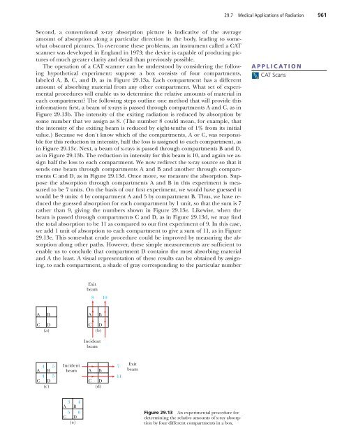
29.7 Medical Applications of Radiation 961Second, a conventional x-ray absorption picture is indicative of the averageamount of absorption along a particular direction in the body, leading to somewhatobscured pictures. To overcome these problems, an instrument called a CATscanner was developed in England in 1973; the device is capable of producing picturesof much greater clarity and detail than previously possible.The operation of a CAT scanner can be understood by considering the followinghypothetical experiment: suppose a box consists of four compartments,labeled A, B, C, and D, as in Figure 29.13a. Each compartment has a differentamount of absorbing material from any other compartment. What set of experimentalprocedures will enable us to determine the relative amounts of material ineach compartment? The following steps outline one method that will provide thisinformation: first, a beam of x-rays is passed through compartments A and C, as inFigure 29.13b. The intensity of the exiting radiation is reduced by absorption bysome number that we assign as 8. (The number 8 could mean, for example, thatthe intensity of the exiting beam is reduced by eight-tenths of 1% from its initialvalue.) Because we don’t know which of the compartments, A or C, was responsiblefor this reduction in intensity, half the loss is assigned to each compartment, asin Figure 29.13c. Next, a beam of x-rays is passed through compartments B and D,as in Figure 29.13b. The reduction in intensity for this beam is 10, and again we assignhalf the loss to each compartment. We now redirect the x-ray source so that itsends one beam through compartments A and B and another through compartmentsC and D, as in Figure 29.13d. Once more, we measure the absorption. Supposethe absorption through compartments A and B in this experiment is measuredto be 7 units. On the basis of our first experiment, we would have guessed itwould be 9 units: 4 by compartment A and 5 by compartment B. Thus, we have reducedthe guessed absorption for each compartment by 1 unit, so that the sum is 7rather than 9, giving the numbers shown in Figure 29.13e. Likewise, when thebeam is passed through compartments C and D, as in Figure 29.13d, we may findthe total absorption to be 11 as compared to our first experiment of 9. In this case,we add 1 unit of absorption to each compartment to give a sum of 11, as in Figure29.13e. This somewhat crude procedure could be improved by measuring the absorptionalong other paths. However, these simple measurements are sufficient toenable us to conclude that compartment D contains the most absorbing materialand A the least. A visual representation of these results can be obtained by assigning,to each compartment, a shade of gray corresponding to the particular numberAPPLICATIONCAT ScansExitbeam810ABABCD(a)CD(b)Incidentbeam4 5A B4 5C D(c)IncidentbeamACBD(d)711Exitbeam3 4A B5 6C D(e)Figure 29.13 An experimental procedure fordetermining the relative amounts of x-ray absorptionby four different compartments in a box.