- Page 1 and 2: NeurologyEdited by Professor Emerit
- Page 3 and 4: That I only found when studying Wil
- Page 5 and 6: fig. 2: origins and distribution of
- Page 7 and 8: Vasodilatation - ØIntestineslongit
- Page 9 and 10: those two modes, its contact with t
- Page 11 and 12: epressive sexual education/impaired
- Page 13 and 14: "The vegetative“ (autonomic) "ner
- Page 15 and 16: 4.1. TechniqueOver the years, lots
- Page 17 and 18: medication which either only battle
- Page 19 and 20: the ANS' level, stress means sympat
- Page 21 and 22: In therapy, we work from the tensio
- Page 23 and 24: Naturally, besides the intensive tr
- Page 25 and 26: neck furthermore intercepts the flo
- Page 27 and 28: Robert A. Dew puts asthma down to t
- Page 29 and 30: Looking at the vernacular gives the
- Page 31 and 32: 5.5. Peptic Ulcer5.5.1. Pulsation o
- Page 33 and 34: appears "openly dependent" or, if c
- Page 35 and 36: During the resolution of the blocks
- Page 37 and 38: Heike S. Buhl M.D., born 1955. Trai
- Page 39 and 40: TitleDiagnosing and Treating Injure
- Page 41 and 42: ody electrolyte strength will gener
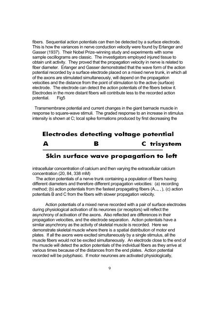
- Page 43 and 44: As presented, the peak of the first
- Page 45: This situation has an important imp
- Page 49 and 50: and active tissue under electrode B
- Page 51 and 52: Action potentials of injured cardia
- Page 53 and 54: a nearby electrode and a distant re
- Page 55 and 56: The distances between the poles of
- Page 57 and 58: ecorded simultaneously the membrane
- Page 59 and 60: When there are conditions of hypoad
- Page 61 and 62: Trauma and injuries are a natural a
- Page 63 and 64: BOOKS1. An Advanced Treatise in Qua
- Page 65 and 66: One of the easiest ways to learn ab
- Page 67 and 68: HOMEOPATHIC TREATMENT OF PAINAbstra
- Page 69 and 70: DESCRIPTION: PAIN - HEAT IMPROVESNU
- Page 71 and 72: BEFOREAFTER4321OOOOOOOOXXXXXXXX0Avg
- Page 73 and 74: tissues to bring about a lowering i
- Page 75 and 76: CAUSES OF HEADACHE PAIN (Vindicate)
- Page 77 and 78: ARTICLES AND STUDIES1. A Practical
- Page 79 and 80: impairment or "disability" evaluati
- Page 81 and 82: criteria:The third step is the comp
- Page 83 and 84: 5. Estimate of the expected date of
- Page 85 and 86: ImpairmentThe Extremities, Spine, a
- Page 87 and 88: 3. Explanation for concluding that
- Page 89 and 90: 3.1b Principles and Methods of Impa
- Page 91 and 92: The range of motion should be recor
- Page 93 and 94: IA is a function of V when Vext = V
- Page 95 and 96: Interphalangeal Joint-Flexion and E
- Page 97 and 98:
Example: Thumb metacarpophalangeal
- Page 99 and 100:
Note: Because the relative value of
- Page 101 and 102:
Determine the length of the finger
- Page 103 and 104:
position (0 cm) to: Lost Retained o
- Page 105 and 106:
If the metacarpophalangeal joint is
- Page 107 and 108:
40 of ring finger 450 of little fin
- Page 109 and 110:
Joint-Radial and Ulnar DeviationMea
- Page 111 and 112:
Flexion IA%=21%Lateral deviation IA
- Page 113 and 114:
extremity.Flexion and ExtensionMeas
- Page 115 and 116:
the upper extremity, and 15% impair
- Page 117 and 118:
Add the impairment values for flexi
- Page 119 and 120:
Internal and External RotationMeasu
- Page 121 and 122:
shoulder motions (flexion/ extensio
- Page 123 and 124:
The peripheral spinal nerves consti
- Page 125 and 126:
obtained history, a thorough medica
- Page 127 and 128:
Example: In an injury, a patient su
- Page 129 and 130:
associated with those particular ne
- Page 131 and 132:
Ulnar or Radial Deviation%Digit Imp
- Page 133 and 134:
Joint Instability% Joint Impairment
- Page 135 and 136:
Example: Implant resection arthropl
- Page 137 and 138:
Intrinsic tightness in the hand may
- Page 139 and 140:
Extensor TendonSubluxation Severity
- Page 141 and 142:
With the patient plantar-flexing th
- Page 143 and 144:
0° 30° 0° 4510° 20° 10° 3020
- Page 145 and 146:
Center the goniometer over the meta
- Page 147 and 148:
Example: If 50% of the distal phala
- Page 149 and 150:
of the toe being tested (Figure 61c
- Page 151 and 152:
Center the goniometer over the late
- Page 153 and 154:
Inversion and Eversion (Subtalar Jo
- Page 155 and 156:
10° active dorsi-flexion 410° act
- Page 157 and 158:
Place the goniometer base as if mea
- Page 159 and 160:
Add the impairment values contribut
- Page 161 and 162:
Place the goniometer base as if mea
- Page 163 and 164:
Hip Joint-Two or More Ranges of Mot
- Page 165 and 166:
3.2f Impairment of the Lower Extrem
- Page 167 and 168:
In evaluating pain that is associat
- Page 169 and 170:
After the individual values for los
- Page 171:
value for loss of function = 15%) 1
- Page 174 and 175:
Principles for Calculating Impairme
- Page 176 and 177:
motion or ankylosis.B. Repeat the a
- Page 178 and 179:
is the greatest angle measured.5. C
- Page 180 and 181:
Example: Occipital flexion measurem
- Page 182 and 183:
possible, again recording both incl
- Page 184 and 185:
Cervical Region-RotationBecause the
- Page 186 and 187:
minimum kyphosis is actually a meas
- Page 188 and 189:
2. With the subject in either the s
- Page 190 and 191:
1. The subject may be seated or sta
- Page 192 and 193:
3.3) Ask the subject to rotate the
- Page 194 and 195:
3.3e Impairments Due to Range of Mo
- Page 196 and 197:
(for automated devices capable of c
- Page 198 and 199:
Example: T12 flexion measurements o
- Page 200 and 201:
3. Record the "0" readings first at
- Page 202 and 203:
3.4 The PelvisThe following shows i
- Page 204 and 205:
Addendum to Chapter 3A. Introductio
- Page 206 and 207:
1. If applicable, use Table 49 (p.
- Page 208 and 209:
B. Repeat the above steps for secon
- Page 210 and 211:
2. Consult the Ankylosis Section of
- Page 212 and 213:
impairment of the whole person.Exam
- Page 214 and 215:
Ankylosis1. Place the goniometer ba
- Page 216 and 217:
6. Add the impairment values contri
- Page 218 and 219:
FINAL IMPAIRMENT RATING FOR WHOLE M
- Page 220 and 221:
Combined value of spinal impairment
- Page 222 and 223:
There is a neutral or relaxed perio
- Page 224 and 225:
SPHENOBASILAR/CRANIAL BASE TORSIONT
- Page 226 and 227:
IMPAIRMENT OF THE NERVOUS SYSTEMThe
- Page 228 and 229:
upper extremities, controlling blad
- Page 230 and 231:
2. Neurological disorder results in
- Page 232 and 233:
4) Neurological: Grades = 5=normal
- Page 234 and 235:
4) Neurological:Sensation: (right/l
- Page 236 and 237:
technique on the part of the physic
- Page 238 and 239:
Direct TechniqueWith the patient co
- Page 240 and 241:
CONTRAINDICATIONSThe only contraind
- Page 242 and 243:
In order to determine the presence
- Page 244 and 245:
IX Glosso- pharyngealPharynx muscle
- Page 246 and 247:
ReflexesMuscle stretch reflexes, cu
- Page 248 and 249:
Abdominal skin reflex (exteroceptiv
- Page 250 and 251:
Sensory symptomsPersistant or episo
- Page 252 and 253:
Pyramidal tract signsEponym Manoeuv
- Page 254 and 255:
doubts of survival7. Psychotic symp
- Page 256 and 257:
· Foramen jugulare syndrome (Siebe
- Page 258:
For cranial nerve stimulation the c
- Page 261 and 262:
Demonstration of specific antibodie
- Page 263 and 264:
4.6.2 Explosive type4.6.3 Postural
- Page 265 and 266:
8.4.2 Caffeine withdrawal headache8
- Page 267 and 268:
Headache: differential diagnosisDes
- Page 269 and 270:
Diagnostic criteria of drug-induced
- Page 271 and 272:
convexity), glioblastoma, oligodend
- Page 273 and 274:
Hypothalamic regulatory hormonesHyp
- Page 275 and 276:
0 = normal1 = stride down to 30-45
- Page 277 and 278:
Parkinsonism: causesAetiology or Pa
- Page 279 and 280:
Paradoxical hyperkinesiaSudden good
- Page 281 and 282:
Meige syndrome(bilateral facial spa
- Page 283 and 284:
ThalamotomyScale for quantification
- Page 285 and 286:
Eating10 = independent (with aids)5
- Page 287 and 288:
InfectionsMeningitisOtitis media, m
- Page 289 and 290:
OculomotorDependent on surgery, var
- Page 291 and 292:
AreflexiaSlowed nerve conduction ve
- Page 293 and 294:
Causes of lumbosacral root disorder
- Page 295 and 296:
Compartment syndromescompartmentsAu
- Page 297:
Lateral root of median nerve (C5-C7
- Page 300 and 301:
Palsy without pronator, teres invol
- Page 302 and 303:
Baker's cyst (in rheumatoid arthrit
- Page 304 and 305:
Gluteal nerve lesionsIntramuscular
- Page 306 and 307:
Differential diagnosis of important
- Page 308 and 309:
2. C8 root syndromeMotor lossAs for
- Page 310 and 311:
Congenital and usually inherited my
- Page 312 and 313:
(iv) Type 7.erythrocytes involved.P
- Page 314 and 315:
Myopathies (polymyositis, dermatomy
- Page 316 and 317:
denervated muscles.)Prostigmine may
- Page 318 and 319:
Polyneuropathy (rarely)Hypothyroidi
- Page 320 and 321:
Multiple sclerosis - CAMBS scaleDis
- Page 322 and 323:
Intrathecal IgG production ≥90Oli
- Page 325 and 326:
THE CV-4 TECHNIQUEThe still point a
- Page 327 and 328:
NEUROLOGY TESTBlood pressure should
- Page 329 and 330:
To find out about the different neu
- Page 331 and 332:
support, clergy support, counseling
- Page 333 and 334:
HYGIENESelf-care deficit (specify l
- Page 335 and 336:
___________________________________
- Page 337 and 338:
ObjectiveHard -formed stoolStrainin
- Page 339 and 340:
Definition: [ failure of the heat t
- Page 341 and 342:
___________________________________
- Page 343 and 344:
Work overloadUnrealistic perception
- Page 345 and 346:
Inappropriate or poorly communicate
- Page 347 and 348:
Decreased skin turgorIncreased puls
- Page 349 and 350:
___________________________________
- Page 351 and 352:
Definition: The client is unable to
- Page 353 and 354:
DEFINING CHARACTERISTICSInternal (i
- Page 355 and 356:
SUPPORTING DATAETIOLOGYLack of expo
- Page 357 and 358:
Satiety immediately after ingesting
- Page 359 and 360:
DEFINING CHARACTERISTICSSubjectiveX
- Page 361 and 362:
Does not defend self-care practices
- Page 363 and 364:
Cognitive perceptualCultural or spi
- Page 365 and 366:
SUPPORTING DATAETIOLOGYEnvironmenta
- Page 367 and 368:
Mechanical factorsShearing forcesPr
- Page 369 and 370:
Changes in postureFrequent yawningN
- Page 371 and 372:
DEFINING CHARACTERISTICSObjectiveSk
- Page 373 and 374:
Toxic reactions to medicationBatter
- Page 375 and 376:
______Self-concept, disturbance in:
- Page 377 and 378:
Bed rest or immobilityDEFINING CHAR
- Page 379 and 380:
Unmet needs[Physiological factors,
- Page 381 and 382:
___________________________________
- Page 383 and 384:
___________________________________
- Page 385 and 386:
Inability to speak in sentencesDoes
- Page 387 and 388:
Decision and actions by family whic
- Page 389 and 390:
ETIOLOGYSituational transition and/
- Page 391 and 392:
Hypotension [postural]Increased pul
- Page 393 and 394:
Central venous pressure changesJugu
- Page 395 and 396:
ObjectiveCryingDevelopmental regres
- Page 397 and 398:
Abnormal blood profile: altered clo
- Page 399 and 400:
Children playing without gates at t
- Page 401 and 402:
Reluctance to attempt movement[C/o
- Page 403 and 404:
SubjectiveReported or observed dysf
- Page 405 and 406:
diagnosis that adjustment to parent
- Page 407 and 408:
Acute Phase:Emotional reactions: an
- Page 409 and 410:
SubjectiveVerbalization of:Change i
- Page 411 and 412:
SUPPORTING DATAETIOLOGYEnvironmenta
- Page 413 and 414:
ETIOLOGYExternal (environmental) fa
- Page 415 and 416:
Interrupted sleep[Falls asleep duri
- Page 417 and 418:
Alteration in behavior or mood evid
- Page 419 and 420:
DEFINING CHARACTERISTICSSubjectiveF
- Page 421 and 422:
6) ORTHOPAEDIC TEST/SIGNS:Noted in
- Page 423 and 424:
Hibbs (positive) (negative) for (rt
- Page 425 and 426:
PROGNOSIS:MODERATE:The prognosis fo
- Page 427 and 428:
RECIPESBETTER BUTTERFor those of yo
- Page 429 and 430:
or flakes. Begin with 1/2 teaspoon
- Page 431 and 432:
in waterfor an elegant main course.
- Page 433 and 434:
This recipe also makes an excellent
- Page 435 and 436:
adding the celery and carrot near t
- Page 437 and 438:
stuffed caps in lightly greased bak
- Page 439 and 440:
TOFU SURPRISES2 tablespoons cold-pr
- Page 441 and 442:
By following these guidelines, you
- Page 443 and 444:
The technique of somatoemtional rec
- Page 445 and 446:
SPINAL IMPAIRMENT RATINGWhole Man1.
- Page 447 and 448:
59. Do you have a loss of interest
- Page 450 and 451:
The technique used to mobilize the
- Page 452 and 453:
The technique for evaluation and tr