
You also want an ePaper? Increase the reach of your titles
YUMPU automatically turns print PDFs into web optimized ePapers that Google loves.
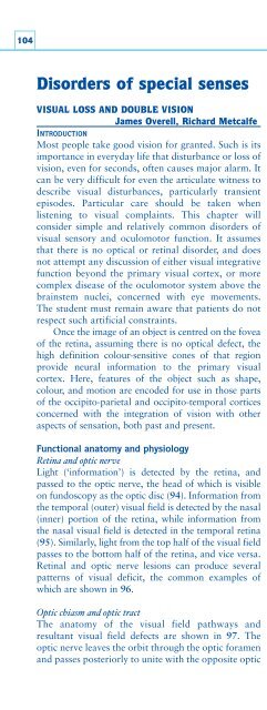
104Disorders of special sensesVISUAL LOSS AND DOUBLE VISIONJames Overell, Richard MetcalfeINTRODUCTIONMost people take good vision for granted. Such is itsimportance in everyday life that disturbance or loss ofvision, even for seconds, often causes major alarm. Itcan be very difficult for even the articulate witness todescribe visual disturbances, particularly transientepisodes. Particular care should be taken whenlistening to visual complaints. This chapter willconsider simple and relatively common disorders ofvisual sensory and oculomotor function. It assumesthat there is no optical or retinal disorder, and doesnot attempt any discussion of either visual integrativefunction beyond the primary visual cortex, or morecomplex disease of the oculomotor system above thebrainstem nuclei, concerned with eye movements.The student must remain aware that patients do notrespect such artificial constraints.Once the image of an object is centred on the foveaof the retina, assuming there is no optical defect, thehigh definition colour-sensitive cones of that regionprovide neural information to the primary visualcortex. Here, features of the object such as shape,colour, and motion are encoded for use in those partsof the occipito-parietal and occipito-temporal corticesconcerned with the integration of vision with otheraspects of sensation, both past and present.Functional anatomy and physiologyRetina and optic nerveLight (‘information’) is detected by the retina, andpassed to the optic nerve, the head of which is visibleon fundoscopy as the optic disc (94). Information fromthe temporal (outer) visual field is detected by the nasal(inner) portion of the retina, while information fromthe nasal visual field is detected in the temporal retina(95). Similarly, light from the top half of the visual fieldpasses to the bottom half of the retina, and vice versa.Retinal and optic nerve lesions can produce severalpatterns of visual deficit, the common examples ofwhich are shown in 96.Optic chiasm and optic tractThe anatomy of the visual field pathways andresultant visual field defects are shown in 97. Theoptic nerve leaves the orbit through the optic foramenand passes posteriorly to unite with the opposite optic94a94b94c94d94 Fundoscopy. a:normal fundus;b: papillitis in apatient with acuteoptic neuritis;c: papilloedema(swelling of theoptic disc) in apatient with raisedintracranialpressure from abrain tumour;d: Optic atrophy ina patient withmultiple sclerosisand multipleprevious episodesof optic neuritis.