
You also want an ePaper? Increase the reach of your titles
YUMPU automatically turns print PDFs into web optimized ePapers that Google loves.
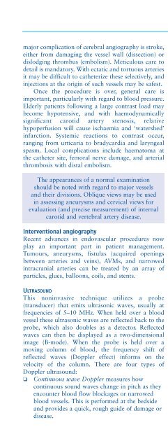
Neurological investigations 39major complication of cerebral angiography is stroke,either from damaging the vessel wall (dissection) ordislodging thrombus (embolism). Meticulous care todetail is mandatory. With ectatic and tortuous arteriesit may be difficult to catheterize these selectively, andinjections at the origin of such vessels may be safest.Once the procedure is over, general care isimportant, particularly with regard to blood pressure.Elderly patients following a large contrast load maybecome hypotensive, and with haemodynamicallysignificant carotid artery stenosis, relativehypoperfusion will cause ischaemia and ‘watershed’infarction. Systemic reactions to contrast occur,ranging from urticaria to bradycardia and laryngealspasm. Local complications include haematoma atthe catheter site, femoral nerve damage, and arterialthrombosis with distal embolism.The appearances of a normal examinationshould be noted with regard to major vesselsand their divisions. Oblique views may be usedin assessing aneurysms and cervical views forevaluation (and precise measurement) of internalcarotid and vertebral artery disease.Interventional angiographyRecent advances in endovascular procedures nowplay an important part in patient management.Tumours, aneurysms, fistulas (acquired openingsbetween arteries and veins), AVMs, and narrowedintracranial arteries can be treated by an array ofparticles, glues, balloons, coils, and stents.ULTRASOUNDThis noninvasive technique utilizes a probe(transducer) that emits ultrasonic waves, usually atfrequencies of 5–10 MHz. When held over a bloodvessel these ultrasonic waves are reflected back to theprobe, which also doubles as a detector. Reflectedwaves can then be displayed as a two-dimensionalimage (B-mode). When the probe is held over amoving column of blood, the frequency shift ofreflected waves (Doppler effect) informs on thevelocity of the column. There are four types ofDoppler ultrasound:❏ Continuous wave Doppler measures howcontinuous sound waves change in pitch as theyencounter blood flow blockages or narrowedblood vessels. This is performed at the bedsideand provides a quick, rough guide of damage ordisease.❏❏❏Duplex Doppler produces a picture of a bloodvessel and the organs that surround it. Acomputer converts the Doppler signals into agraph that provides information about the speedand direction of blood flow through the bloodvessel being examined.Colour Doppler is computer assisted andconverts the Doppler signals into colours thatrepresent the speed and direction of flowthrough the vessel.Power Doppler is a new technique beingdeveloped that is up to five times more sensitivethan colour Doppler and is used to study bloodflow in vessels within solid organs.Ultrasound is used to assess extracranial (carotid)arterial disease (33).Transcranial Doppler ultrasoundTranscranial Doppler ultrasound (TCD) is a safe,reliable, and relatively inexpensive technology formeasuring intracranial blood flow velocities. Waveswith frequencies around 2 MHz are directed towardsintracranial vessels using a handheld probe. Thefrequency shift (Doppler effect) in the reflected soundindicates the velocity of the column of moving blood.33 Carotid Doppler ultrasound showing 70% right internalcarotid stenosis.33