- Page 2:
UnderstandingNEUROLOGYa problem-ori
- Page 14:
CHAPTER 1:HISTORY TAKING ANDPHYSICA
- Page 20:
10These five questions (onset, dura
- Page 24:
12Anatomy of consciousnessConscious
- Page 28:
14The degree of psychomotor activit
- Page 32:
16MemoryMemory is not a unitary fun
- Page 36:
18Difficulties in higher order visu
- Page 40:
20A standardized test such as the M
- Page 44:
22The facial nerve also supplies th
- Page 48:
24Upper limbs❏ Flexion at the elb
- Page 52:
26Two point discriminationAn opened
- Page 56:
28INTRODUCTIONThe evaluation of a c
- Page 60:
30Knowledge of the display and norm
- Page 64:
32ADVANTAGES AND DISADVANTAGES OF M
- Page 68:
34Fluid-attenuated inversion recove
- Page 72:
3625 2625 T1 magnetic resonance ima
- Page 76:
38POSITRON EMISSION TOMOGRAPHYThe p
- Page 80:
40Images are reconstructed from the
- Page 84:
42Other altered conscious statesEEG
- Page 88:
cervical44While VIIIth nerve damage
- Page 92:
46MemoryThere are many different fo
- Page 96:
48passes superiorly above the atlas
- Page 100:
50body disease (tumours and infecti
- Page 104:
52appear clinically not involved, e
- Page 108:
54ElectromyographyIn EMG, a fine bo
- Page 112:
56to proceed to biopsy. It may be o
- Page 116:
58to present with mild vertigo or a
- Page 120:
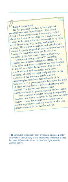
60686969 Computed tomography scan s
- Page 124:
This page intentionally left blank
- Page 128:
64Disorders of consciousnessBLACKOU
- Page 132:
66top down, and so on. Dizziness is
- Page 136:
68EPILEPTIC SEIZURESClassification
- Page 140:
70An understanding of this pathophy
- Page 144:
72The clinical semiology of PNESPNE
- Page 148:
74CASE 2A 27-year-old female presen
- Page 152:
76ACUTE CONFUSIONAL STATESMyfanwy T
- Page 156:
78words beginning with a certain le
- Page 160:
80over days or even weeks. The soma
- Page 164:
8278CASE 1 (continued)An MI screen
- Page 168:
84CASE 2 (continued)Initial investi
- Page 172:
86Disorders of cognitionMEMORY DISO
- Page 176:
88Acute transient disorders of epis
- Page 180:
90asked to recall the three items.
- Page 184: 92CASE 2A 55-year-old female presen
- Page 188: 94SPEECH AND LANGUAGE DISORDERSJohn
- Page 192: 96Dysphonia refers to impaired voca
- Page 196: 98therefore a sign of damage to the
- Page 200: 100can be complemented by functiona
- Page 204: 102CASE 3A 55-year-old male and his
- Page 208: 104Disorders of special sensesVISUA
- Page 212: 106nerve at the optic chiasm. Here
- Page 216: 108may briefly double or the left s
- Page 220: 110Visual fields are assessed by th
- Page 224: 112swelling in the presence of norm
- Page 228: 114CASE 2A 62-year-old male retired
- Page 232: 116102CASE 3 (continued)The visual
- Page 238: Disorders of special senses 1191051
- Page 242: Disorders of special senses121Many
- Page 246: Disorders of special senses123aneur
- Page 250: Disorders of special senses 125Diff
- Page 254: Disorders of special senses 127CLIN
- Page 258: Disorders of special senses129CASE
- Page 262: Disorders of special senses131DIZZI
- Page 266: Disorders of special senses133petro
- Page 270: Disorders of special senses135Table
- Page 274: Disorders of special senses 137Fami
- Page 278: Disorders of special senses 139a b
- Page 282: Disorders of special senses 141dist
- Page 286:
Disorders of special senses 143CLIN
- Page 290:
Disorders of special senses 145CASE
- Page 294:
Disorders of special senses 147REVI
- Page 298:
Disorders of motility 149Functional
- Page 302:
Disorders of motility 151A critical
- Page 306:
Disorders of motility 153C5T2C6 T11
- Page 310:
Disorders of motility 155123a123b12
- Page 314:
Disorders of motility 157124a124b12
- Page 318:
CASE 1 (continued)Disorders of moti
- Page 322:
Disorders of motility 161CASE 3A 57
- Page 326:
Disorders of motility 163TREMOR AND
- Page 330:
Disorders of motility 165SPECIFIC T
- Page 334:
Disorders of motility 167Cerebellar
- Page 338:
Disorders of motility 169CLINICAL A
- Page 342:
Disorders of motility 171CLINICAL S
- Page 346:
Disorders of motility 173CASE 2The
- Page 350:
Disorders of motility 175REVISION Q
- Page 354:
Disorders of motility 177posterior
- Page 358:
Disorders of motility 179Table 39 P
- Page 362:
Disorders of motility 181A positive
- Page 366:
Disorders of motility 183CASE 1 (co
- Page 370:
Disorders of motility 185139139 Mag
- Page 374:
Disorders of sensation 187Disorders
- Page 378:
Disorders of sensation 189existing
- Page 382:
Disorders of sensation 191INSIDIOUS
- Page 386:
Disorders of sensation193Retinal mi
- Page 390:
Disorders of sensation195Raised cre
- Page 394:
Disorders of sensation197Subarachno
- Page 398:
Disorders of sensation199146a147a14
- Page 402:
Disorders of sensation201CLINICAL S
- Page 406:
Disorders of sensation203REVISION Q
- Page 410:
Disorders of sensation205that will
- Page 414:
Disorders of sensation207reflex emp
- Page 418:
Disorders of sensation209Anterior s
- Page 422:
Disorders of sensation211CASE 2A pr
- Page 426:
Disorders of sensation 213REVISION
- Page 430:
Disorders of sensation 215Table 58
- Page 434:
Disorders of sensation 217Other sen
- Page 438:
Disorders of sensation 219Table 60
- Page 442:
Disorders of sensation221Nonorganic
- Page 446:
Disorders of sensation223163 Diagra
- Page 450:
Disorders of sensation225CASE 1 (co
- Page 454:
Disorders of sensation2271661671681
- Page 458:
CHAPTER 4 MULTIPLE CHOICE QUESTIONS
- Page 462:
Multiple choice questions 231DIZZIN
- Page 466:
Multiple choice questions 233f A pa
- Page 470:
Index 235IndexNote: page numbers in
- Page 474:
Index 237electronystagmography 61el
- Page 478:
Index 239oscillopsia 136pain, muscu