- Page 2:
UnderstandingNEUROLOGYa problem-ori
- Page 14: CHAPTER 1:HISTORY TAKING ANDPHYSICA
- Page 20: 10These five questions (onset, dura
- Page 24: 12Anatomy of consciousnessConscious
- Page 28: 14The degree of psychomotor activit
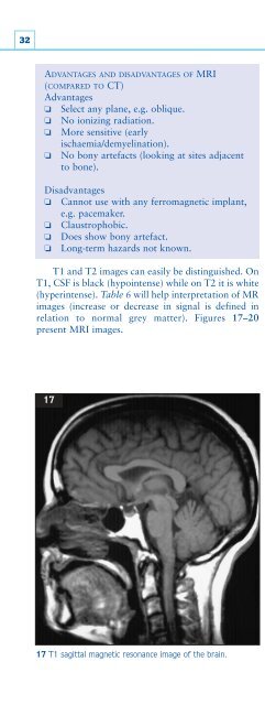
- Page 32: 16MemoryMemory is not a unitary fun
- Page 36: 18Difficulties in higher order visu
- Page 40: 20A standardized test such as the M
- Page 44: 22The facial nerve also supplies th
- Page 48: 24Upper limbs❏ Flexion at the elb
- Page 52: 26Two point discriminationAn opened
- Page 56: 28INTRODUCTIONThe evaluation of a c
- Page 60: 30Knowledge of the display and norm
- Page 66: Neurological investigations 3319a-d
- Page 70: Neurological investigations 35Diffu
- Page 74: Neurological investigations 3727 28
- Page 78: Neurological investigations 39major
- Page 82: Neurological investigations 41Clini
- Page 86: Neurological investigations 43In ad
- Page 90: Neurological investigations 45Orien
- Page 94: Neurological investigations 47SPECI
- Page 98: Neurological investigations 4943Ver
- Page 102: Neurological investigations 51SPINA
- Page 106: Neurological investigations 5351 Di
- Page 110: Neurological investigations 55Repet
- Page 114:
Neurological investigations 57Type
- Page 118:
Neurological investigations 59inves
- Page 122:
Neurological investigations 61Norma
- Page 126:
CHAPTER 3:THE PROBLEMS63DISORDERS O
- Page 130:
Disorders of consciousness 65contra
- Page 134:
Disorders of consciousness 67sinus
- Page 138:
Disorders of consciousness 69exampl
- Page 142:
Disorders of consciousness 71partic
- Page 146:
Disorders of consciousness 73allowi
- Page 150:
Disorders of consciousness75CASE 3A
- Page 154:
Disorders of consciousness 77mainta
- Page 158:
Disorders of consciousness 79Emotio
- Page 162:
Disorders of consciousness 81CLINIC
- Page 166:
Disorders of consciousness 83CASE 2
- Page 170:
Disorders of consciousness 8579CASE
- Page 174:
Disorders of cognition 87and nondom
- Page 178:
Disorders of cognition 89whereas th
- Page 182:
Disorders of cognition 91CLINICAL S
- Page 186:
Disorders of cognition 93CASE 3 (co
- Page 190:
Disorders of cognition 9587 Diagram
- Page 194:
Disorders of cognition 97Conduction
- Page 198:
Disorders of cognition 99with speec
- Page 202:
Disorders of cognition 101CASE 2A 6
- Page 206:
Disorders of cognition 103REVISION
- Page 210:
Disorders of special senses 105Left
- Page 214:
Disorders of special senses 107Past
- Page 218:
Disorders of special senses109occlu
- Page 222:
Disorders of special senses 11199a9
- Page 226:
Disorders of special senses113CLINI
- Page 230:
Disorders of special senses 115CASE
- Page 234:
Disorders of special senses 117103a
- Page 238:
Disorders of special senses 1191051
- Page 242:
Disorders of special senses121Many
- Page 246:
Disorders of special senses123aneur
- Page 250:
Disorders of special senses 125Diff
- Page 254:
Disorders of special senses 127CLIN
- Page 258:
Disorders of special senses129CASE
- Page 262:
Disorders of special senses131DIZZI
- Page 266:
Disorders of special senses133petro
- Page 270:
Disorders of special senses135Table
- Page 274:
Disorders of special senses 137Fami
- Page 278:
Disorders of special senses 139a b
- Page 282:
Disorders of special senses 141dist
- Page 286:
Disorders of special senses 143CLIN
- Page 290:
Disorders of special senses 145CASE
- Page 294:
Disorders of special senses 147REVI
- Page 298:
Disorders of motility 149Functional
- Page 302:
Disorders of motility 151A critical
- Page 306:
Disorders of motility 153C5T2C6 T11
- Page 310:
Disorders of motility 155123a123b12
- Page 314:
Disorders of motility 157124a124b12
- Page 318:
CASE 1 (continued)Disorders of moti
- Page 322:
Disorders of motility 161CASE 3A 57
- Page 326:
Disorders of motility 163TREMOR AND
- Page 330:
Disorders of motility 165SPECIFIC T
- Page 334:
Disorders of motility 167Cerebellar
- Page 338:
Disorders of motility 169CLINICAL A
- Page 342:
Disorders of motility 171CLINICAL S
- Page 346:
Disorders of motility 173CASE 2The
- Page 350:
Disorders of motility 175REVISION Q
- Page 354:
Disorders of motility 177posterior
- Page 358:
Disorders of motility 179Table 39 P
- Page 362:
Disorders of motility 181A positive
- Page 366:
Disorders of motility 183CASE 1 (co
- Page 370:
Disorders of motility 185139139 Mag
- Page 374:
Disorders of sensation 187Disorders
- Page 378:
Disorders of sensation 189existing
- Page 382:
Disorders of sensation 191INSIDIOUS
- Page 386:
Disorders of sensation193Retinal mi
- Page 390:
Disorders of sensation195Raised cre
- Page 394:
Disorders of sensation197Subarachno
- Page 398:
Disorders of sensation199146a147a14
- Page 402:
Disorders of sensation201CLINICAL S
- Page 406:
Disorders of sensation203REVISION Q
- Page 410:
Disorders of sensation205that will
- Page 414:
Disorders of sensation207reflex emp
- Page 418:
Disorders of sensation209Anterior s
- Page 422:
Disorders of sensation211CASE 2A pr
- Page 426:
Disorders of sensation 213REVISION
- Page 430:
Disorders of sensation 215Table 58
- Page 434:
Disorders of sensation 217Other sen
- Page 438:
Disorders of sensation 219Table 60
- Page 442:
Disorders of sensation221Nonorganic
- Page 446:
Disorders of sensation223163 Diagra
- Page 450:
Disorders of sensation225CASE 1 (co
- Page 454:
Disorders of sensation2271661671681
- Page 458:
CHAPTER 4 MULTIPLE CHOICE QUESTIONS
- Page 462:
Multiple choice questions 231DIZZIN
- Page 466:
Multiple choice questions 233f A pa
- Page 470:
Index 235IndexNote: page numbers in
- Page 474:
Index 237electronystagmography 61el
- Page 478:
Index 239oscillopsia 136pain, muscu