- Page 2:
UnderstandingNEUROLOGYa problem-ori
- Page 14:
CHAPTER 1:HISTORY TAKING ANDPHYSICA
- Page 20:
10These five questions (onset, dura
- Page 24:
12Anatomy of consciousnessConscious
- Page 28:
14The degree of psychomotor activit
- Page 32:
16MemoryMemory is not a unitary fun
- Page 36:
18Difficulties in higher order visu
- Page 40:
20A standardized test such as the M
- Page 44:
22The facial nerve also supplies th
- Page 48:
24Upper limbs❏ Flexion at the elb
- Page 52:
26Two point discriminationAn opened
- Page 56:
28INTRODUCTIONThe evaluation of a c
- Page 60:
30Knowledge of the display and norm
- Page 64:
32ADVANTAGES AND DISADVANTAGES OF M
- Page 68:
34Fluid-attenuated inversion recove
- Page 72:
3625 2625 T1 magnetic resonance ima
- Page 76:
38POSITRON EMISSION TOMOGRAPHYThe p
- Page 80:
40Images are reconstructed from the
- Page 84:
42Other altered conscious statesEEG
- Page 88:
cervical44While VIIIth nerve damage
- Page 92:
46MemoryThere are many different fo
- Page 96:
48passes superiorly above the atlas
- Page 100:
50body disease (tumours and infecti
- Page 104:
52appear clinically not involved, e
- Page 108:
54ElectromyographyIn EMG, a fine bo
- Page 112:
56to proceed to biopsy. It may be o
- Page 116:
58to present with mild vertigo or a
- Page 120:
60686969 Computed tomography scan s
- Page 124:
This page intentionally left blank
- Page 128:
64Disorders of consciousnessBLACKOU
- Page 132:
66top down, and so on. Dizziness is
- Page 136:
68EPILEPTIC SEIZURESClassification
- Page 140:
70An understanding of this pathophy
- Page 144:
72The clinical semiology of PNESPNE
- Page 148:
74CASE 2A 27-year-old female presen
- Page 152:
76ACUTE CONFUSIONAL STATESMyfanwy T
- Page 156:
78words beginning with a certain le
- Page 160:
80over days or even weeks. The soma
- Page 164:
8278CASE 1 (continued)An MI screen
- Page 168:
84CASE 2 (continued)Initial investi
- Page 172:
86Disorders of cognitionMEMORY DISO
- Page 176:
88Acute transient disorders of epis
- Page 180:
90asked to recall the three items.
- Page 184:
92CASE 2A 55-year-old female presen
- Page 188:
94SPEECH AND LANGUAGE DISORDERSJohn
- Page 192:
96Dysphonia refers to impaired voca
- Page 196:
98therefore a sign of damage to the
- Page 200:
100can be complemented by functiona
- Page 204:
102CASE 3A 55-year-old male and his
- Page 208:
104Disorders of special sensesVISUA
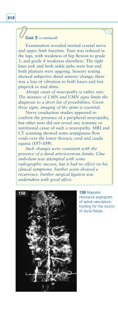
- Page 212:
106nerve at the optic chiasm. Here
- Page 216:
108may briefly double or the left s
- Page 220:
110Visual fields are assessed by th
- Page 224:
112swelling in the presence of norm
- Page 228:
114CASE 2A 62-year-old male retired
- Page 232:
116102CASE 3 (continued)The visual
- Page 236:
118DOUBLE VISIONDiplopia means doub
- Page 240:
120Cranial nervesThree nerves suppl
- Page 244:
122Myasthenia and demyelination are
- Page 248:
124and not by moving their head, c)
- Page 252:
126InvestigationsThese are guided b
- Page 256:
128CASE 2A 72-year-old male with a
- Page 260:
1303 During a focused assessment fo
- Page 264:
132though I am drunk’. Presyncope
- Page 268:
134of these two categories, and to
- Page 272:
136have a toxic cause (for example
- Page 276:
138ExaminationsFocused examinationT
- Page 280:
140heard with that perceived when t
- Page 284:
142InvestigationsThese are guided b
- Page 288:
144CASE 1 (continued)These findings
- Page 292:
146CASE 3A 34-year-old female was r
- Page 296:
148Disorders of motilityWEAKNESSRic
- Page 300:
150release of calcium from the spec
- Page 304:
152obvious limb muscle involvement)
- Page 308:
154❏❏❏Peripheral nerve: sympt
- Page 312:
156Tone, coordination, reflexes, an
- Page 316:
158CLINICAL SCENARIOSThe following
- Page 320:
160CASE 2A 65-year-old male smoker
- Page 324:
162REVISION QUESTIONS1 A lesion of
- Page 328:
164characteristic for that conditio
- Page 332:
166Many additional conditions can h
- Page 336:
168nonrhythmic, involuntary, purpos
- Page 340:
170hesitation, and reduced arm swin
- Page 344:
172CASE 1 (continued)When there is
- Page 348:
174CASE 3A 65-year-old male present
- Page 352:
176POOR COORDINATION Abhijit Chaudh
- Page 356:
178A lesion in the peripheral nerve
- Page 360:
180other type of speech is much slo
- Page 364:
182is a valuable investigation in p
- Page 368:
184CASE 3A 35-year-old female was s
- Page 372: 1867 Ataxic hemiparesis refers to:a
- Page 376: 188can be helpful in clinical pract
- Page 380: 190ExaminationA thorough, thoughtfu
- Page 384: 192Classical migraine is unilateral
- Page 388: 194implicated, particularly over-th
- Page 392: 196Low cerebrospinal fluid pressure
- Page 396: 198Cerebral venous sinus thrombosis
- Page 400: 200Acute hypertensive crisisChronic
- Page 404: 202CASE 2 (continued)She is over-us
- Page 408: 204SPINAL SYMPTOMS: NECK PAIN AND B
- Page 412: 206loss occurs when the cord is com
- Page 416: 208Brown-Séquard syndromeLesions a
- Page 420: 210CLINICAL SCENARIOS155CASE 1A 66-
- Page 426: Disorders of sensation 213REVISION
- Page 430: Disorders of sensation 215Table 58
- Page 434: Disorders of sensation 217Other sen
- Page 438: Disorders of sensation 219Table 60
- Page 442: Disorders of sensation221Nonorganic
- Page 446: Disorders of sensation223163 Diagra
- Page 450: Disorders of sensation225CASE 1 (co
- Page 454: Disorders of sensation2271661671681
- Page 458: CHAPTER 4 MULTIPLE CHOICE QUESTIONS
- Page 462: Multiple choice questions 231DIZZIN
- Page 466: Multiple choice questions 233f A pa
- Page 470: Index 235IndexNote: page numbers in
- Page 474:
Index 237electronystagmography 61el
- Page 478:
Index 239oscillopsia 136pain, muscu