Dental Asia January/February 2019
For more than two decades, Dental Asia is the premium journal in linking dental innovators and manufacturers to its rightful audience. We devote ourselves in showcasing the latest dental technology and share evidence-based clinical philosophies to serve as an educational platform to dental professionals. Our combined portfolio of print and digital media also allows us to reach a wider market and secure our position as the leading dental media in the Asia Pacific region while facilitating global interactions among our readers.
For more than two decades, Dental Asia is the premium journal in linking dental innovators
and manufacturers to its rightful audience. We devote ourselves in showcasing the latest dental technology and share evidence-based clinical philosophies to serve as an educational platform to dental professionals. Our combined portfolio of print and digital media also allows us to reach a wider market and secure our position as the leading dental media in the Asia Pacific region while facilitating global interactions among our readers.

You also want an ePaper? Increase the reach of your titles
YUMPU automatically turns print PDFs into web optimized ePapers that Google loves.
User Report<br />
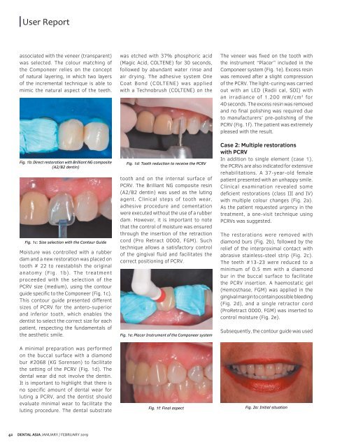
associated with the veneer (transparent)<br />
was selected. The colour matching of<br />
the Componeer relies on the concept<br />
of natural layering, in which two layers<br />
of the incremental technique is able to<br />
mimic the natural aspect of the teeth.<br />
Fig. 1b: Direct restoration with Brilliant NG composite<br />
(A2/B2 dentin)<br />
Fig. 1c: Size selection with the Contour Guide<br />
Moisture was controlled with a rubber<br />
dam and a new restoration was placed on<br />
tooth # 22 to reestablish the original<br />
anatomy (Fig. 1b). The treatment<br />
proceeded with the selection of the<br />
PCRV size (medium), using the contour<br />
guide specific to the Componeer (Fig. 1c).<br />
This contour guide presented different<br />
sizes of PCRV for the antero-superior<br />
and inferior tooth, which enables the<br />
dentist to select the correct size for each<br />
patient, respecting the fundamentals of<br />
the aesthetic smile.<br />
was etched with 37% phosphoric acid<br />
(Magic Acid, COLTENE) for 30 seconds,<br />
followed by abundant water rinse and<br />
air drying. The adhesive system One<br />
Coat Bond (COLTENE) was applied<br />
with a Technobrush (COLTENE) on the<br />
Fig. 1d: Tooth reduction to receive the PCRV<br />
tooth and on the internal surface of<br />
PCRV. The Brilliant NG composite resin<br />
(A2/B2 dentin) was used as the luting<br />
agent. Clinical steps of tooth wear,<br />
adhesive procedure and cementation<br />
were executed without the use of a rubber<br />
dam. However, it is important to note<br />
that the control of moisture was ensured<br />
through the insertion of the retraction<br />
cord (Pro Retract 0000, FGM). Such<br />
technique allows a satisfactory control<br />
of the gingival fluid and facilitates the<br />
correct positioning of PCRV.<br />
Fig. 1e: Placer Instrument of the Componeer system<br />
The veneer was fixed on the tooth with<br />
the instrument “Placer” included in the<br />
Componeer system (Fig. 1e). Excess resin<br />
was removed after a slight compression<br />
of the PCRV. The light-curing was carried<br />
out with an LED (Radii cal, SDI) with<br />
an irradiance of 1.200 mW/cm 2 for<br />
40 seconds. The excess resin was removed<br />
and no final polishing was required due<br />
to manufacturers’ pre-polishing of the<br />
PCRV (Fig. 1f). The patient was extremely<br />
pleased with the result.<br />
Case 2: Multiple restorations<br />
with PCRV<br />
In addition to single element (case 1),<br />
the PCRVs are also indicated for extensive<br />
rehabilitations. A 37-year-old female<br />
patient presented with an unhappy smile.<br />
Clinical examination revealed some<br />
deficient restorations (class III and IV)<br />
with multiple colour changes (Fig. 2a).<br />
As the patient requested urgency in the<br />
treatment, a one-visit technique using<br />
PCRVs was suggested.<br />
The restorations were removed with<br />
diamond burs (Fig. 2b), followed by the<br />
relief of the interproximal contact with<br />
abrasive stainless-steel strip (Fig. 2c).<br />
The teeth #13-23 were reduced to a<br />
minimum of 0.5 mm with a diamond<br />
bur in the buccal surface to facilitate<br />
the PCRV insertion. A haemostatic gel<br />
(Hemosthase, FGM) was applied in the<br />
gingival margin to contain possible bleeding<br />
(Fig. 2d), and a single retractor cord<br />
(ProRetract 0000, FGM) was inserted to<br />
control moisture (Fig. 2e).<br />
Subsequently, the contour guide was used<br />
A minimal preparation was performed<br />
on the buccal surface with a diamond<br />
bur #2068 (KG Sorensen) to facilitate<br />
the setting of the PCRV (Fig. 1d). The<br />
dental wear did not involve the dentin.<br />
It is important to highlight that there is<br />
no specific amount of dental wear for<br />
luting a PCRV, and the dentist should<br />
evaluate minimal wear to facilitate the<br />
luting procedure. The dental substrate<br />
Fig. 1f: Final aspect<br />
Fig. 2a: Initial situation<br />
42<br />
DENTAL ASIA JANUARY / FEBRUARY <strong>2019</strong>