

Dental Asia January/February 2019
For more than two decades, Dental Asia is the premium journal in linking dental innovators and manufacturers to its rightful audience. We devote ourselves in showcasing the latest dental technology and share evidence-based clinical philosophies to serve as an educational platform to dental professionals. Our combined portfolio of print and digital media also allows us to reach a wider market and secure our position as the leading dental media in the Asia Pacific region while facilitating global interactions among our readers.
For more than two decades, Dental Asia is the premium journal in linking dental innovators
and manufacturers to its rightful audience. We devote ourselves in showcasing the latest dental technology and share evidence-based clinical philosophies to serve as an educational platform to dental professionals. Our combined portfolio of print and digital media also allows us to reach a wider market and secure our position as the leading dental media in the Asia Pacific region while facilitating global interactions among our readers.

You also want an ePaper? Increase the reach of your titles
YUMPU automatically turns print PDFs into web optimized ePapers that Google loves.
User Report<br />
Abstract<br />
A 47-year-old male<br />
presented with pain in the<br />
temporomandibular joint (TMJ).<br />
He also had an aesthetic request<br />
since part of his veneer on<br />
the middle upper incisors had<br />
broken off. Fully digitalised work<br />
steps were used to remedy this<br />
issue: the Digital Smile Design<br />
protocol was applied and,<br />
following a minimally invasive<br />
preparation, monolithic veneers<br />
and crowns made of lithium disilicate<br />
ceramic were produced by means of<br />
CAD/CAM. The aim of rehabilitation was<br />
to remedy the loss of bite height as well<br />
as the associated aesthetic and jaw joint<br />
impairments.<br />
Background<br />
The digitalisation of work steps in<br />
dentistry has gained ground in recent<br />
years due to the technical advances<br />
with regards to intraoral scanners and<br />
software programs. This development has<br />
also resulted in improved communication<br />
between the dentist and dental technician.<br />
Digital Smile Design (DSD) is a digital tool<br />
for planning the aesthetic restoration<br />
of facial symmetry. It not only aids<br />
communication between specialists, but<br />
also improves the treatment results which<br />
can be expected. 1 Dynamic documentation<br />
of the smile is an important step in the<br />
2D/3D Digital Smile Design process. The<br />
process can be fully digitalised and also<br />
supports the rehabilitation procedure. The<br />
advantages of video documentation lie in<br />
the fact that this renders documentation,<br />
the Smile Design, the analysis of the facial<br />
symmetry, treatment planning, team<br />
communication and patient education<br />
both more simple and more effective. 2 The<br />
DSD can be converted into a conventional<br />
or virtual diagnostic model to simplify<br />
the subsequent clinical treatments, e.g.<br />
CAD/CAM restoration. 3–7 The combination<br />
of the adhesive technique with lighttransmissive<br />
restoratives makes preparing<br />
for minimally invasive restorative dentistry<br />
interventions simpler. Materials such<br />
as lithium-disilicate ceramic 8–11 boast<br />
Correction of Bite Height:<br />
Fully Digitalised Work Steps,<br />
Integration of the <strong>Dental</strong> Scanner,<br />
Smile Design and CAD/CAM<br />
In this report, a clinical case is represented with<br />
fully digitalised work steps.<br />
By Dr. Miguel Stanley, Dr. Ana Gomes Paz, Dr. Inês Miguel<br />
and Dr. Christian Coachman<br />
similar properties to natural teeth which,<br />
in turn, enabled positive results to be<br />
achieved. 12-13<br />
Intraoral scanners are an important tool<br />
in the digital workflow. These handy<br />
devices allow the impression quality to<br />
be checked directly and, in addition,<br />
the models can be simply transferred,<br />
cost-effectively and quickly via e-mail to<br />
the laboratory. 14 However, there is little<br />
information in the literature about the<br />
ability of intraoral scanners to produce<br />
high-quality impressions. 15–24<br />
Computer-aided design (CAD) software<br />
is invaluable as it controls the fully<br />
automated devices which create the<br />
objects and assemblies in a virtual<br />
environment. 25<br />
In this report, a clinical case is represented<br />
with fully digitalised work steps. Following<br />
minimally invasive preparation, the<br />
Digital Smile protocol and the monolithic<br />
veneers and crowns made from lithiumdisilicate<br />
ceramic remedy the loss of bite<br />
height as well as the associated aesthetic and<br />
jaw joint impairments through CAD/CAM.<br />
Case presentation<br />
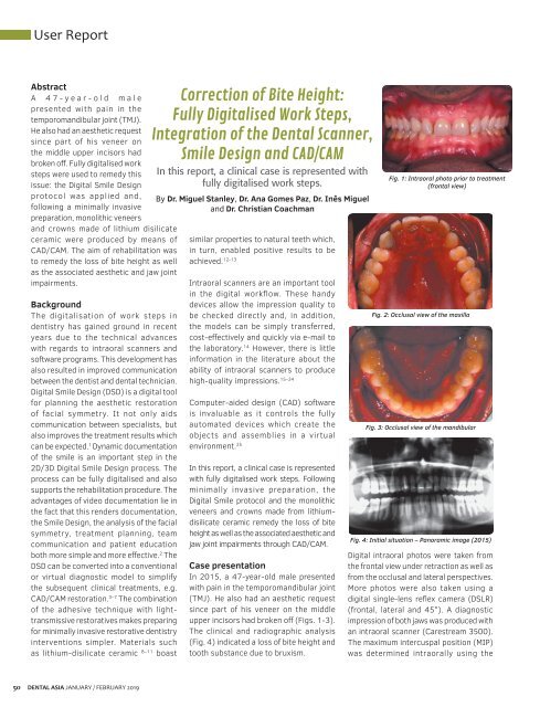
In 2015, a 47-year-old male presented<br />
with pain in the temporomandibular joint<br />
(TMJ). He also had an aesthetic request<br />
since part of his veneer on the middle<br />
upper incisors had broken off (Figs. 1-3).<br />
The clinical and radiographic analysis<br />
(Fig. 4) indicated a loss of bite height and<br />
tooth substance due to bruxism.<br />
Fig. 1: Intraoral photo prior to treatment<br />
(frontal view)<br />
Fig. 2: Occlusal view of the maxilla<br />
Fig. 3: Occlusal view of the mandibular<br />
Fig. 4: Initial situation – Panoramic image (2015)<br />
Digital intraoral photos were taken from<br />
the frontal view under retraction as well as<br />
from the occlusal and lateral perspectives.<br />
More photos were also taken using a<br />
digital single-lens reflex camera (DSLR)<br />
(frontal, lateral and 45°). A diagnostic<br />
impression of both jaws was produced with<br />
an intraoral scanner (Carestream 3500).<br />
The maximum intercuspal position (MIP)<br />
was determined intraorally using the<br />
50<br />
DENTAL ASIA JANUARY / FEBRUARY <strong>2019</strong>


