Dental Asia January/February 2019
For more than two decades, Dental Asia is the premium journal in linking dental innovators and manufacturers to its rightful audience. We devote ourselves in showcasing the latest dental technology and share evidence-based clinical philosophies to serve as an educational platform to dental professionals. Our combined portfolio of print and digital media also allows us to reach a wider market and secure our position as the leading dental media in the Asia Pacific region while facilitating global interactions among our readers.
For more than two decades, Dental Asia is the premium journal in linking dental innovators
and manufacturers to its rightful audience. We devote ourselves in showcasing the latest dental technology and share evidence-based clinical philosophies to serve as an educational platform to dental professionals. Our combined portfolio of print and digital media also allows us to reach a wider market and secure our position as the leading dental media in the Asia Pacific region while facilitating global interactions among our readers.

You also want an ePaper? Increase the reach of your titles
YUMPU automatically turns print PDFs into web optimized ePapers that Google loves.
User Report<br />
Guided Concept in the<br />
Aesthetic Zone<br />
By Dr. Stefen Koubi and MDT Gérald Ubassy<br />
Introduction<br />
The restorative treatment of anterior<br />
teeth often presents a considerable<br />
challenge to general dentists. The aim<br />
is always to obtain the best possible<br />
result. A well-guided approach is<br />
necessary if the visualised outcome is<br />
to be achieved. Precision planning and<br />
a consistent protocol are indispensable.<br />
Modern dentistry has simplified the<br />
ways and means of attaining aesthetic<br />
results. Nevertheless, the success of<br />
the treatment, in the anterior region in<br />
particular, greatly depends on meticulous<br />
planning, which includes a detailed<br />
analysis of the patient’s smile as well as<br />
the fabrication of a working model. This<br />
model is used to plan and reproduce<br />
the shapes and contours of the future<br />
restoration with utmost precision. This<br />
article describes a comparatively simple<br />
treatment technique on the basis of<br />
a clinical case. The initial aesthetic<br />
treatment plan was jointly developed by<br />
the dental technician and the dentist. It<br />
served as a guide or “GPS” for all the<br />
clinical steps which helped the dental<br />
team to successfully “navigate” through<br />
the treatment.<br />
Clinical case presentation<br />
A woman in her forties consulted our<br />
practice due to extreme mobility of her<br />
front teeth, which had caused aesthetic<br />
problems (Figs. 1-2). The teeth had moved<br />
forward and the level of the smile line had<br />
dropped. Due to severe periodontitis, the<br />
upper front teeth showed considerable<br />
gingival recession (Fig. 3). A detailed<br />
examination revealed that the four upper<br />
front teeth could not be saved and had to<br />
be extracted. The treatment plan was to<br />
insert two implants in the sockets of tooth<br />
12 and 22 after the extraction process.<br />
Subsequently, an implant-supported<br />
bridge extending from tooth 12 to 22<br />
would be fabricated and tooth 13 and<br />
23 would be restored with single crowns.<br />
The main objective was to restore the<br />
harmonious appearance of the smile line<br />
and the convex shape of the gingiva. The<br />
patient’s smile was analysed by means<br />
of a photographic record. Furthermore,<br />
impressions of the situation were taken.<br />
Based on this information, a provisional<br />
bridge spanning from tooth 13 to 23 and<br />
incorporating the aesthetic and functional<br />
adjustments would be produced with<br />
PMMA (polymethyl methacrylate) material<br />
(Telio ® CAD).<br />
Fig. 1: The patient was dissatisfied with her smile<br />
Fig. 2: The anterior teeth were damaged due to<br />
periodontitis and moved labially and extruded.<br />
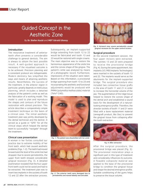
Fig. 3: Intraoral view: severe periodontitis caused<br />
gingival recession for the upper central incisors<br />
Surgical procedure<br />
At the second treatment session, the<br />
four upper incisors were extracted.<br />
The canines 13 and 23 were prepared<br />
to receive the provisional bridge<br />
(Fig. 4). During the same appointment, two<br />
implants (V3, MIS Implants Technologies)<br />
were inserted in the sockets of tooth 12<br />
and 22. The implants would serve as the<br />
abutments for the implant-supported<br />
bridge. The surgical procedure also<br />
included two connective tissue grafts<br />
in the area of tooth 11 and 21 in order<br />
to increase the horizontal volume of the<br />
jaw. The augmentation of the ridge tissue<br />
helped to restore the convex shape of<br />
the dental arch and established a sound<br />
basis for the development of a naturallooking<br />
emergence profile. Therefore, the<br />
alveolar sockets of tooth 11 and 21 were<br />
filled with a bone replacement material<br />
(xenograft product, Bio Oss) to prevent<br />
the gingival tissue from collapsing after<br />
the tooth extraction.<br />
Fig. 4: After extraction<br />
After the surgical procedure, the<br />
provisional bridge was placed (Fig. 5).<br />
The provisional restoration plays a<br />
significant part in this type of treatment<br />
and considerably influences its outcome.<br />
A flowable composite material was<br />
applied to the base of the provisional<br />
bridge to condition the gingival tissue<br />
and shape the desired emergence profile.<br />
Suitable conditioning of the gums helps<br />
to preserve the tissue volume.<br />
46<br />
DENTAL ASIA JANUARY / FEBRUARY <strong>2019</strong>