

Dental Asia January/February 2019
For more than two decades, Dental Asia is the premium journal in linking dental innovators and manufacturers to its rightful audience. We devote ourselves in showcasing the latest dental technology and share evidence-based clinical philosophies to serve as an educational platform to dental professionals. Our combined portfolio of print and digital media also allows us to reach a wider market and secure our position as the leading dental media in the Asia Pacific region while facilitating global interactions among our readers.
For more than two decades, Dental Asia is the premium journal in linking dental innovators
and manufacturers to its rightful audience. We devote ourselves in showcasing the latest dental technology and share evidence-based clinical philosophies to serve as an educational platform to dental professionals. Our combined portfolio of print and digital media also allows us to reach a wider market and secure our position as the leading dental media in the Asia Pacific region while facilitating global interactions among our readers.

Create successful ePaper yourself
Turn your PDF publications into a flip-book with our unique Google optimized e-Paper software.
Behind the Scenes<br />
the teeth can be rotated individually along<br />
their labial axis or in chain mode in the<br />
“Rotate” mode. If, for example, individual<br />
or all four maxillary anterior teeth are<br />
to face in a vestibular direction with the<br />
incisal edge, these can be “grabbed” with<br />
the mouse and moved seamlessly in the<br />
desired direction. If the model analysis is<br />
superimposed, as shown here in Fig. 9, the<br />
set-up can also be adapted individually to<br />
its specifications.<br />
Occlusal and vestibular view<br />
The possibilities of customisation can<br />
also be viewed from the occlusal and<br />
extended additionally. One should make<br />
sure that the anterior and posterior<br />
teeth are located in their regions of the<br />
model analysis. As a rule and as already<br />
mentioned, this is done after the selection<br />
and the command “Set-up: anterior and<br />
posterior teeth”. Figure 10a shows the<br />
posterior teeth exactly in the set-up<br />
region. The mandibular anterior teeth<br />
are rotated slightly along their vertical<br />
axes here. The centre is now shifted<br />
slightly to the right due to the rotation of<br />
tooth 43, which reduces the dental arch<br />
on the right side. The chain mode (see<br />
turquoise bar) then shifts the entire row of<br />
front teeth marginally to the right. Now, of<br />
course, this situation can be customised<br />
additionally.<br />
Fig. 10a: Representation of occlusal placement: here<br />
one can check that the anterior and posterior teeth<br />
are positioned in their regions of the model analysis.<br />
The anterior teeth in the mandible are discreetly<br />
rotated along their vertical axes. The centre is shifted<br />
slightly to the right, which can of course be adjusted<br />
individually with the aid of the chain mode.<br />
The mandibular anterior teeth were<br />
customised somewhat more. This was<br />
done at the patient‘s request and was<br />
agreed in advance. In the process, the<br />
anatomical centre could also be corrected<br />
back to its original position. The result<br />
was that the region 43 to 46 rotated to<br />
vestibular. This now had to be rotated<br />
axially around tooth 44 to lingual.<br />
To recap: the front teeth can be shifted<br />
both in single or chain mode. The<br />
occlusion, which was corrected at this<br />
point in the mandible, is automatically<br />
corrected to the optimum static occlusion<br />
in the maxilla (Fig. 10b).<br />
Fig. 10b: At the patient‘s request, the mandibular<br />
anterior teeth were customised a little more. During<br />
this process, the anatomical centre could also be<br />
corrected back to its original position. The result<br />
was that region 43 to 46 had rotated to vestibular.<br />
Finally, this needed to be rotated axially to lingual<br />
around tooth 44.<br />
The maxillary set-up of this work after<br />
correction: here the anterior teeth<br />
basically remained in an even arch. Only<br />
the tooth axes of the lateral incisors were<br />
inclined along their vestibular tooth axis.<br />
Fig. 11: The set-up in the maxilla after correction.<br />
The anterior teeth were essentially left in an even<br />
arch. Only the tooth axes of the lateral incisors<br />
were inclined along their vestibular tooth axis (see<br />
Figs. 56 and 66). The anterior arch had to be<br />
harmonised evenly in the sagittal step.<br />
In addition, the anterior dental arch had<br />
to be harmonised evenly in its sagittal<br />
step (Fig. 11).<br />
The milling process<br />
Fig. 12a<br />
Fig. 12b<br />
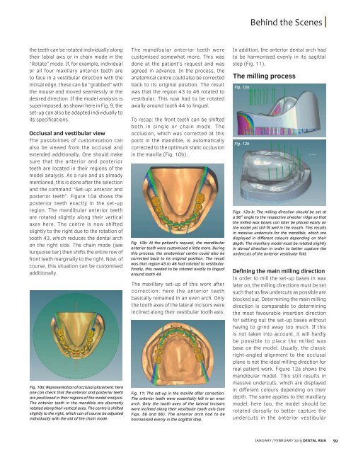
Figs. 12a-b: The milling direction should be set at<br />
a 90° angle to the respective alveolar ridge so that<br />
the milled wax bases can later be placed easily on<br />
the model yet still fit well in the mouth. This results<br />
in massive undercuts for the mandible, which are<br />
displayed in different colours depending on their<br />
depth. The maxillary model must be rotated slightly<br />
in dorsal direction in order to better capture the<br />
undercuts of the anterior vestibular fold.<br />
Defining the main milling direction<br />
In order to mill the set-up bases in wax<br />
later on, the milling directions must be set<br />
such that as few undercuts as possible are<br />
blocked out. Determining the main milling<br />
direction is comparable to determining<br />
the most favourable insertion direction<br />
for setting out the set-up bases without<br />
having to grind away too much. If this<br />
is not taken into account, it will hardly<br />
be possible to place the milled wax<br />
base on the model. Usually, the classic<br />
right-angled alignment to the occlusal<br />
plane is not the ideal milling direction for<br />
real patient work. Figure 12a shows the<br />
mandibular model. This still results in<br />
massive undercuts, which are displayed<br />
in different colours depending on their<br />
depth. The same applies to the maxillary<br />
model: here too, the model should be<br />
rotated dorsally to better capture the<br />
undercuts in the anterior vestibular<br />
JANUARY / FEBRUARY <strong>2019</strong> DENTAL ASIA 59


