

Dental Asia January/February 2019
For more than two decades, Dental Asia is the premium journal in linking dental innovators and manufacturers to its rightful audience. We devote ourselves in showcasing the latest dental technology and share evidence-based clinical philosophies to serve as an educational platform to dental professionals. Our combined portfolio of print and digital media also allows us to reach a wider market and secure our position as the leading dental media in the Asia Pacific region while facilitating global interactions among our readers.
For more than two decades, Dental Asia is the premium journal in linking dental innovators
and manufacturers to its rightful audience. We devote ourselves in showcasing the latest dental technology and share evidence-based clinical philosophies to serve as an educational platform to dental professionals. Our combined portfolio of print and digital media also allows us to reach a wider market and secure our position as the leading dental media in the Asia Pacific region while facilitating global interactions among our readers.

You also want an ePaper? Increase the reach of your titles
YUMPU automatically turns print PDFs into web optimized ePapers that Google loves.
User Report<br />
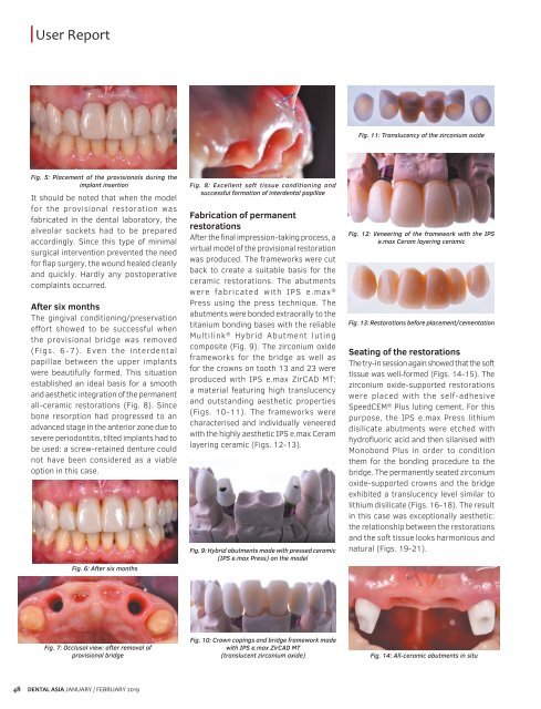
Fig. 11: Translucency of the zirconium oxide<br />
Fig. 5: Placement of the provisionals during the<br />
implant insertion<br />
It should be noted that when the model<br />
for the provisional restoration was<br />
fabricated in the dental laboratory, the<br />
alveolar sockets had to be prepared<br />
accordingly. Since this type of minimal<br />
surgical intervention prevented the need<br />
for flap surgery, the wound healed cleanly<br />
and quickly. Hardly any postoperative<br />
complaints occurred.<br />
After six months<br />
The gingival conditioning/preservation<br />
effort showed to be successful when<br />
the provisional bridge was removed<br />
(Figs. 6-7). Even the interdental<br />
papillae between the upper implants<br />
were beautifully formed. This situation<br />
established an ideal basis for a smooth<br />
and aesthetic integration of the permanent<br />
all-ceramic restorations (Fig. 8). Since<br />
bone resorption had progressed to an<br />
advanced stage in the anterior zone due to<br />
severe periodontitis, tilted implants had to<br />
be used: a screw-retained denture could<br />
not have been considered as a viable<br />
option in this case.<br />
Fig. 6: After six months<br />
Fig. 8: Excellent soft tissue conditioning and<br />
successful formation of interdental papillae<br />
Fabrication of permanent<br />
restorations<br />
After the final impression-taking process, a<br />
virtual model of the provisional restoration<br />
was produced. The frameworks were cut<br />
back to create a suitable basis for the<br />
ceramic restorations. The abutments<br />
were fabricated with IPS e.max ®<br />
Press using the press technique. The<br />
abutments were bonded extraorally to the<br />
titanium bonding bases with the reliable<br />
Multilink ® Hybrid Abutment luting<br />
composite (Fig. 9). The zirconium oxide<br />
frameworks for the bridge as well as<br />
for the crowns on tooth 13 and 23 were<br />
produced with IPS e.max ZirCAD MT:<br />
a material featuring high translucency<br />
and outstanding aesthetic properties<br />
(Figs. 10-11). The frameworks were<br />
characterised and individually veneered<br />
with the highly aesthetic IPS e.max Ceram<br />
layering ceramic (Figs. 12-13).<br />
Fig. 9: Hybrid abutments made with pressed ceramic<br />
(IPS e.max Press) on the model<br />
Fig. 12: Veneering of the framework with the IPS<br />
e.max Ceram layering ceramic<br />
Fig. 13: Restorations before placement/cementation<br />
Seating of the restorations<br />
The try-in session again showed that the soft<br />
tissue was well-formed (Figs. 14-15). The<br />
zirconium oxide-supported restorations<br />
were placed with the self-adhesive<br />
SpeedCEM ® Plus luting cement. For this<br />
purpose, the IPS e.max Press lithium<br />
disilicate abutments were etched with<br />
hydrofluoric acid and then silanised with<br />
Monobond Plus in order to condition<br />
them for the bonding procedure to the<br />
bridge. The permanently seated zirconium<br />
oxide-supported crowns and the bridge<br />
exhibited a translucency level similar to<br />
lithium disilicate (Figs. 16-18). The result<br />
in this case was exceptionally aesthetic:<br />
the relationship between the restorations<br />
and the soft tissue looks harmonious and<br />
natural (Figs. 19-21).<br />
Fig. 7: Occlusal view: after removal of<br />
provisional bridge<br />
Fig. 10: Crown copings and bridge framework made<br />
with IPS e.max ZirCAD MT<br />
(translucent zirconium oxide)<br />
Fig. 14: All-ceramic abutments in situ<br />
48<br />
DENTAL ASIA JANUARY / FEBRUARY <strong>2019</strong>


