Biotechnology in Animal Husbandry - Institut za Stočarstvo
Biotechnology in Animal Husbandry - Institut za Stočarstvo
Biotechnology in Animal Husbandry - Institut za Stočarstvo

Create successful ePaper yourself
Turn your PDF publications into a flip-book with our unique Google optimized e-Paper software.
200<br />
Results and Discussion<br />
V. Gerzilov et al.<br />
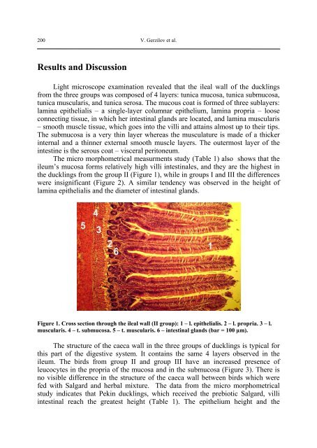
Light microscope exam<strong>in</strong>ation revealed that the ileal wall of the duckl<strong>in</strong>gs<br />
from the three groups was composed of 4 layers: tunica mucosa, tunica submucosa,<br />
tunica muscularis, and tunica serosa. The mucous coat is formed of three sublayers:<br />
lam<strong>in</strong>a epithelialis – a s<strong>in</strong>gle-layer columnar epithelium, lam<strong>in</strong>a propria – loose<br />
connect<strong>in</strong>g tissue, <strong>in</strong> which her <strong>in</strong>test<strong>in</strong>al glands are located, and lam<strong>in</strong>a muscularis<br />
– smooth muscle tissue, which goes <strong>in</strong>to the villi and atta<strong>in</strong>s almost up to their tips.<br />
The submucosa is a very th<strong>in</strong> layer whereas the musculature is made of a thicker<br />
<strong>in</strong>ternal and a th<strong>in</strong>ner external smooth muscle layers. The outermost layer of the<br />
<strong>in</strong>test<strong>in</strong>e is the serous coat – visceral peritoneum.<br />
The micro morphometrical measurments study (Table 1) also shows that the<br />
ileum’s mucosa forms relatively high villi <strong>in</strong>test<strong>in</strong>ales, and they are the highest <strong>in</strong><br />
the duckl<strong>in</strong>gs from the group II (Figure 1), while <strong>in</strong> groups I and III the differences<br />
were <strong>in</strong>significant (Figure 2). A similar tendency was observed <strong>in</strong> the height of<br />
lam<strong>in</strong>a epithelialis and the diameter of <strong>in</strong>test<strong>in</strong>al glands.<br />
Figure 1. Cross section through the ileal wall (II group): 1 – l. epithelialis. 2 – l. propria. 3 – l.<br />
muscularis. 4 – t. submucosa. 5 – t. muscularis. 6 – <strong>in</strong>test<strong>in</strong>al glands (bar = 100 µm).<br />
The structure of the caeca wall <strong>in</strong> the three groups of duckl<strong>in</strong>gs is typical for<br />
this part of the digestive system. It conta<strong>in</strong>s the same 4 layers observed <strong>in</strong> the<br />
ileum. The birds from group II and group III have an <strong>in</strong>creased presence of<br />
leucocytes <strong>in</strong> the propria of the mucosa and <strong>in</strong> the submucosa (Figure 3). There is<br />
no visible difference <strong>in</strong> the structure of the caeca wall between birds which were<br />
fed with Salgard and herbal mixture. The data from the micro morphometrical<br />
study <strong>in</strong>dicates that Pek<strong>in</strong> duckl<strong>in</strong>gs, which received the prebiotic Salgard, villi<br />
<strong>in</strong>test<strong>in</strong>al reach the greatest height (Table 1). The epithelium height and the