Klinische (bio)chemie en methodologie - NVKC
Klinische (bio)chemie en methodologie - NVKC
Klinische (bio)chemie en methodologie - NVKC
You also want an ePaper? Increase the reach of your titles
YUMPU automatically turns print PDFs into web optimized ePapers that Google loves.
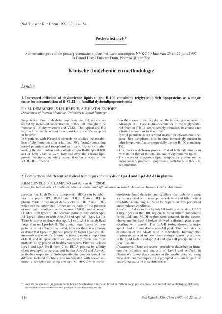
Ned Tijdschr Klin Chem 1997; 22: 114-154<br />
Posterabstracts*<br />
Sam<strong>en</strong>vatting<strong>en</strong> van de posterpres<strong>en</strong>taties tijd<strong>en</strong>s het Lustrumcongres <strong>NVKC</strong> 50 Jaar van 25 tot 27 juni 1997<br />
in Grand Hotel Huis ter Duin, Noordwijk aan Zee<br />
Lipid<strong>en</strong><br />
Subjects with familial dysbetalipoproteinemia (FD) are characterized<br />
by increased conc<strong>en</strong>trations of ß-VLDL thought to be<br />
“remnants” of chylomicrons and VLDL. The typical apo E-2<br />
isoprotein is unable to bind these particles to specific receptors<br />
in the liver.<br />
In 8 pati<strong>en</strong>ts with FD and 6 controls we studied the metabolism<br />
of chylomicrons after a fat load (50 g fat/m2) containing<br />
retinyl palmitate and tocopherol as tracers. Up to 48 h after<br />
loading the distribution and cont<strong>en</strong>ts of apo B-48, apo B-100,<br />
and of both vitamins were followed over the various lipoprotein<br />
fractions, including some flotation classes of the<br />
VLDL+IDL fraction.<br />
<strong>Klinische</strong> (<strong>bio</strong>)<strong>chemie</strong> <strong>en</strong> <strong>methodologie</strong><br />
1. Increased diffusion of chylomicron lipids to apo B-100 containing triglyceride-rich lipoproteins as a major<br />
cause for accumulation of ß-VLDL in familial dysbetalipoproteinemia<br />
P.N.M. DEMACKER, S.J.H. BREDIE, A.F.H. STALENHOEF<br />
Departm<strong>en</strong>t of Internal Medicine, University Hospital Nijmeg<strong>en</strong><br />
From these experim<strong>en</strong>ts we derived the following conclusions:<br />
- Although in FD apo B-48 conc<strong>en</strong>tation in the triglyceriderich<br />
fraction (TRL) is considerably increased, its course after<br />
a limited amount of fat is normal.<br />
- Retinyl palmitate is not a valid marker for chylomicrons because,<br />
like tocopherol, it is in time increasingly pres<strong>en</strong>t in<br />
other lipoprotein fractions especially the apo B-100 containing<br />
TRL.<br />
- This marks a diffusion process; that of both vitamins is an<br />
estimate for that of the total amount of chylomicron lipids.<br />
- The excess of exog<strong>en</strong>ous lipid, temporarily pres<strong>en</strong>t on the<br />
<strong>en</strong>dog<strong>en</strong>ously produced lipoproteins, contributes to ß-VLDL<br />
accumulation.<br />
2. Comparison of differ<strong>en</strong>t analytical techniques of analysis of LpA-I and LpA-I/A-II in plasma<br />
J.H.M LEVELS, R.J. LAMPING and A. van d<strong>en</strong> ENDE<br />
C<strong>en</strong>tre for Hemostasis, Thrombosis, Atherosclerosis and Inflammation Research, Academic Medical C<strong>en</strong>tre, Amsterdam<br />
Introduction. High D<strong>en</strong>sity Lipoprotein (HDL) can be subdivided<br />
in pre-b HDL, HDL2 and HDL3. Mature HDL in<br />
plasma exists in two major d<strong>en</strong>sity classes, HDL2 and HDL3<br />
which can be subdivided further on the basis of the pres<strong>en</strong>ce<br />
of two major apolipoproteins, Apo-AI (28kD) and Apo -AII<br />
(17 kD). Both types of HDL contain particles with either Apo-<br />
AI (LpA-I) alone or with Apo-AI and Apo-AII (LpA-I/A-II).<br />
There is strong evid<strong>en</strong>ce that apoA-I on LpA-I is catabolized<br />
faster than on LpA-I/A-II. The clinical significance of these<br />
particles is not <strong>en</strong>tirely elucidated, however there is a growing<br />
evid<strong>en</strong>ce that LpA-I might be a protective factor against CHD.<br />
Materials and methods. In order to investigate the composition<br />
of HDL and its apo cont<strong>en</strong>t we compared differ<strong>en</strong>t analytical<br />
methods using plasma of healthy volonteers. First we isolated<br />
LpA-I and LpA-I/A-II from 2 ml EDTA plasma by affinity<br />
chromatography using goat-anti-human Apo-AI and Apo-AII<br />
antibodies respectively. Subsequ<strong>en</strong>tly, the composition of the<br />
differ<strong>en</strong>t isolated fractions was investigated with rocket immuno-<br />
electrophoresis using anti apo-AI, HPGC with choles-<br />
terol postcolumn detection and capillary electrophoresis using<br />
a column coated with linear polyacrylamide and filled with a<br />
tris-buffer containing 0.1 % SDS. Separation was performed<br />
under reduced conditions.<br />
Results. LpA-I as well as LpA-I/AII isolates showed on HPGC<br />
a major peak in the HDL region, however minor compon<strong>en</strong>ts<br />
in the LDL and VLDL region were detected. In the electropherogram<br />
the LpA-I isolate showed a distinct peak corresponding<br />
with apo-AI. The LpA-II isolate showed a major<br />
apo-AI and a minor double apo-AII peak. This facilitates the<br />
calculation of the AI/AII ratio in individuals. Immuno-electrophoresis<br />
showed in most cases a single apo-AI precipitate<br />
in the LpAI isolate and apo A-I and apo A-II precipitate in the<br />
Lpa-II isolate.<br />
Conclusions. There are several procedures described in literature<br />
for isolation and analysis of LpA-I and A-I/A-II in<br />
plasma.We found discrepancies in the results obtained using<br />
these differ<strong>en</strong>t techniques. This prompted us to investigate the<br />
underlying cause of these differ<strong>en</strong>ces.<br />
* Voor de pres<strong>en</strong>tatie zijn g<strong>en</strong>ummerde bord<strong>en</strong> beschikbaar van 85 cm breed <strong>en</strong> 240 cm hoog; posters di<strong>en</strong><strong>en</strong> uitsluit<strong>en</strong>d met dubbelzijdig plakband,<br />
dat ter plekke beschikbaar wordt gesteld, te word<strong>en</strong> aangebracht.<br />
114 Ned Tijdschr Klin Chem 1997, vol. 22, no. 3
3. Unusual test for measuring Lp(a) levels in serum: a time-resolved immunofluoroimmunometric assay<br />
M.A.M. BON, G. van der SLUIJS VEER, I. VERMES<br />
Medisch Spectrum Tw<strong>en</strong>te, Hospital Group, Enschede, The Netherlands<br />
Lp(a) is a cholesterol-ester-rich lipoprotein that resembles low<br />
d<strong>en</strong>sity lipoprotein (LDL). Lp(a) contains apolipoprotein B-<br />
100 and glycoprotein(a) which is coval<strong>en</strong>tly bound to the<br />
apoB-100.<br />
The molecular mass of Lp(a) ranges from 300 through 800<br />
Dalton. A strong and indep<strong>en</strong>d<strong>en</strong>t relationship exists betwe<strong>en</strong><br />
the serum conc<strong>en</strong>trations of Lp(a) and the incid<strong>en</strong>ce of atherosclerotic<br />
vascular disease. There are several methods to determine<br />
the Lp(a) levels in serum. Mostly these methods are time<br />
consuming and difficult to perform in large series. In addition,<br />
all of these assays are influ<strong>en</strong>ced by matrix effects.<br />
We attempted to set up an assay to determine the Lp(a) levels<br />
in serum with use of a Time Resolved Fluoroimmunometric<br />
Assay (TRIFMA) as used in the DELFIAtm system (Wallac<br />
Oy, Turku, Finland). In this TRIFMA we used a 70 times<br />
higher dilution factor which diminishes matrix effects. Although<br />
use of an immunometric assay is an unusual application<br />
to measure such high conc<strong>en</strong>trations of circulating protein,<br />
the high dilution factor has b<strong>en</strong>efits.<br />
Enzym<strong>en</strong><br />
4. The activity of dihydropyrimidine dehydrog<strong>en</strong>ase in human blood cells<br />
Dihydropyrimidine dehydrog<strong>en</strong>ase (DPD) is the initial and<br />
rate-limiting <strong>en</strong>zyme in the catabolism of the pyrimidine bases<br />
thymine and uracil. DPD is also responsible for the breakdown<br />
of 5-fluorouracil, thereby limiting the efficacy of the therapy.<br />
Pati<strong>en</strong>ts suffering from a defici<strong>en</strong>cy of this <strong>en</strong>zyme do not exhibit<br />
a characteristic clinical ph<strong>en</strong>otype, although in childr<strong>en</strong><br />
the defici<strong>en</strong>cy of DPD is oft<strong>en</strong> accompanied by a neurological<br />
disorder. The diagnosis of pati<strong>en</strong>ts with a DPD defici<strong>en</strong>cy can<br />
be established by measurem<strong>en</strong>t of the activity of DPD in<br />
leukocytes. So far, it was not known whether DPD is pres<strong>en</strong>t<br />
in all types of human blood cells. Therefore, we purified human<br />
monocytes, lymphocytes, granulocytes, platelets and red<br />
blood cells from healthy donors with elutriation and determined<br />
the activity of DPD with a radiochemical HPLC assay.<br />
Ned Tijdschr Klin Chem 1997, vol. 22, no. 3<br />
Microtiter plates were coated with a sheep anti-human Lp(a)<br />
(Immuno GmbH, Heidelberg, Germany). Anti-human Lp(a)<br />
raised in rabbits (DAKO ITK Diagnostics, Glostrup, D<strong>en</strong>mark)<br />
was labelled with Eu 3+ , using the Wallac labelling<br />
reag<strong>en</strong>t (Wallac Oy, Turku, Finland) and used in the assay to<br />
mark the bound Lp(a).<br />
The lower detection limit of the assay is 2.2 mg/l. Linearity in<br />
serum dilutions is satisfactory. The intra- and inter-assay imprecisions<br />
CV’s were 9% and 10% respectively. The median<br />
of the Lp(a) conc<strong>en</strong>tration found in blood samples of 194<br />
healthy volunteers was 106 mg/l, the 75th perc<strong>en</strong>tile was 296<br />
mg/l, the 90th 547 mg/l and the 95th 738 mg/l Lp(a) respectively.<br />
Comparison with a turbidimetric assay shows the correlation<br />
(r = 0.96, n = 25).<br />
The great advantage of this TRIFMA is the low background,<br />
large working range and the high dilution factor. This<br />
TRIFMA is an assay which is reliable, easy to handle and suitable<br />
for automation in routine use.<br />
A.B.P. van KUILENBURG 1 , H. van LENTHE 1 , M.J. BLOM 1 , E. MUL 2 and A.H. van GENNIP 1<br />
Academic Medical C<strong>en</strong>tre 1 , University of Amsterdam and C<strong>en</strong>tral Laboratory of The Netherlands Red Cross Blood Transfusion<br />
Service 2 , Amsterdam, The Netherlands<br />
Surprisingly, the highest activity was found in monocytes<br />
(13.7 ± 5.5 nmol/mg/h, n = 12) followed by lymphocytes (5.6<br />
± 1.6 nmol/mg/h, n = 14), granulocytes (2.2 ± 0.5 nmol/mg/h,<br />
n = 7) and platelets (1.5 ± 0.9 nmol/mg/h, n = 12). We could<br />
not detect any DPD activity in red blood cells. There appeared<br />
to be a significant inter-pati<strong>en</strong>t variation in the activity of DPD<br />
in the various blood cells. Furthermore, the intra-pati<strong>en</strong>t variation<br />
of the ratio betwe<strong>en</strong> monocytes and lymphocytes proved<br />
to be 2.4 ± 0.6, n = 12). Our observation that the activity of<br />
DPD is not restricted to lymphocytes but is also pres<strong>en</strong>t in<br />
other blood cells (except red blood cells) might provide an explanation<br />
for the large variation in DPD activity measured in<br />
leukocytes from pati<strong>en</strong>ts during treatm<strong>en</strong>t with 5-fluorouracil.<br />
5. Evaluation of diagnosis of falciparum malaria by measurem<strong>en</strong>t of lactate dehydrog<strong>en</strong>ase from Plasmodium<br />
falciparum (pLDH)<br />
N. de JONGE 1 , L.G. VISSER 2 , M.A. van RIJN 1,3 and J.M. PEKELHARING 1,3<br />
CKCL 1 , Ziek<strong>en</strong>huis Ley<strong>en</strong>burg, Afdeling Infectieziekt<strong>en</strong> 2 , AZL, SSDZ 3 , Delft<br />
The diagnosis of malaria still relies on the microscopic examination<br />
of thick smears and blood films. Although inexp<strong>en</strong>sive<br />
in reag<strong>en</strong>ts and equipm<strong>en</strong>t, this method is laborious and<br />
requires high skill. Makler and Hinrichs (1993) rec<strong>en</strong>tly<br />
described the measurem<strong>en</strong>t of Plasmodium specific lactate<br />
dehydrog<strong>en</strong>ase (pLDH) as a method for the diagnosis of falciparum<br />
malaria. This method is based on the ability of pLDH<br />
to use 3-acetyl pyridine NAD (APAD) at a much faster rate<br />
than human red cell LDH (hLDH).<br />
We studied the possible interfer<strong>en</strong>ce of human LDH activity in<br />
the assay. In addition we tested serum samples from pati<strong>en</strong>ts<br />
with pneumonia caused by Pneumocystis carinii and pati<strong>en</strong>ts<br />
with typhoid. The following samples were studied: (1) A<br />
“standard curve” of hLDH. (2) Serum from two pati<strong>en</strong>ts with<br />
microscopically confirmed infection with P. falciparum, with<br />
a parasitemia of 17% and 30%, resp. and hLDH activity of<br />
571 and 693 U/l, resp. (3) Two serum samples with hLDH<br />
activity of 157 U/l and 282 U/l, from pati<strong>en</strong>ts with typhoid. (4)<br />
Two serum samples from pati<strong>en</strong>ts with Pneumocystis carinii<br />
pneumonia (PCP) with hLDH activity of 222 U/l and 273 U/l.<br />
(5) Serum samples of six pati<strong>en</strong>ts with no history of malaria,<br />
PCP, or typhoid, with hLHD activity ranging from 163 - 1433<br />
U/l.<br />
An increased pLDH activity was observed for the non-infected<br />
individuals with increased hLDH activity . A positive, though<br />
not statistically significant relationship was found betwe<strong>en</strong> human<br />
LDH activity and pLDH activity (Spearman’s rho 0.6,<br />
n=12). Positive assay results were found for the P. falciparum<br />
115
infected pati<strong>en</strong>ts, but also for the non-infected individual with<br />
increased hLDH conc<strong>en</strong>tration. Positive reactions were found<br />
for the pati<strong>en</strong>ts with falciparum malaria with a parasitemia of<br />
30% and 17%, respectively. Samples of pati<strong>en</strong>ts with Pneumocystis<br />
carinii or typhoid did not show a positive reaction above<br />
the background level.<br />
Concluding, there is a considerable cross reaction betwe<strong>en</strong><br />
pLDH and hLDH, a finding which undoubtedly lowers the<br />
s<strong>en</strong>sitivity of the assay in the diagnosis of P. falciparum infections<br />
with low parasitemia.<br />
Refer<strong>en</strong>ce<br />
Makler, Hinrichs. Measurem<strong>en</strong>t of the lactate dehydrog<strong>en</strong>ase activity<br />
of P. falciparum as an assessm<strong>en</strong>t of parasitemia. Am J Trop Med<br />
Hyg 1993; 48: 205-10.<br />
6. Uniforme uitslag<strong>en</strong> van <strong>en</strong>zymactiviteit<strong>en</strong> in de (ziek<strong>en</strong>huis)laboratoria in de regio rondom Vliet <strong>en</strong> Oude Rijn<br />
G. STEEN 1 , P. FRANCK 2 , J. SOUVERIJN 3 <strong>en</strong> R. van WERMESKERKEN 4<br />
Klinisch Chemische Laboratoria van Ziek<strong>en</strong>huis Rijnland 1 , Ley<strong>en</strong>burg 2 , AZL 3 <strong>en</strong> Bronovo 4<br />
Uitslag<strong>en</strong> van <strong>en</strong>zymactiviteit<strong>en</strong> gemet<strong>en</strong> in verschill<strong>en</strong>de<br />
laboratoria kunn<strong>en</strong> aanzi<strong>en</strong>lijk variër<strong>en</strong> als gevolg van verschill<strong>en</strong><br />
in meettemperatuur of sam<strong>en</strong>stelling van het<br />
reactiem<strong>en</strong>gsel. In 1994 is met dit projekt aangevang<strong>en</strong> om<br />
vergelijkbaarheid van <strong>en</strong>zymuitslag<strong>en</strong> voor alle 19 deelnem<strong>en</strong>de<br />
laboratoria te bereik<strong>en</strong>. Het gaat om de <strong>en</strong>zym<strong>en</strong> AF,<br />
ALAT, ASAT, Gamma GT, LD, CK <strong>en</strong> Amylase.<br />
Aanpak. Het Rijnland Ziek<strong>en</strong>huis heeft gefungeerd als Regio<br />
Refer<strong>en</strong>tie Laboratorium (RRL). Hier zijn de <strong>en</strong>zymactiviteit<strong>en</strong><br />
gemet<strong>en</strong> van 300 donor<strong>en</strong>/poliklinische patiënt<strong>en</strong> (Ref. W.<br />
RRL). De to<strong>en</strong>malige rapportagetemperatuur was 30°C. De<br />
nieuw te hanter<strong>en</strong> Regio Refer<strong>en</strong>tiewaard<strong>en</strong> (RRW) zijn vastgesteld<br />
a.d.h.v. literatuur, aanbeveling<strong>en</strong>, bijsluiters van<br />
reag<strong>en</strong>skits <strong>en</strong> refer<strong>en</strong>tiewaard<strong>en</strong> van laboratoria of regio’s die<br />
reeds 37°C als rapportagetemperatuur hadd<strong>en</strong>. Het RRL past<br />
zijn meetniveau aan aan het Regioniveau door voor elk <strong>en</strong>zym<br />
de factor te verm<strong>en</strong>igvuldig<strong>en</strong> met de ratio bov<strong>en</strong>gr<strong>en</strong>s<br />
RRW/bov<strong>en</strong>gr<strong>en</strong>s Ref. W. RRL).<br />
Het RRL analyseert 6 pools van verse patiënt<strong>en</strong>sera met e<strong>en</strong><br />
groot bereik aan <strong>en</strong>zymactiveit<strong>en</strong> <strong>en</strong> stuurt deze naar alle<br />
andere laboratoria. Elk laboratorium analyseert de 6 pools <strong>en</strong><br />
berek<strong>en</strong>t de ratio tuss<strong>en</strong> de uitslag<strong>en</strong> van het RRL <strong>en</strong> de zelf<br />
gevond<strong>en</strong> uitslag<strong>en</strong> <strong>en</strong> past zijn meetniveau aan aan het Regioniveau<br />
door voor elk <strong>en</strong>zym zijn factor te verm<strong>en</strong>igvuldig<strong>en</strong><br />
met deze ratio.<br />
Resultat<strong>en</strong>. A.g.v. de uniformering is de regionale spreiding in<br />
de <strong>en</strong>zymuitslag<strong>en</strong> afg<strong>en</strong>om<strong>en</strong> van gemiddeld 36 tot 7 %. De<br />
spreiding in de uitslag<strong>en</strong> van meegezond<strong>en</strong> controlemonsters<br />
was aanzi<strong>en</strong>lijk groter, terwijl de spreiding in de resultat<strong>en</strong> van<br />
BCR-refer<strong>en</strong>tiepreparat<strong>en</strong> gering was. De uitslag<strong>en</strong> van de<br />
meeste <strong>en</strong>zymbepaling<strong>en</strong> kwam<strong>en</strong> goed overe<strong>en</strong> met die van<br />
de BCR-preparat<strong>en</strong>.<br />
Conclusie Het is mogelijk om <strong>en</strong>zymbepaling<strong>en</strong> binn<strong>en</strong> e<strong>en</strong><br />
regio te uniformer<strong>en</strong> m.b.v. pools van verse patiënt<strong>en</strong>sera.<br />
Consolidatie. Door tweemaal per jaar pools rond te stur<strong>en</strong>,<br />
blijv<strong>en</strong> we in staat het bereikte resultaat vast te houd<strong>en</strong>. Aan<br />
laboratoria die per <strong>en</strong>zym meer dan 10 % van het gemiddelde<br />
afwijk<strong>en</strong>, wordt geadviseerd zich aan het regiogemiddelde te<br />
conformer<strong>en</strong>.<br />
7. Lysozyme activity in faecal fluid: refer<strong>en</strong>ce range and a possible dietary effect on baseline levels<br />
J.P.M. WIELDERS 1 , H.C. HOMAN 1 , E. BATMAN 2 and M.H. OTTEN 2<br />
Departm<strong>en</strong>ts of Clinical Chemistry 1 and Internal Medicine 2 , Eemland Hospital, Amersfoort<br />
Introduction. Lysozyme (muramidase EC 3.2.1.17) hydrolyses<br />
the bond betwe<strong>en</strong> N-acetylglucosamine and N-acetyl-muramic<br />
acid. It has be<strong>en</strong> detected in leucocytes and in many <strong>bio</strong>logical<br />
fluids like tears, urine, serum, pleural effusion and faecal fluid.<br />
Lysozyme measurem<strong>en</strong>t in faecal fluid has be<strong>en</strong> suggested for<br />
discriminating and monitoring inflammatory bowel disease.<br />
This study describes the analytical method and the establishm<strong>en</strong>t<br />
of a refer<strong>en</strong>ce range for lysozyme in faecal fluid. Special<br />
att<strong>en</strong>tion has be<strong>en</strong> giv<strong>en</strong> to a possible dietary effect on baseline<br />
levels of lysozyme.<br />
Study protocol and analytical method. A group of 26 healthy<br />
subjects (age 20-50 years), volunteered to collect their stools<br />
for 7 days and to write down their meal constitu<strong>en</strong>ts. At one<br />
day during this period they were asked to eat a very spicy<br />
meal. Stool samples were kept froz<strong>en</strong> untill processing and<br />
analysis.<br />
Lysozyme was recovered by extraction of faeces with<br />
NaCl/Brij solution. After c<strong>en</strong>trifugation lysozyme activity was<br />
measured in the supernatant by monitoring the absorbance<br />
decrease of a Micrococcus Leisodycticus susp<strong>en</strong>sion, using<br />
h<strong>en</strong> egg-white lysozyme as a standard. Results are expressed<br />
as mg HEL/l: H<strong>en</strong> Egg Lysozyme activity equival<strong>en</strong>ts.<br />
Results. The intra assay VC of the analysis was 7.5 % at 10.7<br />
mg HEL/l and 4.4 % at 73 mg HEL/l. From the history of previous<br />
meals, 78 stool samples were considered to be regular,<br />
that is not preceeded by spicy meals in the 3 previous days.<br />
The frequ<strong>en</strong>cy curve showed positive skewness. The onesided<br />
refer<strong>en</strong>ce range for faecal fluid lysozyme was calculated<br />
by the distribution-free method of Bezemer (1981): < 42 mg<br />
KEL/l. For a group of 39 samples excreted within the 2 days<br />
after a spicy meal, we found a mean faecal lysozyme conc<strong>en</strong>tration<br />
of 48 mg KEL/l (s.d. 54 mg KEL/l).<br />
Discussion. The method developed is suitable for determination<br />
of lysozyme in faecal fluid. There seems to be a significant<br />
contribution of diet on the baseline level. Some volunteer’s<br />
faecal lysozyme activity increased, while others did not<br />
respond. Further details will be pres<strong>en</strong>ted at the confer<strong>en</strong>ce.<br />
116 Ned Tijdschr Klin Chem 1997, vol. 22, no. 3
Eiwitt<strong>en</strong><br />
8. Detection and id<strong>en</strong>tification of monoclonal gammopathies by capillary electrophoresis and immunosubtraction<br />
Y.M.C. HENSKENS, J.M. PEKELHARING and G.A.E. PONJEE<br />
Klinisch Chemisch Laboratorium, Diagnostisch C<strong>en</strong>trum SSDZ, Delft<br />
Capillary electrophoresis (CE) is an analytical tool for separating<br />
molecules based on molecular size, electric charge and hydrophobicity.<br />
CE has be<strong>en</strong> suggested as a s<strong>en</strong>sitive and rapid<br />
alternative for conv<strong>en</strong>tional agarose gel electrophoresis (AGE)<br />
in detecting monoclonal gammopathies. The standard method<br />
for id<strong>en</strong>tifying paraproteins, immunofixation electrophoresis<br />
(IFE), can be replaced by immunosubtraction capillary electrophoresis<br />
(IS-CE). IS-CE is performed by immunoprecipitation<br />
of the paraprotein with solid phase bound antibodies<br />
(against IgG, IgM, IgA, k or l) followed by CE separation.<br />
The aim of this prospective study was to compare CE and IS-<br />
CE with the conv<strong>en</strong>tional refer<strong>en</strong>ce methods (AGE <strong>en</strong> IFE) for<br />
detection and id<strong>en</strong>tification of paraproteins. AGE was performed<br />
using home-made gels, coomassie brilliant blue staining<br />
and d<strong>en</strong>sitometric scanning. IFE reag<strong>en</strong>ts were from Dako.<br />
CE was performed on a Beckman P/ACE System 5000 using<br />
UV detection, untreated fused-silica capillaries (27 cm, 50 mm<br />
ID), boric acid running buffer (150 mM, pH 9.9) a running<br />
time of 4.4 min. IS-CE reag<strong>en</strong>ts were from Beckman. Sixtyone<br />
monoclonal bands with conc<strong>en</strong>trations ranging from 0.6 to<br />
50.9 g/l were demonstrated by the routine method out of 468<br />
serum samples. Using CE, fifty-sev<strong>en</strong> paraproteins were detected<br />
and quantified (fig.1) on the electropherogram. Four<br />
paraproteins were not detected by CE of which three were IgG<br />
of 0.6, 1.1 and 2.2 g/l respectively and one was an IgM paraprotein<br />
of 20.3 g/l. Although the above described running conditions<br />
were optimized for paraprotein detection, the IgM band<br />
of 20.3 g/l could only be separated by changing the ionic<br />
str<strong>en</strong>gth (150 to 100 mmol/l) or the pH (9.9 to 10.2) of the<br />
running buffer. In comparison with IFE, fifty-six paraproteins<br />
were typed id<strong>en</strong>tically using IS-CE and only one paraprotein<br />
(IgM kappa, 14,9 g/l) could not be id<strong>en</strong>tified. On the other<br />
Ned Tijdschr Klin Chem 1997, vol. 22, no. 3<br />
hand, a monoclonal IgA band was demonstrated by CE and<br />
IS-CE which had not be<strong>en</strong> detected by AGE. We conclude that<br />
in g<strong>en</strong>eral CE could be a useful method for detection of paraproteins<br />
and that IS-CE is a good alternative for IFE. On the<br />
other hand, all running conditions tried so far appeared to miss<br />
one or more of the paraproteins. Further studies are required to<br />
investigate ionic str<strong>en</strong>gth and pH of the running buffer since<br />
these prove to be the most crucial factors for the CE separation<br />
of paraproteins.<br />
Figure 1. Comparison of the quantitation of 57 paraproteins that could<br />
be detected by agarose gel electrophoresis (AGE) and capillary electrophoresis<br />
(CE); correlation betwe<strong>en</strong> the methods was 0.96.<br />
9. Laboratoriumdiagnostiek van auto-immuun ziekt<strong>en</strong> (AIZ): immuunfluoresc<strong>en</strong>tie (IF) of ELISA?<br />
J. M. M. RONDEEL, W. van GELDER, H. van der LEEDEN 1 <strong>en</strong> R. B. DINKELAAR<br />
Afdeling <strong>Klinische</strong> Chemie <strong>en</strong> Reumatologie 1 , Drechtsted<strong>en</strong> ZH, Dordrecht<br />
Inleiding. In de diagnostiek van AIZ wordt IF steeds meer<br />
vervang<strong>en</strong> door ELISA. Wij vergelek<strong>en</strong> beide techniek<strong>en</strong><br />
m.b.t. aanvrag<strong>en</strong> voor anti-nucleaire antilicham<strong>en</strong> (ANA) <strong>en</strong><br />
extraheerbare nucleaire antig<strong>en</strong><strong>en</strong> (ENA) <strong>en</strong> onderzocht<strong>en</strong> hun<br />
klinische waarde.<br />
Material<strong>en</strong> <strong>en</strong> method<strong>en</strong>. Gedur<strong>en</strong>de 20 wek<strong>en</strong> werd<strong>en</strong> alle<br />
ANA/ENA aanvrag<strong>en</strong> (n=500) onderzocht met IF op Hep-2<br />
cell<strong>en</strong>, Ouchterlony immunodiffusie (OID) <strong>en</strong> met e<strong>en</strong><br />
ANA/ENA scre<strong>en</strong> ELISA (VARELISA; importeur IQP,<br />
Groning<strong>en</strong>). Alle<strong>en</strong> de IF+OID uitslag werd gerapporteerd aan<br />
de kliniek. Bij e<strong>en</strong> positieve uitslag van 1 of meerdere test<strong>en</strong><br />
werd de aanvrager schriftelijk gevraagd naar de red<strong>en</strong> van de<br />
aanvraag, de consequ<strong>en</strong>tie van de uitslag <strong>en</strong> naar de aanwezigheid<br />
van e<strong>en</strong> AIZ bij de patiënt. Wij vergelek<strong>en</strong> 3 strategieën:<br />
IF+OID, ELISA alle<strong>en</strong> <strong>en</strong> IF+ELISA.<br />
Resultat<strong>en</strong>. De gemiddelde leeftijd van de patiënt<strong>en</strong> was 53<br />
(2-92) jr; 64% was vrouw. De meeste aanvrag<strong>en</strong> werd<strong>en</strong> door<br />
de internist (29%), de reumatoloog (21%) <strong>en</strong> de huisarts<br />
(18%) gedaan. Respons op de <strong>en</strong>quête was 80%. De meest g<strong>en</strong>oemde<br />
red<strong>en</strong><strong>en</strong> voor aanvraag war<strong>en</strong>: gewrichtsklacht<strong>en</strong><br />
(37%), follow-up/controle (30%) of afwijk<strong>en</strong>de laboratorium<br />
uitslag<strong>en</strong> (7%). In 66% van de gevall<strong>en</strong> vond de aanvrager dat<br />
de uitslag ge<strong>en</strong> klinische consequ<strong>en</strong>ties had ongeacht de uit-<br />
slag of gebruikte techniek. Van de onderzochte sera was 8%<br />
positief bij IF+OID <strong>en</strong> 11% in de ELISA. In 12% kwam<strong>en</strong><br />
IF+OID <strong>en</strong> ELISA niet met elkaar overe<strong>en</strong>. Er kwam<strong>en</strong> 25<br />
AIZ voor in de geënquêteerde groep (18x reumatoide artritis,<br />
RA). Voorspell<strong>en</strong>de waarde van e<strong>en</strong> positieve (PVW) <strong>en</strong> e<strong>en</strong><br />
negatieve (NVW) uitslag voor AIZ sam<strong>en</strong> met kost<strong>en</strong> voor<br />
reag<strong>en</strong>tia (lijstprijz<strong>en</strong>) <strong>en</strong> arbeidsduur staan vermeld in onderstaande<br />
tabel.<br />
PVW (%) NVW (%) 2 reag<strong>en</strong>tia arbeidsduur<br />
IF+OID 53 1 83 ±Fl 4500,- 209 uur<br />
ELISA 26 61 ±Fl 9000,- 108 uur<br />
IF+ELISA 41 75 ±Fl 6000,- 230 uur<br />
1: p=0.04416 (IF+OID vs ELISA; χ 2 met Bonferroni); 2: p>0.05<br />
Indi<strong>en</strong> RA buit<strong>en</strong> de groep van AIZ werd gehoud<strong>en</strong> bleek de<br />
PVW niet significant te verschill<strong>en</strong> tuss<strong>en</strong> de 3 strategieën.<br />
Conclusies. In de laboratoriumdiagnostiek van AIZ g<strong>en</strong>iet<br />
IF+OID de voorkeur. Opvall<strong>en</strong>d is echter de zeer geringe klinische<br />
consequ<strong>en</strong>tie van deze diagnostiek. De rol van het laboratorium<br />
in de diagnostiek van AIZ verdi<strong>en</strong>t nader onderzoek.<br />
117
10. Vrije lichte ket<strong>en</strong>s in urine<br />
M.J.H. KOCK-JANSEN, C.M.M. LAARAKKERS, A.J.W. BRANTEN 1 , J.F.M. WETZELS 1 <strong>en</strong> I.S. KLASEN<br />
C<strong>en</strong>traal Klinisch Chemisch Laboratorium <strong>en</strong> afdeling Nefrologie 1 , AZN-St Radboud, Nijmeg<strong>en</strong><br />
Kwantitatieve bepaling<strong>en</strong> van vrije lichte ket<strong>en</strong>s word<strong>en</strong> bemoeilijkt<br />
door de specificiteit van de antisera. Antisera teg<strong>en</strong><br />
vrije ket<strong>en</strong>s hebb<strong>en</strong> veelal ook reactiviteit met gebond<strong>en</strong> ket<strong>en</strong>s.<br />
Wij mak<strong>en</strong> gebruik van de sandwich-ELISA methode<br />
waarin zonder voorbewerking monsters kunn<strong>en</strong> word<strong>en</strong> gemet<strong>en</strong>.<br />
Na test<strong>en</strong> van meerdere antisera kwam<strong>en</strong> als de meest specifieke<br />
bepaling<strong>en</strong> uit de bus:<br />
Vrije kappa bepaling: coating met konijn anti-humaan vrije<br />
kappa, detectie met HRP gelabeld konijn anti-humaan totaal<br />
kappa (beide Dako)<br />
Vrije lambda bepaling: coating met anti-humaan vrije lambda,<br />
2e antiserum muis monoclonaal anti-humaan lambda <strong>en</strong> detectie<br />
met HRP gelabeld konijn anti-muis-immunoglobuline<br />
(Alle Dako). Als het 2e antiserum vervang<strong>en</strong> werd door antilicham<strong>en</strong><br />
gericht teg<strong>en</strong> het Fc gedeelte van IgG dan werd ge<strong>en</strong><br />
signaal gevond<strong>en</strong>. Dit wijst erop dat ge<strong>en</strong> intacte immunoglobuline<br />
molecul<strong>en</strong> word<strong>en</strong> meegemet<strong>en</strong>. Omdat ge<strong>en</strong> internationale,<br />
of betrouwbare commerciële standaard bestaat voor<br />
vrije lichte ket<strong>en</strong>s, wordt hier gebruik gemaakt van e<strong>en</strong> zelfge-<br />
11. Vergelijking van de performance van twee ANA-ELISA’s<br />
R. KUSTERS, E.G.M. CRISTEN <strong>en</strong> G. van der SLUIJS VEER<br />
Medisch Spectrum Tw<strong>en</strong>te, Enschede<br />
De firma’s Bayer <strong>en</strong> Gull diagnostics bied<strong>en</strong> beide e<strong>en</strong><br />
ELISA-kit aan voor de scre<strong>en</strong>ing op de aanwezigheid van anti<br />
nucleaire antistoff<strong>en</strong> (ANA). In het laboratorium van het Medisch<br />
Spectrum Tw<strong>en</strong>te (MST) is onderzocht of de ELISAmethode<br />
analytische-, logistieke- of financiële voordel<strong>en</strong> zou<br />
hebb<strong>en</strong> t.o.v. het gebruikelijke onderzoek. In het onderzoek<br />
zijn resultat<strong>en</strong>, verkreg<strong>en</strong> met beide kits, vergelek<strong>en</strong> met de resultat<strong>en</strong><br />
van de standaard procedure, die in het laboratorium<br />
van het MST gevolgd wordt. In deze procedure wordt met behulp<br />
van indirecte immuno-fluoresc<strong>en</strong>tie van humane epitheliale<br />
cell<strong>en</strong> (HEP-2) bepaald of ANA in het patiënt<strong>en</strong>serum<br />
aanwezig zijn. Bov<strong>en</strong>di<strong>en</strong> wordt het fluoresc<strong>en</strong>tiepatroon bepaald<br />
<strong>en</strong> de int<strong>en</strong>siteit van de fluoresc<strong>en</strong>tie geschat. Deze twee<br />
parameters zijn indicatief voor de aard van het vervolgonderzoek.<br />
Voor de test is e<strong>en</strong> selectie gemaakt uit de in de serumbank<br />
van het MST aanwezige, bijzondere monsters, deze zijn<br />
dus niet repres<strong>en</strong>-tatief voor het aanbod van sera in de routine.<br />
De totale overe<strong>en</strong>komst van de ELISA’s met de HEP-2 is redelijk<br />
(70%).<br />
12. De w<strong>en</strong>selijkheid van uniforme calibratie van IgM-reumafactorbepaling<strong>en</strong><br />
De bepaling van de IgM-reumafactor speelt e<strong>en</strong> belangrijke<br />
rol bij de diagnostiek van reumatoïde artritis (R.A.) <strong>en</strong> bij de<br />
voorspelbaarheid van het verloop van deze ziekte. In dit verband<br />
werd door ons tot 1996 gebruik gemaakt van de semikwantitatieve<br />
latex-fixatie test (F. Klein et al. J Clin Pathol<br />
1979; 32: 90). Daarbij werd<strong>en</strong> de resultat<strong>en</strong> uitgedrukt in<br />
IU/ml door toepassing van het Nederlands refer<strong>en</strong>tieserumpreparaat<br />
voor de bepaling van reumafactor<strong>en</strong> van de stichting<br />
Relares. De test werd als positief beschouwd bij e<strong>en</strong> conc<strong>en</strong>tratie<br />
>12,5 IU/ml. In 1996 werd deze semi-kwantitatieve test<br />
i.v.m. e<strong>en</strong> hogere klinische s<strong>en</strong>sitiviteit (77%) <strong>en</strong> e<strong>en</strong> betere<br />
reproduceerbaarheid vervang<strong>en</strong> door de kwantitatieve IgMreumafactor-elisa-test<br />
van het C.L.B. te Amsterdam. Ook bij<br />
deze test werd voor de kalibratie gebruik gemaakt van het bov<strong>en</strong>g<strong>en</strong>oemde<br />
preparaat. Mede t<strong>en</strong>gevolge hiervan werd bij 46<br />
maakt standaardserum. In e<strong>en</strong> pilotexperim<strong>en</strong>t werd met behulp<br />
van deze ELISA bepaling<strong>en</strong> bij e<strong>en</strong> viertal nefrologische<br />
patiënt<strong>en</strong> met proteïnurie de hoeveelheid vrije kappa <strong>en</strong><br />
lambda ket<strong>en</strong>s in bloed <strong>en</strong> urine gemet<strong>en</strong>. Tev<strong>en</strong>s werd hiervan<br />
de ratio berek<strong>en</strong>d <strong>en</strong> werd de selectiviteitsindex bepaald,<br />
berek<strong>en</strong>d als de klaring van IgG gedeeld door de klaring van<br />
transferrine (0,2<br />
laag selectief).<br />
Deze preliminaire resultat<strong>en</strong> gev<strong>en</strong> aan dat de fractionele excretie<br />
van vrije kappa ket<strong>en</strong>s vele mal<strong>en</strong> hoger is dan de fractionele<br />
excretie van vrije lambda ket<strong>en</strong>s. De ratio hiervan lijkt<br />
omgekeerd ev<strong>en</strong>redig te zijn met de selectiviteitsindex. Mogelijke<br />
verklaring<strong>en</strong> hiervoor zijn dat door e<strong>en</strong> grotere polymerisatiegraad<br />
de klaring van vrije lambda ket<strong>en</strong>s minder is dan<br />
voor vrije kappa ket<strong>en</strong>s, of dat er e<strong>en</strong> verschil bestaat in tubulaire<br />
reabsorptie voor vrije kappa <strong>en</strong> lambda ket<strong>en</strong>s. Dit, ev<strong>en</strong>als<br />
e<strong>en</strong> mogelijk verband met de klinische verschill<strong>en</strong> tuss<strong>en</strong><br />
kappa <strong>en</strong> lambda nefropathie, di<strong>en</strong>t nader te word<strong>en</strong> onderzocht.<br />
De ELISA’s kom<strong>en</strong> onderling goed overe<strong>en</strong> (76%). Het zog<strong>en</strong>aamde<br />
dubieuze gebied van uitslag<strong>en</strong> is bij de Malakit breder<br />
dan bij de ANA-scre<strong>en</strong>. ROC-analyse laat zi<strong>en</strong> dat het oppervlak<br />
onder beide curves id<strong>en</strong>tiek is, echter de curves kruis<strong>en</strong><br />
elkaar.<br />
Geconcludeerd wordt dat de ELISA-bepaling<strong>en</strong> wat betreft<br />
s<strong>en</strong>sitiviteit <strong>en</strong> specificiteit bruikbaar zijn voor routine-scre<strong>en</strong>ing<br />
op de aanwezigheid van ANA. Echter, het ontbrek<strong>en</strong> van<br />
aanwijzing<strong>en</strong> voor vervolgonderzoek, wordt door ons laboratorium<br />
als duidelijk nadeel gezi<strong>en</strong>. Hierdoor zou het vervolgonderzoek<br />
t.w. anti-DNA antistoff<strong>en</strong> met TRFIA, ANA-blot<br />
<strong>en</strong> immunodiffusie sterk to<strong>en</strong>em<strong>en</strong>, hetge<strong>en</strong> zowel voor de logistiek<br />
als de kost<strong>en</strong> negatieve gevolg<strong>en</strong> zou hebb<strong>en</strong>. Bov<strong>en</strong>di<strong>en</strong><br />
is het met de nieuwe g<strong>en</strong>eratie HEP-cell<strong>en</strong> (HEP-2000)<br />
mogelijk om in de indirecte fluoresc<strong>en</strong>tie-assay ook het anti-<br />
SSA-antilichaam te detecter<strong>en</strong>. In laboratoria waar het<br />
vervolgonderzoek niet uitgevoerd wordt, zoud<strong>en</strong> de ELISA’s<br />
voor primaire scre<strong>en</strong>ing toegepast kunn<strong>en</strong> word<strong>en</strong>.<br />
R.K.A. van WERMESKERKEN 1 , G.W. VELDHUIZEN 1 , M.J. v.d. ZWAN 1 <strong>en</strong> M.L. WESTEDT 2<br />
Klinisch Chemisch <strong>en</strong> Hematologisch laboratorium 1 <strong>en</strong> Afdeling Reumatologie 2 , Ziek<strong>en</strong>huis Bronovo, D<strong>en</strong> Haag<br />
patiënt<strong>en</strong> met e<strong>en</strong> positieve latex-test e<strong>en</strong> opvall<strong>en</strong>d goed verband<br />
gevond<strong>en</strong> tuss<strong>en</strong> de nieuwe (= y) <strong>en</strong> de oude (= x) onderzoeksresultat<strong>en</strong>:<br />
y = 1,47.x - 11,9, r= 0,90.<br />
Helaas blijkt uit de <strong>en</strong>quête reumafactor<strong>en</strong>, nrs. 104 - 109 van<br />
de SKZL, sectie MCA, dat er nationaal bij e<strong>en</strong> grote verscheid<strong>en</strong>heid<br />
aan methodes, waarvan niet bek<strong>en</strong>d is, wat wordt gemet<strong>en</strong>,<br />
nog onvoldo<strong>en</strong>de sprake is van uniformering van uitslag<strong>en</strong><br />
(voorbeeld: positief serum 104; door ons gevond<strong>en</strong>:<br />
230 IU/ml; laagste gerapporteerde waarde: 4096 IU/l). Het is daarom gew<strong>en</strong>st, dat naar analogie<br />
van de uniforme calibratie van de GlyHb-HbA1c-bepaling, het<br />
op e<strong>en</strong> WHO-standaard gebaseerde refer<strong>en</strong>tieserum zal word<strong>en</strong><br />
gebruikt om tot e<strong>en</strong> betere vergelijkbaarheid van de resultat<strong>en</strong><br />
van verschill<strong>en</strong>de laboratoria te kom<strong>en</strong>.<br />
118 Ned Tijdschr Klin Chem 1997, vol. 22, no. 3
13. Wat is er mis met de vergelijkbaarheid tuss<strong>en</strong> de verschill<strong>en</strong>de ceruloplasminebepaling<strong>en</strong>, ondanks herstandaardisatie?<br />
C. de KAT-ANGELINO <strong>en</strong> I.S. KLASEN<br />
C<strong>en</strong>traal Klinisch Chemisch laboratorium, AZN-St. Radboud, Nijmeg<strong>en</strong><br />
Ondanks herstandaardisatie t.o.v. CRM470 blijft bij de ceruloplasminebepaling<br />
in de rondz<strong>en</strong>ding<strong>en</strong> van de kwaliteitsbewaking<br />
e<strong>en</strong> conc<strong>en</strong>tratieverschil waarneembaar tuss<strong>en</strong> de<br />
diverse firma’s. Om de oorzaak hiervan te achterhal<strong>en</strong> werd<strong>en</strong><br />
verschill<strong>en</strong>de method<strong>en</strong> <strong>en</strong> antisera vergelek<strong>en</strong>, waarbij ook<br />
meerdere standaard<strong>en</strong> werd<strong>en</strong> gebruikt. Regressie lijn<strong>en</strong> werd<strong>en</strong><br />
berek<strong>en</strong>d t.o.v. de Mancini methode met Behring antiserum<br />
(MB).<br />
Opmerkelijk was dat drooggevror<strong>en</strong> standaard<strong>en</strong> in de nefelometrie<br />
hoger werd<strong>en</strong> gemet<strong>en</strong> dan in de Mancini-methode met<br />
hetzelfde antiserum.<br />
Ned Tijdschr Klin Chem 1997, vol. 22, no. 3<br />
Zolang deze verschill<strong>en</strong> blijv<strong>en</strong> bestaan, staan zij het vaststell<strong>en</strong><br />
van cons<strong>en</strong>sus-refer<strong>en</strong>tiewaard<strong>en</strong> in de weg.<br />
E<strong>en</strong> correcte ceruloplasminebepaling, zodat bov<strong>en</strong>staande verschill<strong>en</strong><br />
geminimaliseerd word<strong>en</strong>, di<strong>en</strong>t o.i. gebruik te mak<strong>en</strong><br />
van:<br />
- e<strong>en</strong> buffer die zo min mogelijk aspecifieke reacties geeft<br />
(d.w.z. zo min mogelijk signaal van het monster met de<br />
buffer, zonder antiserum)<br />
- e<strong>en</strong> gezuiverde antiserumfractie i.p.v volserum<br />
- e<strong>en</strong> standaardserum dat in de nefelometrie <strong>en</strong> de Mancini<br />
vergelijkbare waard<strong>en</strong> geeft.<br />
Antiserum buffer standaard methode regressie lijn R2<br />
B (vol serum) B B (droog) nefelometrie BNII 1,16 MB + 0,02 0,990<br />
B (vol serum) O B (droog) nefelometrie BNII 1,12 MB - 0,01 0,991<br />
D (Ig fractie) B B (droog) nefelometrie BNII 0,92 MB + 0,04 0,972<br />
D (Ig fractie) O B (droog) nefelometrie BNII 0,95 MB + 0,01 0,978<br />
D (Ig fractie) O O (serum) nefelometrie BNII 1,00 MB - 0,01 0,977<br />
A (ontvet <strong>en</strong> gestabiliseerd<br />
vol serum)<br />
A A (droog) nefelometrie Array 1,35 MB + 0,01 0,942<br />
D (Ig fractie) O (serum) mancini 0,95 MB + 0,02 0,985<br />
B (vol serum) O (serum) mancini 1.00 MB + 0,00<br />
n= 8 of 9 patiënt<strong>en</strong>sera; A: Beckman; B: Behring; D: Dako; O: eig<strong>en</strong> standaard of buffer<br />
Elektrolyt<strong>en</strong><br />
14. Standardisation of ISE to FAES for ionised sodium and potassium in undiluted serum, plasma or whole<br />
blood by use of IFCC recomm<strong>en</strong>ded refer<strong>en</strong>ce materials<br />
H. BAADENHUIJSEN 1 , N.C. d<strong>en</strong> BOER 2 , C.M. BOERSMA-COBBAERT 3 , W.R. KULPMANN 4 , B.H.A. MAAS 5 and<br />
A.H.J. MAAS 5<br />
University Hospital Sint Radboud 1 Nijmeg<strong>en</strong>; Sint Antonius Hospital 2 Nieuwegein; University Hospital Dijkzigt 3 , Rotterdam; Institut<br />
für <strong>Klinische</strong> Chemie I, Medizinische Hochschule 4 , Hannover; EURO-TROL 5 , Wag<strong>en</strong>ing<strong>en</strong><br />
Ion-selective electrodes (ISEs) respond to ion-activity and<br />
therefore do not s<strong>en</strong>se substance conc<strong>en</strong>tration directly. However,<br />
it is recognised that sodium and potassium in plasma will<br />
continue to be expressed for clinical purposes in terms of substance<br />
conc<strong>en</strong>tration (mmol/l). A conv<strong>en</strong>tion is proposed by<br />
IFCC (SD-WGSE) and NCCLS whereby for routine clinical<br />
purposes results of ISE measurem<strong>en</strong>ts of sodium and potassium<br />
in undiluted plasma should be reported in terms of substance<br />
conc<strong>en</strong>tration. In specim<strong>en</strong>s with normal conc<strong>en</strong>trations<br />
of plasma water, the values will concur with the true total substance<br />
conc<strong>en</strong>tration as determined for example by flame<br />
atomic emission spectrometry (FAES) or ISE measurem<strong>en</strong>ts<br />
on diluted samples. In specim<strong>en</strong>s with abnormal conc<strong>en</strong>trations<br />
of plasma water the results differ. However, under these<br />
circumstances, measurem<strong>en</strong>t of sodium and potassium by ISE<br />
in the undiluted sample will more appropriately reflect the<br />
reactivity of sodium and potassium and are therefore clinically<br />
more relevant than the determination in diluted samples.<br />
An interlaboratory study for the standardisation of direct<br />
sodium and potassium ISE systems to the FAES refer<strong>en</strong>ce<br />
method was organised to evaluate the t<strong>en</strong>tative IFCC Guide-<br />
lines. Ampoules (3 ml, 2 ml sol.) containing blanks, standards<br />
(6 levels), bovine albumin containing solutions (BA, 5 levels)<br />
and human pooled serum (HS, 5 levels) providing a range of<br />
sodium conc<strong>en</strong>trations from 120 to 160 mmol/l and a range of<br />
potassium conc<strong>en</strong>trations from 2.0-6.0 mmol/l were manufactured.<br />
The series of standards were used to check the calibration<br />
and to verify the linearity of the FAES according to the<br />
NIST refer<strong>en</strong>ce method. The BA and HS were analysed with<br />
the FAES and ISE-instrum<strong>en</strong>t according to manufacturer’s<br />
instructions showing a coeffici<strong>en</strong>t of correlation (0.99. The intra-<br />
and inter-laboratory variation for FAES was (1%. For ISE<br />
the intra-laboratory variation was (1% and >1% for the interlaboratory<br />
variation. The ISE results were standardised to<br />
FAES providing a coeffici<strong>en</strong>t of correlation (0.99, an intercept<br />
of 0 and a slope of 1. The standardisation protocol was validated<br />
using pati<strong>en</strong>t serum, plasma and whole blood.<br />
The stability of the HS refer<strong>en</strong>ce material seems to be at least<br />
one year, wh<strong>en</strong> it is kept at -20°C. The BA refer<strong>en</strong>ce material<br />
shows a stability of at least 2 years wh<strong>en</strong> stored in the refrigerator<br />
at 2-8°C.<br />
119
15. Het belang van de meting van iCa bij IC patiënt<strong>en</strong><br />
J.W. JANSSEN 1 , A.T. POST 2 <strong>en</strong> A.P. RIETVELD 2<br />
<strong>Klinische</strong> chemisch hematologisch laboratorium 1 <strong>en</strong> Afdeling int<strong>en</strong>sive care 2 , St. Franciscus Gasthuis te Rotterdam<br />
Wij hebb<strong>en</strong> onderzocht of de meting van geïoniseerd calcium<br />
(iCa) tot dezelfde interpretatie leidt van de calcium status bij<br />
IC patiënt<strong>en</strong> als de (traditionele) meting van het totale calcium<br />
(tCa). Tev<strong>en</strong>s is de waarde van het berek<strong>en</strong>de geïoniseerde<br />
calcium (biCa) voor de interpretatie van de calcium status onderzocht.<br />
Bij 146 IC patiënt<strong>en</strong> zijn 456 calcium bepaling<strong>en</strong> uitgevoerd.<br />
De groep patiënt<strong>en</strong> is onderverdeeld in 5 diagnosegroep<strong>en</strong>: A,<br />
respiratoire insufficiëntie (n=17); B, cardiovasculaire insufficiëntie<br />
(n=32); C, overige interne aando<strong>en</strong>ing<strong>en</strong>, (n=30); D,<br />
gecompliceerde chirurgie (n=44); E, e<strong>en</strong>voudige chirurgie<br />
(n=23). Het tCa (refer<strong>en</strong>tie waard<strong>en</strong> 2,15-2,55 mmol/l) is gemet<strong>en</strong><br />
in heparine plasma <strong>en</strong> het iCa (refer<strong>en</strong>tie waard<strong>en</strong> 1,15-<br />
1,35 mmol/l) is gemet<strong>en</strong> in arterieel volbloed. De formule<br />
voor biCa luidde: biCa = 0,524 {tCa · 2,7 / (1,7 + [Alb] / 42)}.<br />
In alle diagnosegroep<strong>en</strong> wordt e<strong>en</strong> discrepantie in de interpretatie<br />
van de waard<strong>en</strong> van tCa <strong>en</strong> iCa gevond<strong>en</strong>: Bij e<strong>en</strong> normale<br />
tCa waarde wordt in 35% van die monsters e<strong>en</strong> verlaagde<br />
iCa waarde gemet<strong>en</strong> <strong>en</strong> in 2% van die monsters e<strong>en</strong><br />
Endocrinologie<br />
16. Aldosteron/r<strong>en</strong>ine ratio voor de diagnostiek van primair hyperaldosteronisme<br />
Inleiding. De door Hiramatsu et al. (Arch Intern Med 1981;<br />
141: 1589-1593) beschrev<strong>en</strong> aldosteron/r<strong>en</strong>ine ratio voor de<br />
diagnostiek van primair hyperaldosteronisme is door ons<br />
gebruikt bij e<strong>en</strong> dertigtal patiënt<strong>en</strong> met hypert<strong>en</strong>sie <strong>en</strong>/of<br />
hypokaliëmie. De patiënt<strong>en</strong> stopt<strong>en</strong> of reduceerd<strong>en</strong> hun medicatie,<br />
indi<strong>en</strong> mogelijk. Bloed werd zowel staand als ligg<strong>en</strong>d<br />
(30 minut<strong>en</strong> rust<strong>en</strong>) afg<strong>en</strong>om<strong>en</strong>, waarbij één van de vrag<strong>en</strong><br />
was of beide afname protocoll<strong>en</strong> noodzakelijk war<strong>en</strong>. Bij patiënt<strong>en</strong><br />
werd e<strong>en</strong> dexamethason remming (FIH-1) <strong>en</strong>/of e<strong>en</strong><br />
zout belasting uitgevoerd, indi<strong>en</strong> daartoe aanleiding was.<br />
Method<strong>en</strong>. Als r<strong>en</strong>ine bepaling werd de iR<strong>en</strong>ine(IRMA) bepaling<br />
van Nichols Institute gekoz<strong>en</strong>; dit mede n.a.v. verkreg<strong>en</strong><br />
<strong>bio</strong>chemische informatie vanuit het AZ Rotterdam. Het voordeel<br />
van deze bepaling is de grote stabiliteit van iR<strong>en</strong>ine in<br />
EDTA-plasma bij kamertemperatuur, de korte analysetijd <strong>en</strong><br />
het niet nodig zijn van e<strong>en</strong> angiot<strong>en</strong>sine g<strong>en</strong>eratiestap. Serum<br />
verhoogde iCa waarde. Deze verhoogde iCa waarde bij e<strong>en</strong><br />
normale tCa waare wordt alle<strong>en</strong> bij patiënt<strong>en</strong> in diagnose groep<strong>en</strong><br />
B <strong>en</strong> C gevond<strong>en</strong>. Bij e<strong>en</strong> verhoogde tCa waarde wordt in<br />
62% van de monsters e<strong>en</strong> normale iCa waarde gevond<strong>en</strong>, in<br />
35% van de monsters is ev<strong>en</strong>e<strong>en</strong>s het iCa verhoogd <strong>en</strong> in 4%<br />
(e<strong>en</strong> waarde) wordt bij e<strong>en</strong> verhoogd tCa e<strong>en</strong> verlaagd iCa gevond<strong>en</strong>.<br />
Bij verlaagde tCa waard<strong>en</strong> is de correlatie met de iCa<br />
waard<strong>en</strong> beter: In 88% van deze monsters is het iCa ook verlaagd<br />
<strong>en</strong> in 12% is het iCa normaal.<br />
Het gebruik van het biCa, waarbij gecorrigeerd wordt voor het<br />
aanwezige albumine, biedt t.o.v. het tCa ge<strong>en</strong> verbetering.<br />
Ook hier vind<strong>en</strong> wij e<strong>en</strong> discrepantie tuss<strong>en</strong> de waard<strong>en</strong> van<br />
het biCa <strong>en</strong> het iCa. De biCa waarde komt gemiddeld 0,2<br />
mmol/l hoger uit in vergelijking met de iCa waarde. Dit verschil<br />
is het grootst (0,54 mmol/l) in diagnose groep B.<br />
Conclusie. Het gebruik van tCa <strong>en</strong> het biCa leidt bij de behandeling<br />
van IC patiënt<strong>en</strong> vaak tot e<strong>en</strong> overschatting van de calcium<br />
status. Wij bevel<strong>en</strong> voor IC patiënt<strong>en</strong> de meting van iCa<br />
aan.<br />
J.H. SCHADE 1 , D. DIJKSTRA 1 , J.A.M. BERK 1 , A.J. BAKKER 1 , C. HALMA 2 , W.J. FAGEL 2 , L.J.M. de HEIDE 2 <strong>en</strong><br />
J.W. KAPPELLE 2<br />
Stichting Klinisch Chemisch Laboratorium Leeuward<strong>en</strong> 1 <strong>en</strong> MCL-Noord Leeuward<strong>en</strong> 2<br />
17. Glucocorticoid receptors, fibromyalgia and low back pain<br />
The fibromyalgia syndrome (FMS) is a puzzling form of<br />
nonarticular rheumatism, characterized by stiffness and<br />
chronic widespread musculoskeletal pain, accompanied by<br />
fatigue, anxiety, poor sleep, headache, irritable bowel complaints,<br />
subjective swelling of (peri-)articular areas and a<br />
rather typical disturbance of stage 4 (non-REM) sleep. The<br />
syndrome is marked by pain upon pressure at certain sites,<br />
known as t<strong>en</strong>der points. FMS was shown to be a disorder associated<br />
with an altered functioning of the stress response system.<br />
We suggested that negative feedback of cortisol could be<br />
disturbed. Therefore we investigated the properties and func-<br />
aldosteron werd gemet<strong>en</strong> met e<strong>en</strong> RIA met PEG-scheiding<br />
(Abbott).<br />
Resultat<strong>en</strong>. De door ons gehanteerde refer<strong>en</strong>tiewaard<strong>en</strong> voor<br />
aldosteron, iR<strong>en</strong>ine <strong>en</strong> de aldosteron/iR<strong>en</strong>ine ratio zijn gebaseerd<br />
op de gegev<strong>en</strong>s uit Rotterdam. De interassay variatiecoefficiënt<br />
voor aldosteron is
groups (FMS: 6498 ± 252, LBP: 6625 ± 284, controls: 6576 ±<br />
304), but the dissociation constant (Kd) in the FMS (14.5 ±<br />
0.9 nmol/l) and LBP (14.7 ± 1.3 nmol/l) subjects was significantly<br />
higher than in the controls (10.9 ± 0.8 nmol/l) (p
pyle<strong>en</strong> buisjes, ingevror<strong>en</strong> bij -20°C <strong>en</strong> gedistribueerd onder 6<br />
deelnemers, die 8 method<strong>en</strong> gebruikt<strong>en</strong>.<br />
Method<strong>en</strong>. TSH-bepaling<strong>en</strong>:1:Access, Sanofi-Pasteur; 2: ACS-<br />
180, CibaCorning, TSH-1; 3: ACS-180, CibaCorning, TSH-2;<br />
4: AXSYM, Abbott, TSH-II; 5: DELFIA, LKB-Wallac, TSH-<br />
Ultra; 6: Immulite, DPC (3e); 7: ES-700, Boehringer-Mannheim<br />
(2e); 8: Johnson&Johnson,TSH30.<br />
Elke deelnemer heeft de meting<strong>en</strong> verricht in e<strong>en</strong> normale routine<br />
situatie in mei 1996.<br />
Resultat<strong>en</strong>. Voor elke methode werd de tuss<strong>en</strong>-serie precisie<br />
dosis plot (PDP) geconstrueerd, waarbij voor elke pool de gemiddelde<br />
waarde van alle TSH-meting<strong>en</strong> op de x-as werd<br />
uitgezet teg<strong>en</strong> de %CV op de y-as. Uit deze PDP’s bleek dat<br />
de Immulite-methode het best scoorde met e<strong>en</strong> FG
Tumordiagnostiek<br />
23. Kinetic properties of CTP synthetase from HL-60 cells<br />
A.B.P. van KUILENBURG, L. ELZINGA and A.H. van GENNIP<br />
Academic Medical C<strong>en</strong>tre, University of Amsterdam, Lab. G<strong>en</strong>etic Metabolic Diseases<br />
CTP synthetase is g<strong>en</strong>erally regarded as the rate-limiting <strong>en</strong>zyme<br />
in the synthesis of cytosine nucleotides from both de<br />
novo and uridine-salvage pathways. Increased activity of CTP<br />
synthetase has be<strong>en</strong> observed in a variety of malignant cells.<br />
Furthermore, mutations eliminating the allosteric regulation of<br />
CTP synthetase by CTP caused a form of multidrug resistance.<br />
So far, the kinetic properties of CTP synthetase have not be<strong>en</strong><br />
studied in human tumor cells.<br />
The steady-state kinetic properties of CTP synthetase were<br />
studied using homog<strong>en</strong>ates of HL-60 cells. The activity of<br />
CTP synthetase was determined with a non-radiochemical assay<br />
using anion-exchange HPLC.<br />
A profound positive cooperativity was observed for UTP, with<br />
Ned Tijdschr Klin Chem 1997, vol. 22, no. 3<br />
a Hill number of 3.0, under saturating conditions of the other<br />
substrates. In an analogous way only a slight positive cooperativity<br />
was observed for ATP (Hill number = 1.2). Moreover,<br />
negative cooperativity was observed for GTP (Hill number =<br />
0.7). The <strong>en</strong>zyme proved to be still s<strong>en</strong>sitive to inhibition by<br />
CTP.<br />
The observed profound cooperative behavior of CTP synthetase<br />
from HL-60 cells for the substrates UTP, ATP and<br />
GTP is not in line with results obtained by others for the <strong>en</strong>zyme<br />
of bovine liver or rat liver. These latter studies showed<br />
that the <strong>en</strong>zyme follows normal Michaelis-M<strong>en</strong>t<strong>en</strong> kinetics.<br />
The purification and characterization of CTP synthetase from<br />
tumour and human tissues is in progress.<br />
24. The activity of CTP-synthetase is increased in pediatric acute lymphoblastic leukemia<br />
A.C. VERSCHUUR, A.H. van GENNIP, E.J. MULLER, P.A. VOUTE and A.B.P. van KUILENBURG<br />
Academic Medical C<strong>en</strong>tre, University of Amsterdam, Laboratory G<strong>en</strong>etic Metabolic Diseases<br />
Childr<strong>en</strong> suffering from acute lymphoblastic leukemia (ALL)<br />
possess an increased conc<strong>en</strong>tration of cytidine triphosphate<br />
(CTP) in their lymphoblasts compared to resting lymphocytes.<br />
Through studying nucleotide fluxes in a MOLT-3 lymphoblastic<br />
cell line we already showed that the increased CTP conc<strong>en</strong>tration<br />
is the result of an <strong>en</strong>hanced activity of CTP synthetase<br />
(CTPS). We now analyzed the in vitro CTPS activity in lymphoblasts<br />
of childr<strong>en</strong> with ALL. If increased, CTPS might be<br />
inhibited by drugs like cyclop<strong>en</strong>t<strong>en</strong>ylcytosine (CPEC).<br />
We measured the CTPS activity in lymphoblasts of 16 pediatric<br />
pati<strong>en</strong>ts with ALL at diagnosis, compared to proliferating<br />
and quiesc<strong>en</strong>t lymphocytes. The <strong>en</strong>zyme activity was measured<br />
by a non-radiochemical assay using ion-exchange<br />
HPLC.<br />
The mean <strong>en</strong>zyme activity proved to be significantly higher in<br />
lymphoblasts compared to quiesc<strong>en</strong>t lymphocytes (6.9 versus<br />
2.1 nmol CTP/mg protein/hr, p=0.002). The activity in lymphoblasts<br />
seems also higher compared to proliferating lymphocytes<br />
(6.9 versus 5.0, p=0.17). Preliminary results of incubation<br />
experim<strong>en</strong>ts with lymphoblasts show that CPEC is<br />
metabolized to it’s active triphosphate configuration, leading<br />
to a CTP depletion.<br />
Our results provide the first evid<strong>en</strong>ce of an increased CTPS<br />
activity in pediatric ALL. Therefore, inhibiting CTPS by a<br />
drug like CPEC might be promising. CPEC is not only a<br />
pot<strong>en</strong>tial cytostatic drug, but is also capable of <strong>en</strong>hancing the<br />
cytotoxic effect or arabinofuranosyl cytosine.<br />
25. The s<strong>en</strong>sitivity of tumor cells for induction of apoptosis by anticancer drugs: a technique to study optimal<br />
drug choice in vitro<br />
H.J. GUCHELAAR 1 , I. VERMES 2 , R.P. KOOPMANS 3 , C.P.M. REUTELINGSPERGER 4 and C. HAANEN 2<br />
Depts. of Pharmacy 1 and Clin. Pharmacology 3 , Academical Medical C<strong>en</strong>tre Amsterdam, Dept. of Clin. Chemistry, Medical Spectrum<br />
Tw<strong>en</strong>te 2 , Enschede, and Dept. of Biochemistry 4 , State University Maastricht<br />
The induction of apoptosis and necrosis by the anticancer<br />
drugs cladribine (CDA), cytarabine (ARA-C), cisplatin<br />
(CDDP), and 5-fluorouracil (5FU) was studied in a conc<strong>en</strong>tration<br />
range of 10 -4 -10 -9 M in vitro in the human leukemia<br />
cell lines HSB2 and Jurkat using a flowcytometric assay. In<br />
the assay, FITC-labelled Annexin-V is used, which is a s<strong>en</strong>sitive<br />
probe for phosphatidylserine exposure on the outer side of<br />
the membrane on apoptotic- (A) and necrotic (N) cells. Together<br />
with Propidium iodide exclusion by vital- (V) and A cells,<br />
it permits the quantification of V, A and N cells simultaneously.<br />
A time- and dose dep<strong>en</strong>d<strong>en</strong>t decrease of V cells, as well as an<br />
increase of A and N cells was observed upon continuous incubation<br />
with a range of conc<strong>en</strong>trations likely to be reached in<br />
the cellular compartm<strong>en</strong>t (10 -6 -10 -8 M) in vivo.<br />
The data were fit to a differ<strong>en</strong>tial equation model in which V<br />
cells become irreversibly A by a direct pathway (V → A) and<br />
N by an irreversible indirect pathway following the A state (A<br />
→ N). The rate constants of both pathways were estimated and<br />
turned out to be conc<strong>en</strong>tration dep<strong>en</strong>d<strong>en</strong>t. Plots of the rate<br />
constants were fitted to conc<strong>en</strong>tration-effect model, and Emax<br />
(maximum rate at infinite conc<strong>en</strong>tration) and EC50 (conc<strong>en</strong>tration<br />
at half maximum rate) were calculated. It has be<strong>en</strong><br />
shown that apoptosis was induced with increasing s<strong>en</strong>sitivity<br />
in the order CDDP
26. Determination of CTP synthetase activity by a fast and s<strong>en</strong>sitive non-radiochemical assay using anionexchange<br />
HPLC<br />
A.B.P. van KUILENBURG, L. ELZINGA, A.C. VERSCHUUR, A.A. van d<strong>en</strong> BERG, R.J. SLINGERLAND and<br />
A.H. van GENNIP<br />
Academic Medical C<strong>en</strong>ter, University of Amsterdam, Lab. G<strong>en</strong>etic Metabolic Diseases<br />
CTP synthetase is g<strong>en</strong>erally regarded as the rate-limiting <strong>en</strong>zyme<br />
in the synthesis of cytosine nucleotides from both de novo and<br />
uridine-salvage pathways. Increased activity of CTP synthetase<br />
has be<strong>en</strong> observed in a variety of malignant cells. Up to now, no<br />
fast and s<strong>en</strong>sitive assay procedures are available in which the<br />
activity of CTP synthetase can be measured in crude cell<br />
homog<strong>en</strong>ates through the detection of non-radiolabeled CTP.<br />
A non-radiochemical assay procedure of CTP synthetase was<br />
developed in which CTP is detected at 280 nm after separation<br />
of all nucleoside triphosphates with anion-exchange HPLC. A<br />
complete separation was achieved within 11 min and the minimum<br />
amount of CTP which could be accurately determined<br />
proved to be 5 pmol. Therefore, our assay procedure is t<strong>en</strong>-fold<br />
more s<strong>en</strong>sitive compared to the frequ<strong>en</strong>tly used radiochemical<br />
27. Serum CYFRA 21-1 levels in non-small cell lung carcinoma in relation to tumour staging, prognosis and<br />
tumour growth kinetics<br />
H.J. SMIT, L.L.J. van der MAAS, I. VERMES <strong>en</strong> C. HAANEN<br />
Medisch Spectrum Tw<strong>en</strong>te, Enschede<br />
Cyfra 21-1, measuring the soluble fraction of cytokeratin 19,<br />
has be<strong>en</strong> described as a s<strong>en</strong>sitive tumour marker for non-small<br />
cell lung carcinoma (NSCLC). Serum Cyfra 21-1 levels were<br />
measured by an IRMA(CIS) in 57 pati<strong>en</strong>ts with lung disease<br />
(41 NSCLC, 5 small cell lung cancer (SCLC) and 11 pati<strong>en</strong>ts<br />
with b<strong>en</strong>ign diseases). A control group consisted of 9 pati<strong>en</strong>ts<br />
with differ<strong>en</strong>t non-lung tumours. No specificity of the test was<br />
found for NSCLC, as 4 out of 5 SCLC and 4 b<strong>en</strong>ign pulmonary<br />
disease showed elevated Cyfra 21-1 levels. In 17 pati<strong>en</strong>ts<br />
who underw<strong>en</strong>t surgery because of NSCLC serum Cyfra 21-1<br />
levels were determined before and one week after surgery.<br />
Considering staging, stage T2 NSCLC showed higher mean<br />
Cyfra 21-1 levels than stage T1. In stage T2 and T3 there was<br />
a decrease of the mean Cyfra 21-1 level after resection. We<br />
have investigated whether serum Cyfra 21-1 level before<br />
resection is related to the <strong>bio</strong>logical character of the tumour.<br />
Flow cytometric analysis of labelling indices and S-phase time<br />
Hematologie<br />
Herzi<strong>en</strong>ing van pré-operatieve bloedbestellijst<strong>en</strong> beoogt e<strong>en</strong><br />
vermindering in gereserveerde <strong>en</strong> uitgegev<strong>en</strong> / retour aangebod<strong>en</strong><br />
erytrocyt<strong>en</strong>conc<strong>en</strong>trat<strong>en</strong> (EC’s).<br />
Methode. Urologische operaties / ingrep<strong>en</strong> waarbij bloedtransfusies<br />
hebb<strong>en</strong> plaatsgevond<strong>en</strong>, word<strong>en</strong> geëvalueerd aan de<br />
hand van de volg<strong>en</strong>de variabel<strong>en</strong>: transfusiekans %T, gemiddeld<br />
aantal toegedi<strong>en</strong>de e<strong>en</strong>hed<strong>en</strong> EC’s (mEC’s) <strong>en</strong> de BOQ<br />
(Blood Ordering Quotiënt: aantal gereserveerde EC’s / toegedi<strong>en</strong>de<br />
EC’s bij getransfundeerde patiënt<strong>en</strong>).<br />
Aan de hand van gegev<strong>en</strong>s uit de programma’s OPERA <strong>en</strong><br />
BLOEDBANK van het ziek<strong>en</strong>huisinformatiesysteem (ZIS)<br />
zijn over 1995, na sam<strong>en</strong>voeging van verrichting<strong>en</strong>, de<br />
bov<strong>en</strong>g<strong>en</strong>oemde variabel<strong>en</strong> berek<strong>en</strong>d, 24 <strong>en</strong> 48 uur postoperatief.<br />
Bij de evaluatie is als richtlijn aangehoud<strong>en</strong>: 1) het bestell<strong>en</strong><br />
van EC’s bij e<strong>en</strong> %T van > 30%; 2) bij e<strong>en</strong> %T van < 30%<br />
e<strong>en</strong> T&S (Type and Scre<strong>en</strong>) <strong>en</strong> 3) bij e<strong>en</strong> %T van 0% e<strong>en</strong><br />
assays. The assay was linear with time and protein conc<strong>en</strong>tration,<br />
although at low protein conc<strong>en</strong>tration a lag phase was<br />
observed. An amount of 2 x 10⁶ cells was already suffici<strong>en</strong>t to<br />
determine the specific activity of CTP synthetase in HL-60<br />
cells, lymphocytes and in lymphoblasts obtained from pediatric<br />
pati<strong>en</strong>ts suffering from acute lymphoblastic leukemia.<br />
The mean specific activity of CTP synthetase in lymphoblasts<br />
(6.6 ± 2.8 nmol/mg/h) was approximately 3.5-fold higher than<br />
that observed in lymphocytes from healthy donors (1.9 ± 0.8<br />
nmol/mg/h). Furthermore, we observed that the specific activity<br />
of CTP synthetase in lymphoblasts from a pati<strong>en</strong>t obtained<br />
at diagnosis (10 nmol/mg/h) was almost 4-fold higher<br />
than that observed in the lymphocytes obtained at remission<br />
(2.7 nmol/mg/h).<br />
in cell susp<strong>en</strong>sions of these tumours after in vivo labeling with<br />
iododeoxyuridine provides information on tumour cell proliferation<br />
capacity (T pot; pot<strong>en</strong>tial tumour doubling time).<br />
The relation betwe<strong>en</strong> T pot and Cyfra 21-1 level is not clear: 7<br />
pati<strong>en</strong>ts with long pot<strong>en</strong>tial doubling time (> 5 days) showed<br />
high (∆ 3 ng/ml), but of 10 pati<strong>en</strong>ts with short pot<strong>en</strong>tial doubling<br />
time (< 5 days) showed 6 a low (
29. Klinisch-chemische toepassing van capillaire electroforese met gebruik van e<strong>en</strong> dynamische coating<br />
C.J.A. DOELMAN, C.W.M. SIEBELDER, W.A. NIJHOF, C.W. WEYKAMP <strong>en</strong> TH.J. PENDERS<br />
Streekziek<strong>en</strong>huis Kon. Beatrix, Winterswijk<br />
Capillaire Elektroforese (CE) is e<strong>en</strong> relatief nieuwe scheidingstechniek,<br />
waarbij gebruik wordt gemaakt van de ladingsdichtheid<br />
van het molecuul. Bij CE kunn<strong>en</strong> zeer hoge resoluties<br />
bereikt word<strong>en</strong> door e<strong>en</strong> combinatie van elektroforese <strong>en</strong><br />
elektro<strong>en</strong>dosmose (1). Sinds de komst van commerciële apparatuur<br />
is de ontwikkeling van CE raz<strong>en</strong>dsnel gegaan <strong>en</strong> zijn er<br />
vele toepassing<strong>en</strong> beschrev<strong>en</strong>.<br />
Hemoglobine derivat<strong>en</strong> <strong>en</strong> variant<strong>en</strong> kunn<strong>en</strong> met e<strong>en</strong>voudige<br />
capillaire zone elektroforese word<strong>en</strong> gescheid<strong>en</strong>, echter daarvoor<br />
is e<strong>en</strong> hoge pH vereist. E<strong>en</strong> nieuwe methode voor het<br />
scheid<strong>en</strong> van hemoglobine variant<strong>en</strong> (HbC, HbS) <strong>en</strong> derivat<strong>en</strong><br />
(HbA1c) is door ons uitvoerig getest. Deze scheiding (bij pH<br />
4,5) maakt gebruik van e<strong>en</strong> zog<strong>en</strong>aamde dynamische coating,<br />
waarbij het capillair voor de scheiding gecoat wordt met behulp<br />
van e<strong>en</strong> eiwit, waarbij e<strong>en</strong> stabiele elektro<strong>en</strong>dosmotische<br />
flow wordt verkreg<strong>en</strong>. Hemoglobine derivat<strong>en</strong> <strong>en</strong> variant<strong>en</strong><br />
word<strong>en</strong> door binding aan e<strong>en</strong> polysaccharide binn<strong>en</strong> 5 minut<strong>en</strong><br />
gescheid<strong>en</strong> op deze kolom.<br />
Deze techniek is voor de HbA1c bepaling uitermate geschikt.<br />
30. New hemoglobin-based refer<strong>en</strong>ce material suitable for all types of Hemoximeters<br />
Dedicated instrum<strong>en</strong>ts (hemoximeters) for the simultaneous<br />
measurem<strong>en</strong>t of the clinically relevant hemoglobin derivatives,<br />
i.e. oxyhemoglobin (O2Hb), deoxyhemoglobin (HHb), carboxyhemoglobin<br />
(COHb) and methemoglobin (MetHb), are<br />
based on multiwavel<strong>en</strong>gth spectrophotometry. Dye-based materials,<br />
commonly used as calibrators for total Hb and for<br />
quality control of some of the Hb-derivatives, are selected for<br />
use on a specific instrum<strong>en</strong>t because of the dep<strong>en</strong>d<strong>en</strong>cy on<br />
wavel<strong>en</strong>gths and optical design. H<strong>en</strong>ce there is a need for refer<strong>en</strong>ce<br />
material which may be used for all types of hemoximeters.<br />
We evaluated HEMOXITROLTM, a lyophilized bovine hemoglobin<br />
matrix containing predefined fractions of hemoglobin<br />
derivatives (FO2Hb, FHHb, FCOHb and FMetHb) and<br />
predefined total hemoglobin conc<strong>en</strong>tration (ctHb) and oxyg<strong>en</strong><br />
saturation (sO2).<br />
Continuous spectra of single Hb-derivatives of human and<br />
bovine hemoglobin and of HEMOXITROLTM were compared<br />
on a HP-8450A spectrophotometer to establish the<br />
absorptivities at ( = 480 – 650 nm with intervals of 2 nm; in<br />
betwe<strong>en</strong> wavel<strong>en</strong>gths were obtained by interpolation.<br />
Arterial and v<strong>en</strong>ous HEMOXITROLTM samples were<br />
analysed with the HP-8450A in two modes : first using human<br />
Ned Tijdschr Klin Chem 1997, vol. 22, no. 3<br />
Er vindt ge<strong>en</strong> interfer<strong>en</strong>tie plaats met gecarbamyleerd, geacetyleerd<br />
of foetaal hemoglobine. Bov<strong>en</strong>di<strong>en</strong> beïnvloed<strong>en</strong> de hemoglobine<br />
variant<strong>en</strong> HbS <strong>en</strong> HbC de HbA1c-bepaling niet.<br />
De variatie van de bepaling is bij e<strong>en</strong> laag (4,3%), midd<strong>en</strong><br />
(7,0%) <strong>en</strong> hoog (10,5%) perc<strong>en</strong>tage HbA1c voor de binn<strong>en</strong>run<br />
variatie respectievelijk ,.7%, 2,9% <strong>en</strong> 1,4% <strong>en</strong> voor de tuss<strong>en</strong>run<br />
variatie respectievelijk 3,7%, 3,3% <strong>en</strong> 1,9%.<br />
Deze methode correleert uitstek<strong>en</strong>d met de DCCT refer<strong>en</strong>tiemethode<br />
Bio-Rex 70 HPLC waarbij e<strong>en</strong> vergelijking van CE<br />
HbA1c = -1,41 + 1,02 HPLC HbA1c (r=0,98) gevond<strong>en</strong> is. De<br />
CE methode bleek zeer goed te standaardiser<strong>en</strong> op de DCCT<br />
refer<strong>en</strong>tiemethode met behulp van 3 SKZL calibrator<strong>en</strong>.<br />
Literatuur<br />
1. Li SFY. Capillary electrophoresis. J Chromatogr Library. Vol 52.<br />
Amsterdam, Elsevier, 1993.<br />
2. Doelman CJA, Siebelder CWM, Nijhof WA, Weykamp CW, Janss<strong>en</strong>s<br />
J, P<strong>en</strong>ders TJ. Capillary electrophoresis system for HbA1c<br />
determinations evaluated. Clin Chem, in press.<br />
B.H.A. MAAS 1 , R.A.J. ERNST 1 , A.H.J. MAAS 1 , N. FOGH-ANDERSEN 2 , E.K.A. WINCKERS 3 , A. BUURSMA 4 and<br />
W.G. ZIJLSTRA 4<br />
Euro-Trol B.V. NL 1 , Wag<strong>en</strong>ing<strong>en</strong>; Herlev Hospital 2 , Herlev; University Hospital 3 Utrecht; Beatrix Childr<strong>en</strong>s Clinic 4 , Groning<strong>en</strong><br />
31. Flowcytometrie van intracellulaire cytokin<strong>en</strong><br />
Cytokin<strong>en</strong> zijn polypeptid<strong>en</strong>, die in leukocyt<strong>en</strong> word<strong>en</strong> geproduceerd<br />
<strong>en</strong> die na vrijkom<strong>en</strong> (release) uit de cel kunn<strong>en</strong> aanhecht<strong>en</strong><br />
aan specifieke receptor<strong>en</strong> van naburige cell<strong>en</strong>. Op<br />
deze wijze gev<strong>en</strong> cytokin<strong>en</strong> signal<strong>en</strong>, die de intercellulaire<br />
communicatie verzorg<strong>en</strong> <strong>en</strong> het gedrag van andere cell<strong>en</strong> beïnvloed<strong>en</strong>.<br />
Cytokin<strong>en</strong> spel<strong>en</strong> e<strong>en</strong> belangrijke rol bij de orkestratie<br />
van immunologische reacties, ontstekingsreacties, auto-immuun<br />
f<strong>en</strong>om<strong>en</strong><strong>en</strong>, het groeigedrag van weefsels <strong>en</strong> tumor<strong>en</strong>,<br />
het immuniteitsbeloop na HIV-infectie <strong>en</strong> bij de ontwikkeling<br />
van AIDS.<br />
absorptivities, and second, bovine absorptivities, in order to<br />
observe non-human behaviour. The outcome was compared<br />
with results of four types of routine analysers (Radiometer<br />
ABL520/OSM3, Instrum<strong>en</strong>tation Laboratory IL282/IL482,<br />
Ciba-Corning CCD2500/270 and AVL912) and computer<br />
models simulating the routine analysers at the instrum<strong>en</strong>t-specific<br />
wavel<strong>en</strong>gths. The absorptivities of human hemoglobin or<br />
Hemoxitrol were used and the absorbances were measured<br />
with the HP-8450A.<br />
To evaluate the practical usefulness of HEMOXITROLTM a<br />
field study has be<strong>en</strong> carried out in five major hospitals and a<br />
quality assessm<strong>en</strong>t trial in 40 laboratories.<br />
Based on continuous spectra, it can be concluded that HEMO-<br />
XITROLTM derivatives for hemoglobin are virtually id<strong>en</strong>tical<br />
with those of human hemoglobin, except for small methemoglobin<br />
differ<strong>en</strong>ces. Results based on routine analysers and<br />
computer models show but small differ<strong>en</strong>ces wh<strong>en</strong> the human<br />
mode (or default mode) was used on bovine hemoglobin. Also<br />
the problem of absorptivities at room temperature used to simulate<br />
the outcome of analysers at 37 o C may be of influ<strong>en</strong>ce as<br />
well as pH.<br />
The stability of HEMOXITROLTM was found to be at least<br />
one year, wh<strong>en</strong> it is kept at 2-8 o C (2 years at -20 o C).<br />
A.M.T. van LEEUWEN 1 , Chr.H.H. t<strong>en</strong> NAPEL 1 , F.M.F.G. OLTHUIS 1 <strong>en</strong> C.G. FIGDOR 2<br />
Laboratorium Medisch Spectrum Tw<strong>en</strong>te 1 , Enschede <strong>en</strong> Afdeling Tumorimmunologie 2 Katholieke Universiteit, Nijmeg<strong>en</strong><br />
Dit onderzoek betreft de cytokineproductie in CD4+ lymfocyt<strong>en</strong><br />
bij gezond<strong>en</strong> <strong>en</strong> bij e<strong>en</strong> groep pati<strong>en</strong>t<strong>en</strong> met HIV-infectie.<br />
Het is bek<strong>en</strong>d dat bij e<strong>en</strong> voortschrijd<strong>en</strong>de HIV-infectie het<br />
aantal CD4+ cell<strong>en</strong> vermindert <strong>en</strong> het immuunapparaat deficiënt<br />
wordt. In de literatuur zijn aanwijzing<strong>en</strong> dat als gevolg<br />
van HIV infectie, e<strong>en</strong> verschuiving plaatsvindt van de aanvankelijk<br />
dominante Th1-populatie (de cellulaire immuniteit stimuler<strong>en</strong>de,<br />
IFN <strong>en</strong> IL2 producer<strong>en</strong>de CD4+ cell<strong>en</strong>) naar de<br />
Th2-populatie (de humorale immuniteit stimuler<strong>en</strong>de, IL4 <strong>en</strong><br />
IL10 producer<strong>en</strong>de CD4+ cell<strong>en</strong>). De verhouding Th1 tot Th2<br />
125
lymfocyt<strong>en</strong> is derhalve e<strong>en</strong> parameter voor het beloop van e<strong>en</strong><br />
HIV infectie <strong>en</strong> bij de beoordeling van het effect van e<strong>en</strong> antiretrovirale<br />
behandeling.<br />
Vol bloed wordt gestimuleerd met phorbolester <strong>en</strong> e<strong>en</strong> calcium-ionofoor.<br />
De geproduceerde cytokin<strong>en</strong> word<strong>en</strong> in het<br />
Golgi-apparaat van de lymfocyt<strong>en</strong> geaccumuleerd door de release<br />
te blokker<strong>en</strong> middels incubatie met Brefeldin-A. Na<br />
membraankleuring van de CD4+ cell<strong>en</strong> met anti-CD4, word<strong>en</strong><br />
de cell<strong>en</strong> gefixeerd <strong>en</strong> gepermeabiliseerd, waarna kleuring van<br />
de cytokin<strong>en</strong> plaatsvindt met fluorescer<strong>en</strong>d gelabelde humane<br />
monoclonale antilicham<strong>en</strong>. (anti-IFN, anti-IL2, anti-IL4, anti-<br />
IL10). Vervolg<strong>en</strong>s word<strong>en</strong> in de CD4+ cell<strong>en</strong> de betreff<strong>en</strong>de<br />
32. Integriteit van CD34+ cell<strong>en</strong> vóór <strong>en</strong> na selectie met immunobeads<br />
Onderzocht werd het cytaferese materiaal verkreg<strong>en</strong> bij<br />
patiënt<strong>en</strong>, die na e<strong>en</strong> int<strong>en</strong>sieve chemotherapie weg<strong>en</strong>s maligniteit<br />
e<strong>en</strong> autologe reïnfusie van hematopoietische cell<strong>en</strong><br />
ontving<strong>en</strong>. De f<strong>en</strong>otypering van de CD34+ cell<strong>en</strong> was bij<br />
zev<strong>en</strong> evalueerbare patiënt<strong>en</strong> als volgt:<br />
95 % : CD13+,CD38+,HLA-DR+<br />
10-50 % : CD7+<br />
0 % : CD90+,CD38-,HLA-Dr-<br />
34. Utilisation of a cryptic splice site induced by a novel mutation in the human red cell type pyruvate kinase<br />
g<strong>en</strong>e causing severe nonspherocytic hemolytic anemia<br />
W. W. van SOLINGE, R. van WIJK and R. J. KRAAIJENHAGEN<br />
Clinical Laboratory, Eemland Hospital, Amersfoort<br />
Background. Mutations in the liver (L) and red cell (R) type<br />
pyruvate kinase g<strong>en</strong>e are associated with pyruvate kinase defici<strong>en</strong>cy,<br />
a disorder causing hereditary nonspherocytic hemolytic<br />
anemia. Molecular studies help to establish a correct diagnosis<br />
and make pr<strong>en</strong>atal diagnosis possible. In addition, these studies<br />
-wh<strong>en</strong> ext<strong>en</strong>ded to RNA analysis- focus on understanding the<br />
pathog<strong>en</strong>esis of the disease.<br />
Pati<strong>en</strong>ts and Methods. DNA from a family with known pyruvate<br />
kinase defici<strong>en</strong>cy was isolated. In addition, total RNA<br />
was isolated from reticulocytes. Glycolytic <strong>en</strong>zyme activities<br />
were measured. PCR, Reverse Transcription PCR and DNA<br />
sequ<strong>en</strong>cing were used to analyse the PKLR g<strong>en</strong>es.<br />
Results. A novel mutation in the PKLR g<strong>en</strong>e was detected at<br />
position +1 in IVS-5 (G to A). A second mutation was detected<br />
in this family at nucleotide 1436 (G to A). Two family members<br />
were compound heterozygotes, sev<strong>en</strong> were heterozygotes<br />
and three individuals were unaffected. PK activity correlated<br />
35. Pr<strong>en</strong>atal diagnosis of pyruvate kinase defici<strong>en</strong>cy by DNA analysis<br />
Background. Mutations in the red cell type pyruvate kinase<br />
g<strong>en</strong>e are associated with pyruvate kinase defici<strong>en</strong>cy, an important<br />
cause of nonspherocytic hemolytic anemia. In utero, measurem<strong>en</strong>t<br />
of relevant <strong>en</strong>zyme activity is not possible, because<br />
the red cell type PK <strong>en</strong>zyme is not expressed at this stage of<br />
developm<strong>en</strong>t. For a reliable diagnosis, fetal DNA needs to be<br />
analysed for mutations in the PK g<strong>en</strong>e.<br />
Par<strong>en</strong>ts who have a severely affected (blood transfusion dep<strong>en</strong>d<strong>en</strong>t)<br />
PK defici<strong>en</strong>t child, oft<strong>en</strong> request pr<strong>en</strong>atal diagnosis<br />
in subsequ<strong>en</strong>t pregnancies. We describe here the first pr<strong>en</strong>atal<br />
diagnosis in The Netherlands for pyruvate kinase defici<strong>en</strong>cy.<br />
Pati<strong>en</strong>ts and Methods. The proband is a severely PK-defici<strong>en</strong>t<br />
boy of 3 years old. DNA was isolated from white blood cells<br />
from proband and both his par<strong>en</strong>ts. A chorionic villi <strong>bio</strong>psy<br />
was performed 10 weeks after gestation and DNA extracted<br />
according to standard procedures. Mutations were analysed<br />
using PCR and direct DNA-sequ<strong>en</strong>cing.<br />
Ned Tijdschr Klin Chem 1997, vol. 22, no. 3<br />
with the number of mutated alleles. An aberrant RNA transcript<br />
was detected in individuals with the IVS-5+1 mutation,<br />
containing an extra sequ<strong>en</strong>ce of 51 nucleotides of intron 5.<br />
Conclusions. As a result of the novel mutation in the PKLR<br />
g<strong>en</strong>e the 3’-splice site of exon 5 is skipped and a cryptic splice<br />
site in intron 5 is used instead. This g<strong>en</strong>erates a transcript<br />
which, if translated, codes for a PK protein with an insert of<br />
17 aminoacids. At pres<strong>en</strong>t, we configure the 17 additional<br />
aminoacids into a 3D model of human red cell pyruvate kinase<br />
to better understand the effect of this mutation. The 1436 mutation<br />
involves the last nucleotide of exon 10 and thus also involves<br />
a splice site. No abberant RNA transcripts induced by<br />
this second mutation have be<strong>en</strong> detected to date. In addition,<br />
the 1436 mutation leads to the substitution of an arginine by a<br />
histidine residue. Both mutations are associated with PK defici<strong>en</strong>cy.<br />
Co-inheritance of the IVS-5+1 mutation with the 1436<br />
mutation results in severe PK-defici<strong>en</strong>cy.<br />
W. W. van SOLINGE 1 , R. J. KRAAIJENHAGEN 1 , R. van WIJK 1 , .R J. SINKE 2 , H. K. PLOOS van AMSTEL 2 and<br />
G. RIJKSEN 3<br />
Clinical Laboratory, Eemland Hospital 1 , Amersfoort; Clinical G<strong>en</strong>etics C<strong>en</strong>tre 2 Utrecht and Departm<strong>en</strong>t of Hematology, University<br />
Hospital 3 , Utrecht<br />
36. Molecular diagnosis of haemochromatosis<br />
Background. Haemochromatosis is an autosomal recessive<br />
disorder in the iron metabolism. Rec<strong>en</strong>tly, the id<strong>en</strong>tification of<br />
a single point mutation in the HLA-H g<strong>en</strong>e was reported, responsible<br />
for the disease in its hereditary form. This mutation<br />
causes the substitution of a cysteine residue by a tyrosine<br />
residue at position 282. In addition a polymorphic site was<br />
described at position 63 (histidine to aspartate). We wished to<br />
develop the molecular tools to detect these mutations, in order<br />
to evaluate the test in relevant pati<strong>en</strong>ts in our hospitals.<br />
Materials and Methods. PCR-primers were synthesized with<br />
M13 sequ<strong>en</strong>ces added. DNA was isolated from pati<strong>en</strong>ts and<br />
amplified using PCR. Restriction <strong>en</strong>zyme digestion and sequ<strong>en</strong>cing<br />
was used to id<strong>en</strong>tify mutations. Pati<strong>en</strong>t samples were<br />
either from known haemochromatosis pati<strong>en</strong>ts or from pati<strong>en</strong>ts<br />
in which the disease was listed in the differ<strong>en</strong>tial diagnosis.<br />
Results. The proband was found to be a compound heterozygote.<br />
The paternal allele exhibited a Glu241Stop nons<strong>en</strong>se<br />
mutation caused by a G to T conversion at position 721. The<br />
maternal allele showed a Arg510Gln miss<strong>en</strong>se mutation due to<br />
a G to A conversion at position 1529.<br />
PK activity was < 2.0 U/gHb (refer<strong>en</strong>ce range 8.4-14.4<br />
U/gHb).The fetus was shown to be a compound heterozygote<br />
for the described mutations.<br />
Conclusions. Two disease causing mutations in the PK g<strong>en</strong>e<br />
were id<strong>en</strong>tified. Both mutations were published before and<br />
known to be associated with PK defici<strong>en</strong>cy. The fetus was a<br />
compound heterozygote and thus severely affected by the<br />
disease. Pr<strong>en</strong>atal diagnosis for PK defici<strong>en</strong>cy is now possible,<br />
using molecular techniques. If par<strong>en</strong>ts decide to carry the<br />
pregnancy to term, the DNA test is useful for clinicians to<br />
anticipate problems that may occur in the newborn.<br />
W.W. van SOLINGE 1,2 , R.J. KRAAIJENHAGEN 1 , B.B. van der MEIJDEN 1 , J.P.M. WIELDERS 1 and D.W. SWINKELS 2<br />
Clinical Laboratory, Eemland Hospital 1 , Amersfoort and Departm<strong>en</strong>t of Clinical Chemistry 2 , University Hospital Nijmeg<strong>en</strong> St Radboud,<br />
Nijmeg<strong>en</strong><br />
Results:<br />
G<strong>en</strong>otypes Pati<strong>en</strong>ts Controls<br />
C282Y / H63D n<br />
++ – 7 0<br />
+- +- 2 0<br />
+- – 1 2<br />
– +- 3 1<br />
– – 5 24<br />
Total 18 27<br />
More data will be available at the time of the confer<strong>en</strong>ce.<br />
Discussion. The molecular analysis of mutations in the g<strong>en</strong>e<br />
for haemochromatosis is now available. In all pati<strong>en</strong>ts previ-<br />
127
ously diagnosed to have haemochromatosis, the mutation at<br />
position 282 was detected in homozygous form. If the diagnosis<br />
can be confirmed on the molecular level, liver <strong>bio</strong>psies<br />
37. Prev<strong>en</strong>tie immunisatie door c-, E- of K antige<strong>en</strong> bij vrouw<strong>en</strong> < 45 jaar<br />
Eind april 1996 werd poliklinisch bloed van e<strong>en</strong> 1 dag oude<br />
baby aangebod<strong>en</strong> voor bilirubine. Resultaat: 299 µmol/l. Moeder<br />
<strong>en</strong> zoontje werd<strong>en</strong> opg<strong>en</strong>om<strong>en</strong>. Hij kreeg onmiddellijk<br />
fototherapie, daar bilirubine na 3 uur reeds 322 mmol/l was.<br />
Verder onderzoek (directe antiglobulinetest (DAT): 4+, Hb:<br />
9,0 mmol/l, LDH: 1160 U/l) wees op e<strong>en</strong> hemolytische anemie<br />
van de pasgebor<strong>en</strong>e (oorzaak: anti-c). Door 4 dag<strong>en</strong> fototherapie<br />
daalde bilirubine tot 148 µmol/l.<br />
De moeder was tijd<strong>en</strong>s haar zwangerschap niet onderzocht op<br />
irregulaire antistoff<strong>en</strong> omdat zij D-positief is. Dit was haar<br />
derde kind.<br />
Bloedgroepserologisch onderzoek (februari 1997) van moeder,<br />
vader <strong>en</strong> kinder<strong>en</strong> resulteerde in:<br />
Stolling<br />
moeder vader 1e kind 2e kind 3e kind<br />
Bloedgr./rh. A/pos B/pos B/pos O/pos B/pos<br />
Rh.f<strong>en</strong>otype CCDee CcDee CcDee CcDee CcDee<br />
Irr.antistof anti-c<br />
38. E<strong>en</strong> turbidimetrische methode voor Von Willebrand Factor<br />
J.H. SCHADE, D.A.G. BOYMANS, A. ZEINSTRA, R. de JONG <strong>en</strong> A.J. BAKKER<br />
Stichting Klinisch Chemisch Laboratorium, Leeuward<strong>en</strong><br />
Inleiding. Von Willebrand Factor kan word<strong>en</strong> gemet<strong>en</strong> via de<br />
ristocetine cofactor test (VWF:Rcof) of de bepaling van Von<br />
Willebrand antige<strong>en</strong> (VWF:Ag). VWF:Ag wordt bepaald<br />
m.b.v. rocketelektroforese (Laurell) of e<strong>en</strong> ELISA. E<strong>en</strong> bruikbare<br />
turbidimetrische of nefelometrische methode is niet<br />
beschrev<strong>en</strong>. De gemiddelde conc<strong>en</strong>tratie van VWF:Ag van 8<br />
mg/l in plasma stelt bijzonder hoge eis<strong>en</strong> aan affiniteit <strong>en</strong><br />
specificiteit van het antilichaam voor turbidimetrie.<br />
Method<strong>en</strong>. De Laurell methode wordt uitgevoerd met zelfgemaakte<br />
gels met Dako antilichaam (A 0082). Voor turbidimetrie<br />
wordt vooraf VWF:Ag geïsoleerd door e<strong>en</strong> 60 minut<strong>en</strong><br />
dur<strong>en</strong>de incubatie bij 4 °C m.b.v. e<strong>en</strong> PEG-6000 precipitatie<br />
(eindconc<strong>en</strong>tratie 6.25%). Na c<strong>en</strong>trifuger<strong>en</strong> bij 200xg wordt<br />
het eiwitpellet in PBS-T (0,05 mol/l fosfaat buffer, pH=7,5,<br />
0,01% Twe<strong>en</strong>-80) gesolubiliseerd, waarna de antig<strong>en</strong>e conc<strong>en</strong>tratie<br />
wordt gemet<strong>en</strong> met de ELAN (Merck) <strong>en</strong> het Dako antiserum<br />
(A 0082). E<strong>en</strong> standaardreeks wordt gemaakt uitgaande<br />
may not be necessary. The diagnosis can now be made at an<br />
early age, making treatm<strong>en</strong>t possible before excessive iron<br />
storage takes place.<br />
E. van VOORST tot VOORST 1 , M. FOKKERT 1 , J. KOLKMAN - de VROOMEN 1 , S. KOEKKOEK - MEIJER 1 ,<br />
M. WESTERVOORDE 1 <strong>en</strong> F. van de LOGT 2<br />
Laboratorium 1 <strong>en</strong> Kinderg<strong>en</strong>eeskunde 2 , Ziek<strong>en</strong>huis De Weez<strong>en</strong>land<strong>en</strong>, Zwolle<br />
Retrospectief onderzoek van positieve DAT bij pasgebor<strong>en</strong><strong>en</strong><br />
(1-1-1995 t/m 10-2-1997, totaal 32) leverde op 7 x: zwakke<br />
reactie, ge<strong>en</strong> verder onderzoek gedaan, klinisch irrelevant; 18<br />
x: IgG anti-A of IgG anti-B; 5 x: anti-D; 1x anti-D èn anti-E;<br />
1x anti-c (zie bov<strong>en</strong>).<br />
De moeder met anti-D èn anti-E bleek zeker 5 keer zwanger te<br />
zijn geweest. Bloedgroepserologisch onderzoek van haar <strong>en</strong><br />
haar echtg<strong>en</strong>oot (hun drie kinder<strong>en</strong> zijn niet onderzocht): moeder:<br />
A/neg, rhesusf<strong>en</strong>otype ccdee; vader: O/pos, rhesusf<strong>en</strong>otype<br />
ccDEE.<br />
Conclusie:<br />
a) onderzoek naar irregulaire antistoff<strong>en</strong> tijd<strong>en</strong>s zwangerschap<br />
(3e <strong>en</strong> 7-8e maand) di<strong>en</strong>t, ook bij D-positiviteit, te gebeur<strong>en</strong>,<br />
b) immunisatie door het c- respectievelijk E-antige<strong>en</strong> (<strong>en</strong> D-)<br />
niet te voorkom<strong>en</strong> was gezi<strong>en</strong> de rhesusf<strong>en</strong>otypes van de<br />
ouders <strong>en</strong><br />
c) ons ziek<strong>en</strong>huis de aanbeveling van het College voor de<br />
Bloedtransfusie om vrouw<strong>en</strong> tot 45 jaar uitsluit<strong>en</strong>d erytrocyt<strong>en</strong><br />
toe te di<strong>en</strong><strong>en</strong> die compatibel zijn m.b.t. de antig<strong>en</strong><strong>en</strong><br />
c, E <strong>en</strong> K, terecht niet, mede om kost<strong>en</strong>-bat<strong>en</strong> analytische<br />
red<strong>en</strong><strong>en</strong> (ca 150 vrouw<strong>en</strong>/jaar), heeft ingevoerd.<br />
van poolplasma van 80 mannelijke <strong>en</strong> vrouwelijke donor<strong>en</strong><br />
(100%). Patiënt<strong>en</strong> plasma wordt onverdund <strong>en</strong> 1+1 verdund<br />
gemet<strong>en</strong>. De conc<strong>en</strong>tratie van totaal eiwit wordt door de voorbewerking<br />
gereduceerd van 65 naar 8 g/l. Tev<strong>en</strong>s wordt e<strong>en</strong><br />
kruisreager<strong>en</strong>de compon<strong>en</strong>t, die alle<strong>en</strong> in vrouwelijk plasma<br />
aantoonbaar is, door deze procedure geëlimineerd. De VWF:<br />
Rcof wordt gemet<strong>en</strong> m.b.v. e<strong>en</strong> BioData 4-kanaals aggregatiemeter<br />
<strong>en</strong> Behring bloedplaatjes.<br />
Resultat<strong>en</strong>. Regressieanalyse, uitgevoerd volg<strong>en</strong>s Passing/<br />
Bablok geeft de volg<strong>en</strong>de resultat<strong>en</strong>: VWF:AgELAN =<br />
0,975xVWF: AgLaurell + 12,6; n=40; R=0,58 <strong>en</strong> VWF:Ag-<br />
ELAN = 0,80xVWF:Rcof + 32,7; n=40; R=0,63.<br />
Conclusie. De turbidimetrische bepaling voor VWF:Ag lijkt<br />
e<strong>en</strong> alternatief voor de Laurell methode te kunn<strong>en</strong> zijn. Tev<strong>en</strong>s<br />
is deze methode mogelijk geschikt te mak<strong>en</strong> voor het collage<strong>en</strong><br />
bind<strong>en</strong>d vermog<strong>en</strong> van VWF.<br />
39. Performance and quality control concerning measurem<strong>en</strong>t of Lysis Onset Time on a Thrombolytic Assessm<strong>en</strong>t<br />
System<br />
P.C.M. BARTELS and M. SCHOORL<br />
Departm<strong>en</strong>t of Clinical Chemistry, Hematology and Immunology, Medical C<strong>en</strong>tre Alkmaar<br />
A Thrombolytic Assessm<strong>en</strong>t System (TAS) has be<strong>en</strong> designed<br />
for point of care testing. Guidelines and criteria for quality assurance<br />
should be provided as a service from the laboratory to<br />
the clinical departm<strong>en</strong>t.<br />
Measurem<strong>en</strong>t of Lysis Onset Time (LOT) is applicated in<br />
order to establish the effect of treatm<strong>en</strong>t during thrombolytic<br />
therapy in subjects with a myocard infarct. The principle of<br />
measurem<strong>en</strong>t is based on establishm<strong>en</strong>t of the lytic state of a<br />
blood sample after ‘in vivo’ administration of streptokinase<br />
(SK).<br />
128 Ned Tijdschr Klin Chem 1997, vol. 22, no. 3
Within day precision studies resulted in coeffici<strong>en</strong>ts of variation<br />
of approximately 23 % and 10 % for control plasma<br />
specim<strong>en</strong>s and blood samples, respectively.<br />
The time interval elapsed before actual measurem<strong>en</strong>t is a very<br />
critical pre-analytical factor. According to instructions of the<br />
manufacturer blood samples should be tested within 5 minutes<br />
after v<strong>en</strong>apuncture.<br />
Inaccuracy due to detoriation of blood samples as well as control<br />
specim<strong>en</strong>s has be<strong>en</strong> estimated. In blood samples which<br />
Vol bloed flowcytometrie wordt algeme<strong>en</strong> gebruikt om de<br />
expressie te bestuder<strong>en</strong> van oppervlakte antig<strong>en</strong><strong>en</strong> op diverse<br />
celtyp<strong>en</strong>, waaronder bloedplaatjes. Desondanks is er relatief<br />
weinig aandacht besteed aan de optimale omstandighed<strong>en</strong> om<br />
bloed te fixer<strong>en</strong>. Dit is wel belangrijk, omdat fixatie de expressie<br />
van antig<strong>en</strong><strong>en</strong> stabiliseert. Het doel van deze studie was het<br />
vaststell<strong>en</strong> van het effect van to<strong>en</strong>em<strong>en</strong>de conc<strong>en</strong>traties formaldehyde<br />
(0% tot 1% [w/v] fc) op de expressie van diverse<br />
antig<strong>en</strong><strong>en</strong> op het oppervlak van zowel rust<strong>en</strong>de als met ADP<br />
(10 mM)-gestimuleerde bloedplaatjes, tot maximaal 48 uur na<br />
stimulatie. Bloed werd afg<strong>en</strong>om<strong>en</strong> bij 7 gezonde vrijwilligers<br />
<strong>en</strong> de expressie werd gemet<strong>en</strong> met vol bloed flowcytometrie.<br />
Bij niet-gefixeerde bloedplaatjes nam de expressie toe van het<br />
glycoproteïne (GP) Ib, CD62 (P-selectine) <strong>en</strong> CD63 (GMP55),<br />
ev<strong>en</strong>als de fibrinoge<strong>en</strong>-bindingsconformatie van het GPIIb/<br />
IIIa-complex (PAC-1), zowel op rust<strong>en</strong>de als met ADP-gestimuleerde<br />
bloedplaatjes. De <strong>en</strong>ige uitzondering was PAC-1 op<br />
Ned Tijdschr Klin Chem 1997, vol. 22, no. 3<br />
have be<strong>en</strong> ‘in vitro’ <strong>en</strong>riched to a conc<strong>en</strong>tration of 1000 Units<br />
SK/l increases in LOT are demonstrated amounting to 15-30<br />
% within 5 minutes storage and 35-65 % within 15 minutes.<br />
Control plasma samples demonstrated an increase in LOT<br />
results of 15-20 % and 25-40 % respectively. Our results are<br />
not within specifications which had be<strong>en</strong> supplied by the<br />
manufacturer. More detailed Operator’s Instructions and further<br />
critical evaluation of quality control results are needed to<br />
optimize performance of this test methodology.<br />
40. De mate van aanwezigheid van antig<strong>en</strong><strong>en</strong> op het bloedplaatjesoppervlak is sterk afhankelijk van het gebruikte<br />
fixatief<br />
R.J. BERCKMANS, R. NIEUWLAND, M.C.L. SCHAAP <strong>en</strong> A. STURK<br />
Afdeling <strong>Klinische</strong> Chemie, Academisch Ziek<strong>en</strong>huis, Leid<strong>en</strong><br />
41. Lage proteïne S conc<strong>en</strong>traties: methode of pilgebruik?<br />
M.H. BEUNIS <strong>en</strong> P. VLOT<br />
St. Franciscus Gasthuis, Rotterdam<br />
Sinds de introductie van het trombofiliepakket in 1995 bepal<strong>en</strong><br />
we de conc<strong>en</strong>traties van proteïne C (Nodia proteïne C),<br />
proteïne S (Laurell kit, Hel<strong>en</strong>a), Antitrombine III (Nodia,<br />
coagmate) <strong>en</strong> de APC resist<strong>en</strong>tie ratio op verzoek van de arts<br />
bij patiënt<strong>en</strong> die zich pres<strong>en</strong>ter<strong>en</strong> of zich gepres<strong>en</strong>teerd hebb<strong>en</strong><br />
met diep v<strong>en</strong>euze trombose. Ter controle bepal<strong>en</strong> we ook de<br />
PTT-INR <strong>en</strong> de APTT. Bij de interpretatie van de resultat<strong>en</strong><br />
viel het ons op dat de proteïne S -conc<strong>en</strong>traties veel vaker verlaagd<br />
war<strong>en</strong> dan we op grond van de literatuur verwachtt<strong>en</strong>,<br />
nl. in 49 van de 152 monsters met e<strong>en</strong> normale PTT-INR.<br />
We hebb<strong>en</strong> ons afgevraagd of we de bepaling juist uitvoerd<strong>en</strong>.<br />
Afgezi<strong>en</strong> van de soms technische problem<strong>en</strong> met de Laurelltechniek<br />
was de controle (standaard humaan plasma, Behringwerke)<br />
stabiel <strong>en</strong> kwam overe<strong>en</strong> met de briefjeswaarde: x ± sd<br />
= 79 ± 5 %.<br />
Bij e<strong>en</strong> vergelijking met e<strong>en</strong> ELISA-test (Organon Teknika)<br />
war<strong>en</strong> de proteïne S-conc<strong>en</strong>traties weliswaar iets hoger, maar<br />
nog steeds werd bij vele monsters e<strong>en</strong> verlaagde proteïne Sconc<strong>en</strong>tratie<br />
gevond<strong>en</strong>: in de Laurell techniek war<strong>en</strong> 31/69<br />
monsters lager dan 75%, in de ELISA 21/69.<br />
Was de proteïne S dan fysiologisch of t<strong>en</strong> gevolge van het<br />
ziekteproces verlaagd ?<br />
Proteïne S lijkt ge<strong>en</strong> negatief acute fase eiwit <strong>en</strong> is ook niet<br />
sterker verlaagd dan proteïne C of antitrombine bij diffuse intravasale<br />
stolling. Rec<strong>en</strong>te literatuur geeft aan dat met name de<br />
derde g<strong>en</strong>eratie anticonceptiepil invloed heeft op verschill<strong>en</strong>de<br />
stollingsparameters.<br />
gestimuleerde bloedplaatjes die afnam tijd<strong>en</strong>s opslag, waarschijnlijk<br />
door het bind<strong>en</strong> van fibrinoge<strong>en</strong> uit bloed. Bij de<br />
hoogst gebruikte conc<strong>en</strong>tratie formaldehyde, 1%, bleef de expressie<br />
van CD62, CD63 <strong>en</strong> ‘ PAC-1’ constant op rust<strong>en</strong>de <strong>en</strong><br />
gestimuleerde bloedplaatjes. De expressie van GPIb daar<strong>en</strong>teg<strong>en</strong><br />
nam sterk af in de tijd <strong>en</strong> de efficiëntie van de flowcytometer<br />
om bloedplaatjes te herk<strong>en</strong>n<strong>en</strong> in vol bloed nam af met<br />
e<strong>en</strong> factor 4. De telefficiëntie bleek afhankelijk te zijn van de<br />
gebruikte conc<strong>en</strong>traties formaldehyde <strong>en</strong> albumine. Zowel de<br />
expressie van de onderzochte antig<strong>en</strong><strong>en</strong> op rust<strong>en</strong>de <strong>en</strong> gestimuleerde<br />
bloedplaatjes als de telefficiëntie van de flow<br />
cytometer war<strong>en</strong> optimaal tot 48 uur na fixatie met 0,2% formaldehyde<br />
[w/v]. Sam<strong>en</strong>vatt<strong>en</strong>d kan word<strong>en</strong> gezegd dat het<br />
gebruikte perc<strong>en</strong>tage fixatief de mate van expressie van antig<strong>en</strong><strong>en</strong><br />
sterk beïnvloedt <strong>en</strong> daarmee de resultat<strong>en</strong> van klinische<br />
studies.<br />
Retrospectief hebb<strong>en</strong> we onze resultat<strong>en</strong> van monsters met e<strong>en</strong><br />
INR < 1,3 <strong>en</strong> e<strong>en</strong> APTT < 40 geëvalueerd: De resultat<strong>en</strong> zijn<br />
gegev<strong>en</strong> in onderstaande tabel:<br />
Conclusie:<br />
Er is e<strong>en</strong> groot verschil tuss<strong>en</strong> immunochemische proteïne S<br />
bepaling<strong>en</strong>;<br />
Bij vrouw<strong>en</strong> word<strong>en</strong> meer verlaagde waard<strong>en</strong> gevond<strong>en</strong> dan<br />
bij mann<strong>en</strong>;<br />
Hangt het verhoogd trombose risico bij orale anticonceptie sam<strong>en</strong><br />
met e<strong>en</strong> verlaagde proteïne S conc<strong>en</strong>tratie?<br />
Tabel.1. Evaluatie proteïne C <strong>en</strong> proteïne S uitslag<strong>en</strong><br />
Proteïne C Proteïne S<br />
M V M V<br />
Aantal 61 91 61 91<br />
Gem. 89 99 91 81<br />
SD 16 22 23 23<br />
42. E<strong>en</strong> patiënte met e<strong>en</strong> ernstige, verworv<strong>en</strong> Factor X-deficiëntie<br />
A. CASTEL 1 , E.W.H. WAALDER 1 , E. de JAGER 2 <strong>en</strong> R. BIEGER 2<br />
Klinisch Chemisch <strong>en</strong> Hematologisch Laboratorium 1 <strong>en</strong> Afdeling Interne G<strong>en</strong>eeskunde 2 , Ziek<strong>en</strong>huis Bronovo, D<strong>en</strong> Haag<br />
In februari 1996 werd in ons ziek<strong>en</strong>huis e<strong>en</strong> 69-jarige vrouw<br />
opg<strong>en</strong>om<strong>en</strong> met spontane haematom<strong>en</strong> <strong>en</strong> pijnklacht<strong>en</strong> in de<br />
extremiteit<strong>en</strong>. In het verled<strong>en</strong> had zij ondermeer e<strong>en</strong> app<strong>en</strong>dectomie<br />
<strong>en</strong> uterusextirpatie ondergaan zonder bloedingscomplicaties.<br />
De APTT <strong>en</strong> de PTT war<strong>en</strong> beide zeer sterk verl<strong>en</strong>gd<br />
(>80 sec.) <strong>en</strong> er was ge<strong>en</strong> reactie op het toedi<strong>en</strong><strong>en</strong> van vitamine<br />
K. Coumarine-derivat<strong>en</strong> war<strong>en</strong> in serum niet aantoonbaar.<br />
De perc<strong>en</strong>tages Normotest <strong>en</strong> Factor V war<strong>en</strong> respectievelijk<br />
7% <strong>en</strong> 68%. M<strong>en</strong>gproev<strong>en</strong> wez<strong>en</strong> niet op het<br />
voorkom<strong>en</strong> van e<strong>en</strong> circuler<strong>en</strong>d anticoagulans De uitslag<strong>en</strong><br />
van het overige stollingsonderzoek war<strong>en</strong> als volgt: Factor<br />
VIIIc: >130%, Factor VIIRAg: >130%, Factor IX: 67%, Factor<br />
II: >100%, Factor X:
45. Comparison of the Partial Thromboplastin Time (PTT) on the ELECTRA 1600C and the STA using Recombi-<br />
PlasTin and Thrombotest reag<strong>en</strong>ts<br />
P. LANGESLAG, L. van LEEUWEN and P.S.H. KUPPENS<br />
St. Jans Gasthuis, Weert<br />
Background. The ELECTRA 1600C(Hemoliance) as well as<br />
the STA(STAGO) are fully automated random access analysers,<br />
both featuring bi-directional exchange with a host computer.<br />
Aim. Actually our laboratory uses the reag<strong>en</strong>t Thrombotest<br />
(Nycomed) on a six channels Lode (Lode) to control Oral Anticoagulation<br />
Therapy (OAT). Changing to a differ<strong>en</strong>t instrum<strong>en</strong>t<br />
as well as changing to a factor s<strong>en</strong>sitive Recombinant<br />
PTT reag<strong>en</strong>t at the same time does effect OAT. This study<br />
illustrates the variance in Thrombotest betwe<strong>en</strong> the Electra<br />
1600C and STA as well as the variance introduced by switching<br />
to a factor s<strong>en</strong>sitive reag<strong>en</strong>t, RecombiPlasTin (ORTHO).<br />
Material & Methods. PTT’s were determined using the STA<br />
in combination with Thrombotest reag<strong>en</strong>t and the ELECTRA<br />
1600C in combination with Thrombotest and RecombiPlasTin<br />
reag<strong>en</strong>t in 10 normal subjects and 71 pati<strong>en</strong>ts receiving Oral<br />
Anticoagulant Therapy (OAT). The PTT’s were determined<br />
and expressed in International Normalised Ratios(for concurr<strong>en</strong>t<br />
warfarin therapy). Citrated samples from normal subjects<br />
and anticoagulated pati<strong>en</strong>ts were analysed within 2 hours.<br />
Results. The mean +/- 2 standard deviations of normal PTT’s<br />
using Thrombotest on the STA was 37-43 sec. The normal<br />
value using the ELECTRA 1600C for Thrombotest was 37-43<br />
sec, wh<strong>en</strong> using RecombiPlasTin the normal value on<br />
ELECTRA 1600C was 11.4-13.0 sec and for the STA 11.3-<br />
13.1 sec. The ISI value used for RecombiPLasTin was 1.00<br />
and for Thrombotest (Nycomed) 1.01. The mean INR value for<br />
Ned Tijdschr Klin Chem 1997, vol. 22, no. 3<br />
Thrombotest on STA was 2.74 compared to the mean value on<br />
the ELECTRA 1600C 2.85. The mean INR value for RecombiPlasTin<br />
using the ELECTRA 1600 C and STA for the<br />
Thrombotest reag<strong>en</strong>t was 0.976 with Standard Error= 0.1710<br />
INR.<br />
The correlation coeffici<strong>en</strong>t calculated for ELECTRA 1600C<br />
and STA for the Thrombotest reag<strong>en</strong>t was 0.976 with Standard<br />
Error=0.1710 INR. The correlation coeffici<strong>en</strong>t calculated for<br />
the ELECTRA 1600C Thrombotest versus RecombiPlasTin<br />
was 0.947 with Standard Error=0.2558 INR.<br />
From these results, the variations at INR 3.0, introduced by<br />
instrum<strong>en</strong>t change and instrum<strong>en</strong>t related in-precision together<br />
were 5.7%. Variations at INR 3.0, introduced by switching<br />
from Thrombotest to RecombiPlasTin together with instrum<strong>en</strong>t<br />
related in-precision on the ELECTRA 1600C were 8.5%.<br />
Day to day precision on ELECTRA 1600C for RecombiPlas-<br />
Tin was 3.7% and on STA 2.8%.<br />
Day to day precision on ELECTRA for the Thrombotest was<br />
8.9% and on STA 7.5%.<br />
Conclusion. All the methods compared on both automated systems<br />
give good correlations and good precisions.<br />
Switching instrum<strong>en</strong>ts and reag<strong>en</strong>ts at the same time may<br />
cause OAT adjustm<strong>en</strong>ts. The results from this study show that<br />
at INR of 3.0, OAT treatm<strong>en</strong>t will not change in at least 90%<br />
of the tested samples wh<strong>en</strong> switching from Thrombotest to<br />
RecombiPlasTin and switching to the ELECTRA 1600C or to<br />
the STA at the same time.<br />
46. Antithrombine productie door de humane hepatoma cellijn HepG2 wordt verlaagd door interleukines 6 <strong>en</strong><br />
1ß: Antithrombine gedraagt zich als e<strong>en</strong> negatief acuut fase eiwit<br />
R. W. L. M. NIESSEN 1,2 , R. J. LAMPING 2 , C. E. HACK 4 , M. H. PRINS 3 , M. PETERS 1 , J. J. M. de VIJLDER 1 ,<br />
J. W. t<strong>en</strong> CATE 2 <strong>en</strong> A. STURK 5<br />
Emma Kinder Ziek<strong>en</strong>huis AMC 1 , Afdeling Hemostase, Trombose, Atherosclerose <strong>en</strong> Ontsteking<strong>en</strong> 2 , Afdeling Epidemiologie <strong>en</strong> Biostatistiek<br />
3 , AMC, Amsterdam. Afdeling Autoimmuunziekte 4 , C<strong>en</strong>traal Laboratorium van de Nederlandse Bloed Transfusie Di<strong>en</strong>st,<br />
Amsterdam. Afdeling <strong>Klinische</strong> Chemie 5 , AZL, Leid<strong>en</strong><br />
Achtergrond. Patiënt<strong>en</strong> met sepsis <strong>en</strong> patiënt<strong>en</strong> na e<strong>en</strong> grote<br />
operatie hebb<strong>en</strong> verlaagde antitrombine plasma niveaus. In<br />
zulke patiënt<strong>en</strong> word<strong>en</strong> verhoogde niveaus van interleukine 6<br />
(IL-6) waarg<strong>en</strong>om<strong>en</strong>. Van IL-6 is het bek<strong>en</strong>d dat het e<strong>en</strong> inducer<br />
is van positieve <strong>en</strong> negatieve acuut fase reacties. In deze<br />
studie is de relatie bestudeerd tuss<strong>en</strong> de antitrombine productie<br />
door de humane hepatoma cellijn HepG2 <strong>en</strong> de acuut fase reactie.<br />
Aan de hand van de interleukin<strong>en</strong> IL-6 <strong>en</strong> IL-1ß is bekek<strong>en</strong><br />
of antitrombine productie beïnvloed kan word<strong>en</strong> in deze cellijn.<br />
Methode. De humane hepatoma cellijn HepG2 werd gedur<strong>en</strong>de<br />
48 uur behandeld met kweekmedium van de IL-6 producer<strong>en</strong>de<br />
humane galblaas carcinoma T24 cell<strong>en</strong>, recombinant<br />
humaan IL-6, IL-1ß, of combinaties van de laatste twee. Als<br />
controle op de inductie van de positieve <strong>en</strong> negatieve acuut<br />
fase reactie zijn respectievelijk de conc<strong>en</strong>traties van fibrinoge<strong>en</strong><br />
<strong>en</strong> prealbumine (transthyretine) bepaald.<br />
De conc<strong>en</strong>traties van antithrombine, fibrinoge<strong>en</strong> <strong>en</strong> prealbumine<br />
in het kweekmedium zijn met behulp van ELISAs bepaald.<br />
Stimulatie ratios zijn berek<strong>en</strong>d door de hoeveelheid geproduceerd<br />
eiwit in de aanwezigheid van de interleukin<strong>en</strong> te<br />
del<strong>en</strong> door de hoeveelheid in de situatie van afwezigheid van<br />
deze interleukin<strong>en</strong>.<br />
Resultat<strong>en</strong>. De HepG2 cell<strong>en</strong> reager<strong>en</strong> op het T24 kweekmedium<br />
met e<strong>en</strong> verhoogde produktie van fibrinoge<strong>en</strong> (7,4 maal),<br />
<strong>en</strong> verlaagde produktie van prealbumine (0,4 maal) <strong>en</strong> antitrombine<br />
(0,4 maal). Behandeling van HepG2 cell<strong>en</strong> met IL-6<br />
of IL-1ß resulteert in e<strong>en</strong> dosis afhankelijke verhoging van fibrinoge<strong>en</strong><br />
conc<strong>en</strong>tratie (met e<strong>en</strong> maximaal effect van resp. 2,9<br />
maal <strong>en</strong> 2,8 maal), <strong>en</strong> e<strong>en</strong> dosis afhankelijke verlaging in prealbumine<br />
(met e<strong>en</strong> maximaal effect van resp. 0,7 maal <strong>en</strong> 0,6<br />
maal) <strong>en</strong> antitrombine conc<strong>en</strong>traties (met e<strong>en</strong> maximaal effect<br />
van resp. 0,8 maal <strong>en</strong> 0,6 maal). Gelijktijdige behandeling van<br />
de HepG2 cell<strong>en</strong> met IL-6 (1000 pg/ml of 2500 pg/ml) <strong>en</strong> IL-<br />
1ß (25 pg/ml), leidt tot e<strong>en</strong> sterkere afname in prealbumine<br />
(resp. 0,8 <strong>en</strong> 0,6 maal) <strong>en</strong> antitrombine conc<strong>en</strong>tratie (resp. 0,7<br />
<strong>en</strong> 0,6 maal) vergelek<strong>en</strong> met de <strong>en</strong>kelvoudige interleukine behandeling<br />
bij deze conc<strong>en</strong>traties.<br />
Conclusies. Deze in vitro resultat<strong>en</strong> wijz<strong>en</strong> erop dat antitrombine<br />
zich gedraagt als e<strong>en</strong> negatief acuut fase eiwit, vergelijkbaar<br />
met prealbumine. Deze resultat<strong>en</strong> zoud<strong>en</strong> mogelijk de<br />
verlaagde antitrombine plasma niveaus kunn<strong>en</strong> verklar<strong>en</strong>,<br />
welke waarg<strong>en</strong>om<strong>en</strong> word<strong>en</strong> in patiënt<strong>en</strong> na grote operatie <strong>en</strong><br />
in patiënt<strong>en</strong> met sepsis.<br />
131
47. Evaluatie van point-of-care bepaling van stollingsparameters op de coronary care unit (CCU)<br />
A. LEYTE 1 , E.-J. van d<strong>en</strong> DOOL 1 , F. BERENDS 1 , R. ADAMS 2 , E. van de HOEFF 2 , R.J. de WINTER 2 <strong>en</strong> R.J.G. PETERS 2<br />
Academisch Medisch C<strong>en</strong>trum, Universiteit van Amsterdam, Afdeling <strong>Klinische</strong> Chemie 1 <strong>en</strong> Cardiologie 2 , Amsterdam<br />
In de diagnostiek van stollingsstoorniss<strong>en</strong> <strong>en</strong> ter monitoring<br />
van antistollingstherapie wordt op grote schaal gebruik gemaakt<br />
van de geactiveerde partiele tromboplastinetijd (aPTT)<br />
<strong>en</strong> de protrombinetijd (PT)/INR. In e<strong>en</strong> klinische situatie kan<br />
de relatief lange turnaround time van deze stollingsbepaling<strong>en</strong><br />
indi<strong>en</strong> uitgevoerd op e<strong>en</strong> c<strong>en</strong>traal laboratorium problem<strong>en</strong> oplever<strong>en</strong>,<br />
bijvoorbeeld in situaties waar hoge dosering<strong>en</strong> heparine<br />
geïndiceerd zijn. Sinds <strong>en</strong>ige tijd zijn er diverse system<strong>en</strong><br />
voor point-of-care stollingsbepaling<strong>en</strong> beschikbaar die in<br />
zulke gevall<strong>en</strong> voor e<strong>en</strong> snel resultaat zoud<strong>en</strong> kunn<strong>en</strong> zorg<strong>en</strong>.<br />
In dit kader hebb<strong>en</strong> het Laboratorium voor Algem<strong>en</strong>e <strong>Klinische</strong><br />
Chemie <strong>en</strong> de afdeling Cardiologie van het AMC gezamelijk<br />
e<strong>en</strong> verk<strong>en</strong>n<strong>en</strong>de studie opgezet met als doel voldo<strong>en</strong>de<br />
gegev<strong>en</strong>s te verzamel<strong>en</strong> voor e<strong>en</strong> onderbouwde selectie van<br />
e<strong>en</strong> coagulometer voor gebruik op de CCU <strong>en</strong> catheterisatiekamer.<br />
Al in e<strong>en</strong> vroeg stadium blek<strong>en</strong> meters die gebruik<br />
maakt<strong>en</strong> van volbloed zonder anticoagulans niet te voldo<strong>en</strong>.<br />
De verdere evaluatie betrof dan ook uitsluit<strong>en</strong>d het “Thrombolytic<br />
Assessm<strong>en</strong>t System” (TAS) (Cardiovascular Diagnostics<br />
Inc.), e<strong>en</strong> draagbare analyzer die in citraatbloed of citraat-<br />
48. Glycoproteine Ib verdwijnt van het bloedplaatjesoppervlak in pericardiaal bloed tijd<strong>en</strong>s hartoperaties<br />
K.N. MAQUELIN 1 , R.J. BERCKMANS 2 , R. NIEUWLAND 2 , M.C.L. SCHAAP 2 , K. t<strong>en</strong> HAVE 1 , L. EIJSMAN 1 ,<br />
C.R.H. WILDEVUUR 1 <strong>en</strong> A. STURK 2<br />
Afdeling Thoraxchirurgie 1 , Onze Lieve Vrouwe Gasthuis, Amsterdam; Afdeling <strong>Klinische</strong> Chemie 2 , Academisch Ziek<strong>en</strong>huis, Leid<strong>en</strong><br />
Glycoproteïne (GP) Ib is e<strong>en</strong> receptor, uniek gelocaliseerd op<br />
bloedplaatjes, voor von Willebrand Factor. Dit eiwit vormt<br />
e<strong>en</strong> schakel tuss<strong>en</strong> geëxposeerde adhesieve eiwitt<strong>en</strong> in het<br />
sub<strong>en</strong>dotheel, zoals diverse typ<strong>en</strong> collag<strong>en</strong><strong>en</strong>, <strong>en</strong> bloedplaatjes.<br />
Het GPIb is dan ook één van de belangrijkste adhesie receptor<strong>en</strong><br />
op het bloedplaatjesoppervlak. Tijd<strong>en</strong>s (op<strong>en</strong>) hartoperaties<br />
wordt bloed geactiveerd door het extracorporele<br />
circuit <strong>en</strong> door het wondveld. Rec<strong>en</strong>te studies hebb<strong>en</strong> aangetoond<br />
dat de locale activatie van bloed waarschijnlijk bijdraagt<br />
aan systemische bloed activatie <strong>en</strong> aan post-operatief bloedverlies.<br />
Bloedplaatjes in de systemische circulatie van de patiënt<br />
hebb<strong>en</strong> e<strong>en</strong> geringe verminderde expressie van het GPIb<br />
(15%-40%). Aang<strong>en</strong>om<strong>en</strong> wordt dat deze verminderde expressie<br />
leidt tot e<strong>en</strong> vermindert adhesief vermog<strong>en</strong> om aan de beschadigde<br />
vaatwand te adherer<strong>en</strong> <strong>en</strong> dit is mogelijk één van de<br />
oorzak<strong>en</strong> van post-operatieve bloeding<strong>en</strong>. Over activatie van<br />
bloedplaatjes in het wondveldbloed daar<strong>en</strong>teg<strong>en</strong>, dat zich verzamelt<br />
in het pericardium, is niets bek<strong>en</strong>d. Deze activatie werd<br />
49. Micropartikels, ontstaan in vivo, zijn procoagulant in vitro<br />
Stimulatie van bloedplaatjes in vitro resulteert in het verlies<br />
van membraan asymmetrie waardoor fosfatidylserine (PS),<br />
e<strong>en</strong> aminofosfolipide, verschijnt op de buit<strong>en</strong>kant van de<br />
plasma membraan. Membran<strong>en</strong> die PS exposer<strong>en</strong>, katalyser<strong>en</strong><br />
de vorming van t<strong>en</strong>ase- <strong>en</strong> protrombinase complex<strong>en</strong> die leid<strong>en</strong><br />
tot het ontstaan van trombine. Gestimuleerde bloedplaatjes<br />
snoer<strong>en</strong> ook blaasjes af, micropartikels, die net als geactiveerde<br />
bloedplaatjes e<strong>en</strong> hoge expressie hebb<strong>en</strong> van PS.<br />
Algeme<strong>en</strong> is bek<strong>en</strong>d dat micropartikels, afkomstig van bloedplaatjes,<br />
in verhoogde conc<strong>en</strong>traties circuler<strong>en</strong> in patiënt<strong>en</strong> die<br />
plasma stollingsparameters kan bepal<strong>en</strong>. Hierbij wordt gebruik<br />
gemaakt van droge <strong>chemie</strong> voor de stollingsactivatie <strong>en</strong> paramagnetische<br />
ijzeroxide deeltjes voor de (mechanische) stollingsdetectie.<br />
In eerste instantie zijn er method<strong>en</strong>vergelijking<strong>en</strong><br />
uitgevoerd, waarvoor pati<strong>en</strong>t<strong>en</strong>monsters deels op de<br />
CCU werd<strong>en</strong> geanalyseerd (TAS aPTT bepaling<strong>en</strong> in citraatbloed)<br />
<strong>en</strong> deels op het klinisch chemisch laboratorium (TAS<br />
aPTT <strong>en</strong> PT/INR bepaling<strong>en</strong> in citraatbloed <strong>en</strong> -plasma, <strong>en</strong> de<br />
huidige bepaling<strong>en</strong> van het laboratorium als refer<strong>en</strong>tiemethod<strong>en</strong>).<br />
Bov<strong>en</strong>di<strong>en</strong> zijn er zowel voor de aPTT als voor de PT<br />
gegev<strong>en</strong>s voor het vaststell<strong>en</strong> van refer<strong>en</strong>tiewaard<strong>en</strong> <strong>en</strong> voor<br />
het berek<strong>en</strong><strong>en</strong> van imprecisieparameters verzameld. De eerste<br />
ervaring<strong>en</strong> bevestigd<strong>en</strong> het gemak van het werk<strong>en</strong> met snelle<br />
<strong>en</strong> e<strong>en</strong>voudige stollingsbepaling<strong>en</strong> <strong>en</strong> de analytische resultat<strong>en</strong><br />
zijn waar het de PT/INR betrof veelbelov<strong>en</strong>d te noem<strong>en</strong>. Aan<br />
de andere kant illustrer<strong>en</strong> deze resultat<strong>en</strong> zowel de valkuil<strong>en</strong><br />
van dec<strong>en</strong>traal test<strong>en</strong> in het algeme<strong>en</strong> als de meer specifieke<br />
problematiek sam<strong>en</strong>hang<strong>en</strong>d met de aPTT als niet gestandaardiseerde<br />
bepaling.<br />
daarom onderzocht met vol bloed dubbel-label flowcytometrie<br />
in bloed dat werd afg<strong>en</strong>om<strong>en</strong> bij 6 patiënt<strong>en</strong> die e<strong>en</strong> hartoperatie<br />
onderging<strong>en</strong>. Dit bloed werd op verschill<strong>en</strong>de tijdstipp<strong>en</strong><br />
afg<strong>en</strong>om<strong>en</strong> uit e<strong>en</strong> v<strong>en</strong>euze lijn (systemisch bloed) <strong>en</strong> rechtstreeks<br />
uit het pericardium. De expressie van GPIb nam af met<br />
15% (start operatie) <strong>en</strong> 8% af (einde operatie) in het systemische<br />
bloed, terwijl op deze tijdstipp<strong>en</strong> de afname in het pericard<br />
bloed 54% <strong>en</strong> 43% bedroeg. In pericardiaal bloed werd,<br />
ev<strong>en</strong>als in systemisch bloed, ge<strong>en</strong> to<strong>en</strong>ame gezi<strong>en</strong> van de activatie-merkers<br />
CD62 (P-selectine; α-granula) <strong>en</strong> CD63 (GP53;<br />
lysosomale granula) <strong>en</strong> de fibrinoge<strong>en</strong>-bindings conformatie<br />
van het GPIIb/IIIa complex (PAC-1), maar wel werd<strong>en</strong> hoge<br />
conc<strong>en</strong>traties micropartikels aangetroff<strong>en</strong>. GPIb verdwijnt dus<br />
tijd<strong>en</strong>s hartoperaties in geringe mate van het bloedplaatjesoppervlak<br />
in systemisch bloed <strong>en</strong> in veel sterkere mate in pericard<br />
bloed. Of deze afname (patho)fysiologische consequ<strong>en</strong>ties<br />
heeft is nog onbek<strong>en</strong>d.<br />
R. NIEUWLAND 1 , R.C. ROTTEVEEL-EIJKMAN 2 , K.N. MAQUELIN 3 , R.J. BERCKMANS 1 , K.J. ROOZENDAAL 2 ,<br />
L. EIJSMAN 4 , C.E. HACK 5 <strong>en</strong> A. STURK 1<br />
Afdeling <strong>Klinische</strong> Chemie 1 , Academisch Ziek<strong>en</strong>huis Leid<strong>en</strong>; Afdeling Hematologie, Onze Lieve Vrouwe Gasthuis 2 , Amsterdam;<br />
Afdeling Thoraxchirurgie, Onze Lieve Vrouwe Gasthuis 3 , Amsterdam; Afdeling Cardiopulmonale Chirurgie 4 , AZVU, Amsterdam;<br />
Afdeling Autoimmuunziekt<strong>en</strong> 5 , C<strong>en</strong>traal Laboratorium voor de Bloedtransfusie, Amsterdam<br />
lijd<strong>en</strong> aan diverse ziekt<strong>en</strong> die gepaard gaan met e<strong>en</strong> verhoogde<br />
kans op trombose. Desondanks is vrijwel niets bek<strong>en</strong>d over de<br />
functie(s) van deze in vivo ontstane micropartikels. In de huidige<br />
studie is met vol bloed dubbel-label flow cytometrie in 6<br />
patiënt<strong>en</strong> die e<strong>en</strong> op<strong>en</strong>hartoperatie onderging<strong>en</strong>, aangetoond<br />
dat aan het einde van de operatie systemisch bloed 1,6 x 10 9 /l<br />
(mediaan; range 0,4 - 8,9) micropartikels bevatte die afkomstig<br />
war<strong>en</strong> van bloedplaatjes. Bloed, rechtstreeks afg<strong>en</strong>om<strong>en</strong><br />
uit het pericardium <strong>en</strong> afkomstig uit het wondveld, bevatte<br />
14,8 x 10 9 /l (mediaan; range 9,7 - 27,4) van deze microparti-<br />
132 Ned Tijdschr Klin Chem 1997, vol. 22, no. 3
kels. Daarnaast kond<strong>en</strong> ook micropartikels van erytrocyt<strong>en</strong> <strong>en</strong><br />
monocyt<strong>en</strong> word<strong>en</strong> aangetoond. Zowel het plasma als de hieruit<br />
geïsoleerde micropartikels blek<strong>en</strong> in staat het ontstaan van<br />
trombine in gedefibrineerd normaal plasma sterk te stimuler<strong>en</strong>.<br />
Deze stollingsbevorder<strong>en</strong>de activiteit werd niet beïnvloed<br />
in factor XII-deficiënt plasma of door e<strong>en</strong> antilichaam, OT-2,<br />
gericht teg<strong>en</strong> factor XII/XIIa. In factor VII-defici<strong>en</strong>t plasma<br />
Ned Tijdschr Klin Chem 1997, vol. 22, no. 3<br />
werd het ontstaan van trombine echter sterk vertraagd<br />
(p10% tuss<strong>en</strong><br />
ope<strong>en</strong>volg<strong>en</strong>de waard<strong>en</strong> als normaal werd beschouwd. Bij<br />
complicaties werd er bij 7/8 e<strong>en</strong> afwijking in dit CRP-verloop<br />
gevond<strong>en</strong> (s<strong>en</strong>sitiviteit 88%) terwijl bij e<strong>en</strong> normaal postoperatief<br />
beloop het CRP-patroon normaal was bij 29/30 patiënt<strong>en</strong><br />
(specificiteit 97%). De FbDP-conc<strong>en</strong>tratie stijgt in ongecompliceerde<br />
gevall<strong>en</strong> na operatie van gemiddeld 399 ±200<br />
ng/ml tot 2003±1540 ng/ml op dag 5 na operatie (spreiding<br />
718-8747 ng/ml), <strong>en</strong> blijft daarna op dat niveau.<br />
Conclusie. de FbDP-conc<strong>en</strong>tratie stijgt na operatie <strong>en</strong> blijft<br />
hoog. In deze patiënt<strong>en</strong>groep kunn<strong>en</strong> ge<strong>en</strong> conclusies word<strong>en</strong><br />
getrokk<strong>en</strong> met betrekking tot de diagnostische waarde voor<br />
trombose. Het CRP-patroon is bij deze patiënt<strong>en</strong> e<strong>en</strong> gevoelige<br />
maat voor postoperatieve complicaties.<br />
Van deze 35 patiënt<strong>en</strong> blek<strong>en</strong> 17 person<strong>en</strong> heterozygoot te zijn<br />
voor de FV Leid<strong>en</strong> puntmutatie.<br />
Bij één patiënt waarbij de puntmutatie heterozygoot aangetoond<br />
werd, was de APC resist<strong>en</strong>tie 2,1.<br />
Uit deze gegev<strong>en</strong>s kan geconcludeerd word<strong>en</strong> dat over het algeme<strong>en</strong><br />
de APC resist<strong>en</strong>tietest e<strong>en</strong> goede scre<strong>en</strong>ingsparameter<br />
is ter uitsluiting van e<strong>en</strong> FV Leid<strong>en</strong>. Echter slechts bij 49%<br />
van de APC resist<strong>en</strong>te patiënt<strong>en</strong> was sprake van e<strong>en</strong> FV Leid<strong>en</strong><br />
mutatie.<br />
Figuur 1. APC resist<strong>en</strong>tie <strong>en</strong> de FV Leid<strong>en</strong> puntmutatie<br />
133
52. Geautomatiseerde bepaling van complem<strong>en</strong>t activiteit mbv liposome immunoassay<br />
A.W. van TOORENENBERGEN, B. de GRAAF <strong>en</strong> B. van ZELST<br />
Afdeling <strong>Klinische</strong> Chemie, Academisch Ziek<strong>en</strong>huis Rotterdam<br />
Onderzoek naar de functionele activiteit van het complem<strong>en</strong>t<br />
systeem is van belang bij de diagnostiek van auto-immuun<br />
ziekt<strong>en</strong> <strong>en</strong> recidiver<strong>en</strong>de bacteriële infecties. De CH50 is e<strong>en</strong><br />
test om de “klassieke route” van het complem<strong>en</strong>t systeem te<br />
onderzoek<strong>en</strong>. Deze test wordt traditioneel uitgevoerd door na<br />
te gaan hoeveel serum van e<strong>en</strong> patiënt vereist is om e<strong>en</strong> standaard<br />
hoeveelheid ges<strong>en</strong>sibiliseerde erytrocyt<strong>en</strong> te lyser<strong>en</strong>.<br />
Deze procedure is relatief arbeidsint<strong>en</strong>sief.<br />
Rec<strong>en</strong>t is e<strong>en</strong> nieuwe test (liposome immunoassay; Wako,<br />
Japan) beschikbaar gekom<strong>en</strong>, waarmee de activiteit van de<br />
“klassieke route” van het complem<strong>en</strong>t systeem mbv routine<br />
(klinisch chemische) analyse apparatuur gemet<strong>en</strong> kan word<strong>en</strong>.<br />
Deze methode is gebaseerd op lysis van G6PDH- bevatt<strong>en</strong>de,<br />
De “Thrombolytic Assessm<strong>en</strong>t System” analyzer (TAS) is e<strong>en</strong><br />
draagbaar apparaatje, waarmee bed side stollingstest<strong>en</strong> kunn<strong>en</strong><br />
word<strong>en</strong> uitgevoerd. Wanneer e<strong>en</strong> druppel citraat bloed of<br />
plasma in het reactiekamertje van e<strong>en</strong> testkaartje wordt gebracht<br />
word<strong>en</strong> de paramagnetische deeltjes <strong>en</strong> de reag<strong>en</strong>tia<br />
gereconstitueerd <strong>en</strong> begint de test automatisch te lop<strong>en</strong> omdat<br />
er e<strong>en</strong> verandering optreedt in e<strong>en</strong> lichtsignaal, opgevang<strong>en</strong><br />
door e<strong>en</strong> fotodetector. De beweging van de paramagnetische<br />
deeltjes door het snel aan- <strong>en</strong> uitzett<strong>en</strong> van e<strong>en</strong> electromagneet<br />
produceert e<strong>en</strong> signaal. Verandering<strong>en</strong> in de snelheid van deze<br />
beweging kunn<strong>en</strong> hierdoor word<strong>en</strong> gevolgd <strong>en</strong> stol- of lysistijd<strong>en</strong><br />
in second<strong>en</strong> verkreg<strong>en</strong>.<br />
De APTT, PTT <strong>en</strong> heparin managem<strong>en</strong>t time (HMT) werd<strong>en</strong><br />
onderzocht alsook de gebruikersvri<strong>en</strong>delijkheid. De APTT <strong>en</strong><br />
PTT werd<strong>en</strong> vergelek<strong>en</strong> met de Electra 1000C (MLA) <strong>en</strong> de<br />
HMT met de activating clotting time (ACT) op de Hemochron<br />
401(3 apparaatjes). Regressie-lijn<strong>en</strong>(y=ax + b, y=TAS,<br />
x=MLA/ACT) <strong>en</strong> correlatie-coëfficiënt<strong>en</strong> werd<strong>en</strong> vlg. Passing<br />
Bablok berek<strong>en</strong>d. Ook werd<strong>en</strong> 2 lotnummers van de test<strong>en</strong><br />
vergelek<strong>en</strong>.<br />
Conclusie. De TAS is e<strong>en</strong> zeer gebruikersvri<strong>en</strong>delijk apparaatje.<br />
De APTT <strong>en</strong> PT voldo<strong>en</strong>, de HMT di<strong>en</strong>t nog verder onderzocht<br />
te word<strong>en</strong> (gebeurt binn<strong>en</strong>kort). Tuss<strong>en</strong> de lotnummers zijn<br />
(geringe) verschill<strong>en</strong> geconstateerd. Deze di<strong>en</strong><strong>en</strong> voorkom<strong>en</strong> te<br />
word<strong>en</strong>, gezi<strong>en</strong> de mogelijk toekomstige gebruikers.<br />
Gezi<strong>en</strong> de relatief hoge incid<strong>en</strong>tie van hyperhomocysteïnemie<br />
<strong>en</strong> de in de rec<strong>en</strong>te literatuur beschrev<strong>en</strong> sterke associatie met<br />
zowel arterieel als v<strong>en</strong>eus vaatlijd<strong>en</strong>, word<strong>en</strong> patiënt<strong>en</strong> tbv de<br />
dd. steeds vaker onderworp<strong>en</strong> aan e<strong>en</strong> onderzoek naar hyperhomocysteïnemie<br />
Het onderzoek bestaat uit e<strong>en</strong> nuchtere<br />
homocysteïnebepaling in het bloed, waarna de patiënt met<br />
methionine wordt belast <strong>en</strong> na 6 uur wederom e<strong>en</strong> homocysteïnebepaling<br />
wordt verricht. Door de methioninebelasting stijg<strong>en</strong><br />
de homocysteïnespiegels na 6 uur bij gezonde vrijwilligers<br />
tot ± 3 x de basaalwaarde. Gezi<strong>en</strong> deze sterke stijging bij<br />
deze patiënt<strong>en</strong> met e<strong>en</strong> vaatlijd<strong>en</strong>, rijst de vraag of deze combinatie<br />
niet zou kunn<strong>en</strong> leid<strong>en</strong> tot stolactivatie. Dit zou met<br />
name het geval kunn<strong>en</strong> zijn wanneer er reeds afwijking<strong>en</strong> in<br />
de stollingscascade aanwezig zijn (b.v. APC-resist<strong>en</strong>tie)<br />
Method<strong>en</strong> .Patiënt<strong>en</strong> die om red<strong>en</strong> van e<strong>en</strong> vaatlijd<strong>en</strong> e<strong>en</strong> hy-<br />
DNP-gelabelde liposom<strong>en</strong>, welke met anti-DNP antistoff<strong>en</strong><br />
zijn gecoat.<br />
Wij vergelek<strong>en</strong> resultat<strong>en</strong> van de liposome immunoassay met<br />
die van de CH50 (volg<strong>en</strong>s Mayer) <strong>en</strong> e<strong>en</strong> hemolyse-in-gel<br />
bepaling van totaal complem<strong>en</strong>t (Kallestadt).<br />
Inmiddels is de complem<strong>en</strong>t activiteit in 30 sera gemet<strong>en</strong> mbv<br />
bov<strong>en</strong>g<strong>en</strong>oemde 3 tests. 3 sera toond<strong>en</strong> bij alle 3 methodes e<strong>en</strong><br />
sterk verlaagde complem<strong>en</strong>t activiteit.<br />
De liposome immunoassay, uitgevoerd op de Hitachi 911, had<br />
e<strong>en</strong> binn<strong>en</strong>-run vc van < 1%. De voorlopige resultat<strong>en</strong> suggerer<strong>en</strong><br />
dat de liposome immunoassay e<strong>en</strong> zeer tijdsbespar<strong>en</strong>d<br />
alternatief voor de Mayer- <strong>en</strong> lysis-in-gel methode kan zijn.<br />
53. De draagbare “Thrombolytic Assessm<strong>en</strong>t System” analyzer: evaluatie van <strong>en</strong>kele bed side stollingstest<strong>en</strong><br />
E. van VOORST tot VOORST 1 , A. EDELAAR 1 <strong>en</strong> W. SIEMONS 2<br />
Laboratorium 1 <strong>en</strong> Afdeling Thoraxanesthesiologie 2 , Ziek<strong>en</strong>huis De Weez<strong>en</strong>land<strong>en</strong>, Zwolle<br />
54. Leidt hyperhomocysteïnemie als gevolg van e<strong>en</strong> methioninebelastingstest tot verhoogde stollingsneiging?<br />
A. WOLTHUIS <strong>en</strong> J.W.J. van WERSCH<br />
Haematologisch Laboratorium, De Wever Ziek<strong>en</strong>huis, Heerl<strong>en</strong><br />
Resultat<strong>en</strong><br />
Vergelijking van 2 method<strong>en</strong>:<br />
Bepaling n range a b R<br />
APTT 63 22-103 1,4 1,4 0,930<br />
PTT 58 12-70 1,3 -7,5 0,960<br />
HMT* 34 123-805 0,5 129,5 0,855<br />
*In de plot van de HMT/ACT word<strong>en</strong> twee populaties gezi<strong>en</strong>, met<br />
verschill<strong>en</strong>de regressielijn<strong>en</strong>. Uitsplitsing van de resultat<strong>en</strong> per Hemochron<br />
geeft ev<strong>en</strong>e<strong>en</strong>s verschill<strong>en</strong>de lijn<strong>en</strong>.<br />
Vergelijking van 2 lotnummers:<br />
Bepaling n range a b R) R<br />
APTT 10 30-65 1,0 4,1 0,970<br />
PTT 10 13-78 0,79 2,4 0,993<br />
HMT 11 180-490 1,1 26,9 0,991<br />
perhomocysteïnemie-onderzoek onderging<strong>en</strong> (n=43), werd<strong>en</strong><br />
zowel nuchter als 6 uur na belasting, gescre<strong>en</strong>d op TAT, PAP<br />
<strong>en</strong> APC-resist<strong>en</strong>tie. Tev<strong>en</strong>s werd middels e<strong>en</strong> nieuwe test het<br />
proteïne C complex geëvalueerd (ProC Global).<br />
Bij 28 van de 43 patiënt<strong>en</strong> werd na belasting e<strong>en</strong> normale homocysteïneconc<strong>en</strong>tratie<br />
gevond<strong>en</strong> terwijl bij 15 patiënt<strong>en</strong> e<strong>en</strong><br />
verhoogde waarde werd vastgesteld.<br />
Bij selectie op APC-resist<strong>en</strong>tie werd<strong>en</strong> 35 patiënt<strong>en</strong> normaal<br />
bevond<strong>en</strong> <strong>en</strong> 6 patiënt<strong>en</strong> dubieus dan wel afwijk<strong>en</strong>d (ratio <<br />
0,8).<br />
Resultat<strong>en</strong> <strong>en</strong> discussie. In ge<strong>en</strong> van de g<strong>en</strong>oemde patiënt<strong>en</strong>groep<strong>en</strong><br />
vond e<strong>en</strong> significante verandering in de bepaalde parameters<br />
plaats na methioninebelasting.<br />
M<strong>en</strong> di<strong>en</strong>t echter wel te bed<strong>en</strong>k<strong>en</strong> dat e<strong>en</strong> aantal van deze patiënt<strong>en</strong><br />
wel degelijk uitslag<strong>en</strong> had die stollingsactivatie na<br />
134 Ned Tijdschr Klin Chem 1997, vol. 22, no. 3
methioninebelasting ded<strong>en</strong> vermoed<strong>en</strong>. Overig<strong>en</strong>s, het effect<br />
van homocysteïnemie op de stolling zou mede gezi<strong>en</strong> de gesuggereerde<br />
pathofysiologie (<strong>en</strong>dotheel- schade, factor V <strong>en</strong><br />
factor VIII verhoging, competitieve remming van de thrombine-thrombomoduline-binding)<br />
op e<strong>en</strong> langere termijn dan de<br />
hier aangehoud<strong>en</strong> 6 uur evid<strong>en</strong>t kunn<strong>en</strong> word<strong>en</strong>.<br />
Moleculaire Biologie<br />
55. G<strong>en</strong>otypering van het Cytochroom P450 2C19 systeem<br />
M.A.M. BON, E. FALCONI, I. VERMES <strong>en</strong> F.A.J.T.M. van d<strong>en</strong> BERGH<br />
Medisch Spectrum Tw<strong>en</strong>te, Enschede<br />
E<strong>en</strong> belangrijke metabole weg in de afbraak van lipofiele stoff<strong>en</strong>,<br />
waaronder veel g<strong>en</strong>eesmiddel<strong>en</strong>, is het cytochroom P450<br />
<strong>en</strong>zym systeem (CYP450). Van e<strong>en</strong> aantal iso<strong>en</strong>zym<strong>en</strong> zijn<br />
g<strong>en</strong>etische polymorfism<strong>en</strong> bek<strong>en</strong>d, gepaard gaand met e<strong>en</strong><br />
veranderde metabole capaciteit. T<strong>en</strong> gevolge hiervan hebb<strong>en</strong><br />
z.g. poor metabolizers (PM f<strong>en</strong>otype) te hoge bloedspiegels<br />
van bepaalde antidepressiva, neuroleptica, ß-blokkers <strong>en</strong> opiat<strong>en</strong><br />
<strong>en</strong> e<strong>en</strong> verhoogd risico op bijwerking<strong>en</strong>. Het CYP2C19<br />
iso<strong>en</strong>zym hydroxyleert S-mef<strong>en</strong>ytoïne <strong>en</strong> is tev<strong>en</strong>s betrokk<strong>en</strong><br />
bij de afbraak van tricyclische antidepressiva zoals amitryptiline<br />
<strong>en</strong> clomipramine, <strong>en</strong> voorts o.a. van barbiturat<strong>en</strong>, diazepam,<br />
<strong>en</strong> fluoxetine. Van CYP2C19 zijn twee mutaties (M1 <strong>en</strong><br />
M2) beschrev<strong>en</strong> die aanleiding zijn voor het PM f<strong>en</strong>otype. De<br />
frequ<strong>en</strong>tie van het polymorfisme is afhankelijk van het ras,<br />
waarbij CYP 2C19 M1 in Aziat<strong>en</strong> bij 13-23% van de bevolking<br />
voorkomt, in het Kaukasische ras 3-5% <strong>en</strong> in het negroïde<br />
ras 4%. M2 is tot nu toe alle<strong>en</strong> gesignaleerd bij Aziat<strong>en</strong>.<br />
Getracht is om e<strong>en</strong> assay op te zett<strong>en</strong> voor de mutatie M1,<br />
e<strong>en</strong> G 681 → A substitutie in exon 5.<br />
56. BglI restrictie analyse van HLA-B27 PCR fragm<strong>en</strong>t<strong>en</strong><br />
G. de KORT, C. POSTMA, A.P. SPAANS <strong>en</strong> P.H.F. FRANCK<br />
C<strong>en</strong>traal Klinisch Chemisch Laboratorium, Ziek<strong>en</strong>huis Ley<strong>en</strong>burg, D<strong>en</strong> Haag<br />
Spondylitis ankylopoetica (SA) is e<strong>en</strong> vorm van reumatoide<br />
artritis die leidt tot aantasting van de ruggewervels (ziekte van<br />
Bechterew). Indi<strong>en</strong> na klinisch <strong>en</strong> röntg<strong>en</strong>ologisch onderzoek<br />
onzekerheid bestaat over de diagnose, dan kan de uitslag<br />
HLA-B27 van de bepaling behulpzaam zijn bij het stell<strong>en</strong> van<br />
e<strong>en</strong> diagnose. Verder is geblek<strong>en</strong> dat e<strong>en</strong> HLA-B27 positieve<br />
SA patiënt e<strong>en</strong> slechtere prognose heeft dan één die negatief<br />
is.<br />
De procedure voor de HLA-B27 DNA-typering is snel, nauwkeurig<br />
<strong>en</strong> relatief e<strong>en</strong>voudig. Het DNA van de patiënt wordt<br />
geïsoleerd uit bloed. E<strong>en</strong> gedeelte van het DNA wordt verm<strong>en</strong>igvuldigd<br />
met de zgn. PCR (Polymerase Chain Reaction)<br />
Techniek. Het geamplificeerde DNA wordt nader geanaly-<br />
57. Bezint eer ge begint: pitfalls in RT-PCR studies<br />
M.A.M. BON, I. VERMES, L.A.P. BALLERING <strong>en</strong> F.A.J.T.M. van d<strong>en</strong> BERGH<br />
Medisch Spectrum Tw<strong>en</strong>te, Enschede<br />
We hebb<strong>en</strong> getracht om assays op te zett<strong>en</strong> waarbij vroegtijdig<br />
micrometastases in perifeer bloed, lymfeklier<strong>en</strong> <strong>en</strong> be<strong>en</strong>merg<br />
aangetoond kunn<strong>en</strong> word<strong>en</strong>. De tumorcell<strong>en</strong> zijn afkomstig<br />
van het mamma- respectievelijk het prostaatcarcinoom, de<br />
meest voorkom<strong>en</strong>de maligniteit bij vrouw<strong>en</strong> respectievelijk<br />
mann<strong>en</strong>. De RT-PCR reacties zijn ontwikkeld om de expressie<br />
van de mammatumormarkers cytokeratine-19 (K19) <strong>en</strong> mucine-1<br />
(Muc-1), <strong>en</strong> de prostaattumormarkers PSA (prostaat<br />
Ned Tijdschr Klin Chem 1997, vol. 22, no. 3<br />
Hoewel er vooralsnog ge<strong>en</strong> duidelijke casuïstiek over bestaat,<br />
zou gezi<strong>en</strong> de associatie tuss<strong>en</strong> hyperhomocysteïnemie <strong>en</strong><br />
vaatlijd<strong>en</strong> <strong>en</strong> de gesuggereerde pathofysiologie, e<strong>en</strong> methionine-belastingsproef<br />
luxer<strong>en</strong>d kunn<strong>en</strong> werk<strong>en</strong> bij patiënt<strong>en</strong><br />
met e<strong>en</strong> thrombotisch risico. Het hier gepres<strong>en</strong>teerde onderzoek<br />
geeft hier echter ge<strong>en</strong> aanwijzing<strong>en</strong> voor.<br />
DNA wordt geïsoleerd uit volbloed m.b.v de Qiag<strong>en</strong> blood kit<br />
(Westburg). Vervolg<strong>en</strong>s kan het specifieke M1-bevatt<strong>en</strong>d CYP<br />
2C19 g<strong>en</strong>fragm<strong>en</strong>t geamplificeerd word<strong>en</strong>. Dit levert e<strong>en</strong> 169bp<br />
produkt op waarvan de sequ<strong>en</strong>tie (Dr.W.van Soling<strong>en</strong>,<br />
Eemland Zkh, Amersfoort) overe<strong>en</strong>komt met de literatuur.<br />
T<strong>en</strong>gevolge van de puntmutatie bezit het restrictie-<strong>en</strong>zym<br />
Sma-I e<strong>en</strong> herk<strong>en</strong>ningsplaats in het wildtype doch niet in<br />
het gemuteerde g<strong>en</strong>fragm<strong>en</strong>t. Omgekeerd knipt het restrictie<strong>en</strong>zym<br />
Mva-I het gemuteerde fragm<strong>en</strong>t in brokstukk<strong>en</strong> ter<br />
grootte van 49 <strong>en</strong> 120 bp maar niet het wildtype. Toepassing<br />
van beide restrictie-analyses resulteert in e<strong>en</strong>duidige g<strong>en</strong>otypering.<br />
Ofschoon op 12% PAGE gel in sommige gevall<strong>en</strong> e<strong>en</strong><br />
tweede fragm<strong>en</strong>t detecteerbaar is ter hoogte van 170 bp, leidt<br />
tweezijdige restrictie-analyse niet tot interpretatie-problem<strong>en</strong>.<br />
Scre<strong>en</strong>ing van e<strong>en</strong> normale populatie (n=106) levert e<strong>en</strong> allel<br />
frekw<strong>en</strong>tie voor de mutatie op van 16,5 ±0,5%, overe<strong>en</strong>kom<strong>en</strong>d<br />
met e<strong>en</strong> homozygootfrekw<strong>en</strong>tie voor de CYP 2C19 M1<br />
mutatie van 3%. Dit correleert goed met de in de literatuur gevond<strong>en</strong><br />
waarde van 3-5% in het Kaukasische ras.<br />
seerd d.m.v. gel-elektroforese. Hierbij geeft e<strong>en</strong> HLA-B27 positieve<br />
PCR e<strong>en</strong> fragm<strong>en</strong>t van 136 bp te zi<strong>en</strong>. Nadere controle<br />
van dit fragm<strong>en</strong>t wordt veelal niet uitgevoerd. In onze og<strong>en</strong><br />
di<strong>en</strong>t dit wel te geschied<strong>en</strong>. Wij hebb<strong>en</strong> het HLA-B27 fragm<strong>en</strong>t<br />
dan ook onderzocht op de mogelijkheid om e<strong>en</strong> restrictie-fragm<strong>en</strong>t<br />
analyse uit te voer<strong>en</strong>. Hieruit kwam naar vor<strong>en</strong><br />
dat BglI het meest geschikte <strong>en</strong>zym voor e<strong>en</strong> dergelijke bepaling<br />
is. Indi<strong>en</strong> het 136 bp fragm<strong>en</strong>t afkomstig is van het HLA-<br />
B27 g<strong>en</strong> <strong>en</strong> wordt geknipt met BglI, dan word<strong>en</strong> er twee fragm<strong>en</strong>t<strong>en</strong><br />
geg<strong>en</strong>ereerd: e<strong>en</strong> van 80 bp <strong>en</strong> e<strong>en</strong> van 56 bp. Deze<br />
restrictie-<strong>en</strong>zym digestie is volg<strong>en</strong>s ons ess<strong>en</strong>tieel voor e<strong>en</strong><br />
kwalitatief goede HLA-B27 test.<br />
specifiek antige<strong>en</strong>) <strong>en</strong> PSM prostaat specifiek membraan<br />
antige<strong>en</strong>) te detecter<strong>en</strong>. Voor de optimalisatie zijn positieve<br />
mamma- <strong>en</strong> prostaat-carcinoma cellijn<strong>en</strong> gebruikt.<br />
Totaal RNA wordt geïsoleerd m.b.v de RNAzolB kit (Campro<br />
Sci<strong>en</strong>tific, Ve<strong>en</strong><strong>en</strong>daal, the Netherlands). Voor de synthese<br />
van het cDNA wordt gebruik gemaakt van de random- of de<br />
specifieke priming methode. Vervolg<strong>en</strong>s kunn<strong>en</strong> de specifieke<br />
tumormarker sequ<strong>en</strong>ties geamplificeerd word<strong>en</strong>. De detectie<br />
135
van de PCR-product<strong>en</strong> vindt plaats op e<strong>en</strong> polyacrylamidegel<br />
na kleuring met EtBr.<br />
In dit onderzoek wordt duidelijk dat RNase niet de <strong>en</strong>ige pitfall<br />
is bij RNA-werkzaamhed<strong>en</strong> zoals vaak b<strong>en</strong>adrukt wordt in<br />
de literatuur. De verschill<strong>en</strong>de stapp<strong>en</strong> in de procedure van<br />
RNA-isolatie, cDNA synthese, PCR <strong>en</strong> detectie spel<strong>en</strong> ieder<br />
e<strong>en</strong> kritische rol.<br />
Pitfalls in deze RT-PCR studies betreff<strong>en</strong> de RNA-integriteit<br />
in paraffine coupes, target cDNA dat niet de vereiste l<strong>en</strong>gte<br />
bezit, stor<strong>en</strong>de co-amplificatie van g<strong>en</strong>omisch DNA <strong>en</strong> t<strong>en</strong>slotte<br />
niet-specifieke expressie van de tumormarker Muc-1.<br />
Deze laatste pitfall werd bek<strong>en</strong>d na check<strong>en</strong> van de gevoelig-<br />
Divers<strong>en</strong><br />
58. Bed-side Glucose Monitoring in de Neonatologie<br />
Up to 5% of the oxyg<strong>en</strong> consumed by the mitochondrial electron<br />
transport chain, results in formation of superoxide (O2-•).<br />
In the pres<strong>en</strong>t study we investigated which reactive oxyg<strong>en</strong><br />
species are subsequ<strong>en</strong>tly produced by NADH-oxidising submitochondrial<br />
particles (SMPs) in the abs<strong>en</strong>ce or pres<strong>en</strong>ce of<br />
exog<strong>en</strong>ous iron complex.<br />
Production of reactive oxyg<strong>en</strong> species was induced by addition<br />
of NADH to rot<strong>en</strong>one-inhibited SMPs of rat skeletal muscle,<br />
in the abs<strong>en</strong>ce or pres<strong>en</strong>ce of iron chelate (ADP-Fe 3+ ). The<br />
ESR spin trapping technique with DMPO was used for detection.<br />
Rot<strong>en</strong>one-inhibited SMPs supplem<strong>en</strong>ted with NADH<br />
produced hydroxyl radicals (OH•; detected as DMPO-OH<br />
adduct). This OH• production was confirmed by addition of<br />
ethanol, yielding the DMPO-CH3CHOH adduct. Hydroxyl<br />
radical production was completely inhibited by superoxide<br />
dismutase (S.O.D). as well as catalase, while addition of hydrog<strong>en</strong><br />
peroxide (H2O2) greatly <strong>en</strong>hanced OH• production.<br />
These results strongly suggest that transition-metal complexes<br />
in the SMP membrane are involved in the catalytic cleavage of<br />
heid van de RT-PCR t.o.v de huidige immunohistochemische<br />
techniek<strong>en</strong>. Om uiteindelijk aan te kunn<strong>en</strong> ton<strong>en</strong> dat de<br />
RT-PCR gevoeliger is, werd<strong>en</strong> er verdunningsexperim<strong>en</strong>t<strong>en</strong><br />
uitgevoerd met behulp van positieve cell<strong>en</strong> van de mammacarcinoom-<br />
<strong>en</strong> de prostaatcarcinoomcellijn<strong>en</strong>. Uit de verdunningsexperim<strong>en</strong>t<strong>en</strong><br />
m.b.t de s<strong>en</strong>sitiviteit van de Muc-1 RT-<br />
PCR bleek dat in alle jonge gezonde vrijwilligers die<br />
gehanteerd werd<strong>en</strong> als negatieve controle e<strong>en</strong> lage expressie<br />
van het Muc-1 g<strong>en</strong> te detecter<strong>en</strong> was. De conclusie die hieruit<br />
volgt is dat er e<strong>en</strong> ‘cut-off’waarde gesteld di<strong>en</strong>t te word<strong>en</strong> <strong>en</strong><br />
dat de detectie van tumorg<strong>en</strong><strong>en</strong> di<strong>en</strong>t te geschied<strong>en</strong> m.b.v e<strong>en</strong><br />
kwantitatieve RT-PCR.<br />
H.J. HUIJGEN 1 , M. OFFRINGA 2 , M. NINTEMAN 1 , J.L.S. DOLS 1 <strong>en</strong> G.T.B. SANDERS 1<br />
Academisch Medisch C<strong>en</strong>trum, Universiteit van Amsterdam, afdeling <strong>Klinische</strong> Chemie 1 <strong>en</strong> Neonatologie 2 , Amsterdam<br />
Sinds <strong>en</strong>kele jar<strong>en</strong> wordt in het AMC in overleg met de Afdeling<br />
<strong>Klinische</strong> Chemie op verpleegafdeling<strong>en</strong> gebruik gemaakt<br />
van e<strong>en</strong> bedside glucosemeter. Voor het gebruik in de neonatologie<br />
bleek deze meter ongeschikt vanwege de hoge hematocriet<br />
(Ht) bij pasgebor<strong>en</strong><strong>en</strong>. Toch is snelle herk<strong>en</strong>ning van ontregeling<br />
van het glucose metabolisme bij zieke à terme <strong>en</strong><br />
premature pasgebor<strong>en</strong><strong>en</strong> zeer belangrijk; hypoglycaemie komt<br />
geregeld voor. Daarom hebb<strong>en</strong> wij e<strong>en</strong> studie gestart waarin<br />
e<strong>en</strong> nieuwe glucosemeter (Accutr<strong>en</strong>d S<strong>en</strong>sor, Boeringer Mannheim/Lilly<br />
BV) bij deze specifieke patiënt<strong>en</strong> populatie is uitgetest.<br />
Opzet. Bij 35 zieke pasgebor<strong>en</strong><strong>en</strong> (zwangerschapduur 27-41<br />
wek<strong>en</strong>) werd na de hielprik volbloed aangebracht op de glucosestrip.<br />
Tev<strong>en</strong>s werd 25 ml fluoride bloed direct onteiwit<br />
voor onze standaard glucose bepaling (glucose-dehydrog<strong>en</strong>ase/NAD)<br />
<strong>en</strong> werd de Ht uit hetzelfde monster volg<strong>en</strong>s de<br />
c<strong>en</strong>trifugatie methode bepaald.<br />
Resultat<strong>en</strong>. Van 30 patiënt<strong>en</strong> war<strong>en</strong> alle drie parameters<br />
beschikbaar. Accutr<strong>en</strong>d glucose: gem 4,6; min 2,8; max 8,9<br />
mmol/l; standaard glucose: gem 4,2; min 1,7; max 7,6 mmol/l;<br />
Ht: gem 55; min 24; max 76%. Gemiddeld verschil 0,4<br />
mmol/l; gepaarde t-test p=0,005. Vijf maal werd e<strong>en</strong> “Error”<br />
melding waarg<strong>en</strong>om<strong>en</strong>: 3 maal buit<strong>en</strong> de specificaties van de<br />
fabrikant (20%
60. Stabiliteit van klinisch-chemische parameters in verschill<strong>en</strong>de Li-heparine gelbuiz<strong>en</strong><br />
S.C. ENDENBURG 1 , J. HESSELS 1 , H.J.H. KREUTZER 2 <strong>en</strong> M.P.W.M. PAANAKKER 2<br />
Klinisch Chemisch Laboratorium, Tw<strong>en</strong>teborg Ziek<strong>en</strong>huis 1 , Almelo <strong>en</strong> Klinisch Chemisch Laboratorium, Bosch Medic<strong>en</strong>trum 2 , locatie<br />
Groot Ziek<strong>en</strong>gasthuis, ‘s-Hertog<strong>en</strong>bosch<br />
De stabiliteit van klinisch-chemische parameters in Li-heparine<br />
plasma is over e<strong>en</strong> periode van zev<strong>en</strong> dag<strong>en</strong> onderzocht.<br />
Hiervoor is in twee verschill<strong>en</strong>de klinisch-chemische laboratoria<br />
bloed afg<strong>en</strong>om<strong>en</strong> in gelbuiz<strong>en</strong> van drie verschill<strong>en</strong>de firma’s.<br />
In het onderzoek zijn zowel test<strong>en</strong> uitgevoerd in plasma<br />
dat op de gel is bewaard als in plasma dat direct van de cell<strong>en</strong><br />
is gescheid<strong>en</strong>. In elk monster zijn op tijdstipp<strong>en</strong> 0, 6, 24, 72,<br />
186 uur de volg<strong>en</strong>de test<strong>en</strong> uitgevoerd: ALAT, albumine, AF,<br />
amylase, ASAT, bilirubine-direct, bilirubine-totaal, calcium,<br />
chloride, cholesterol, CK, eiwit, fosfaat, gGT, glucose, kalium,<br />
kreatinine, LD, natrium, triglycerid<strong>en</strong>, uraat <strong>en</strong> ureum. Het<br />
plasma is op tijdstipp<strong>en</strong> tuss<strong>en</strong> 0 <strong>en</strong> 6 uur bij kamertempera-<br />
61. Determination of pheomelanin by measurem<strong>en</strong>t of aminohydroxy-ph<strong>en</strong>ylalanine isomers with high performance<br />
liquid chromatography<br />
E.G.W.M. LENTJES 1 , A.M. KOLB 1 , N.P.M. SMIT 2 , A. SCHOTHORST 2 , B.J. VERMEER 2 and S. PAVEL 2<br />
Departm<strong>en</strong>ts of Clinical Chemistry 1 and Dermatology 2 , University Hospital, Leid<strong>en</strong><br />
The colour of human skin mainly dep<strong>en</strong>ds on the quantity,<br />
quality and distribution of the pigm<strong>en</strong>t melanin. There are two<br />
types of melanin: black or dark-brown eumelanin and lightbrown<br />
or red pheomelanin. Eumalanin pigm<strong>en</strong>t appears chemically<br />
resistant and it absorbs a wide range of ultraviolet (UV)<br />
radiation and visible light. Pheomelanin is a heteropolymeric<br />
pigm<strong>en</strong>t consisting of ph<strong>en</strong>olic and sulphur containing b<strong>en</strong>zothiazine<br />
units. This melanin is less stable and its UV absorbing<br />
capacity is less pronounced. In addition, UV irradiation of this<br />
pigm<strong>en</strong>t has be<strong>en</strong> shown to lead to g<strong>en</strong>eration of free radicals.<br />
The pres<strong>en</strong>ce of a relatively high conc<strong>en</strong>tration of pheomelanin<br />
is possibly connected with an increased risk of skin cancer.<br />
The analysis of pheomelanin in <strong>bio</strong>logical samples is based on<br />
a chemical degradation of the melanin polymer and HPLC and<br />
measurem<strong>en</strong>t of specific degradation products. Hydroiodic hydrolysis<br />
provides 4-amino-3-hydroxyph<strong>en</strong>ylalanin (AHP) and<br />
3-amino-L-tyrosine (AT) which are detected with an electrochemical<br />
detector.The samples are hydrolysed overnight at<br />
Ned Tijdschr Klin Chem 1997, vol. 22, no. 3<br />
tuur bewaard terwijl voor de overige meting<strong>en</strong> de buiz<strong>en</strong> bij<br />
4°C zijn bewaard. Vóór iedere meting zijn de buiz<strong>en</strong> gec<strong>en</strong>trifugeerd.<br />
Met behulp van uitgebreide statistische analyse kan<br />
het volg<strong>en</strong>de word<strong>en</strong> geconcludeerd: 1. er is ge<strong>en</strong> significant<br />
verschil gevond<strong>en</strong> tuss<strong>en</strong> buiz<strong>en</strong> van verschill<strong>en</strong>de firma’s; 2.<br />
voor ALAT, ASAT, fosfaat, glucose, kalium <strong>en</strong> LDH bleek<br />
e<strong>en</strong> significant verschil tuss<strong>en</strong> plasma dat direct op de gel is<br />
bewaard <strong>en</strong> plasma dat in e<strong>en</strong> aparte buis is overgegot<strong>en</strong>; 3.<br />
indi<strong>en</strong> het plasma direct van de cell<strong>en</strong> is gescheid<strong>en</strong>, zijn de<br />
onderzochte parameters t<strong>en</strong>minste zev<strong>en</strong> dag<strong>en</strong> stabiel met uitzondering<br />
van ALAT, ASAT, bilirubine <strong>en</strong> LDH.<br />
130°C, for sixte<strong>en</strong> hours, and AT and AHP are th<strong>en</strong> extracted<br />
from the hydrolysates by ion exchange chromatography, and<br />
separated and quantitated by HPLC and electrochemical detection.<br />
Separation of both isomers has not be<strong>en</strong> described before.<br />
The method shows good reproducibility; total imprecision<br />
range from 0.3 to 4% for melanoma cell cultures. The linearity<br />
of the method was shown from 0-490 ng AT and 0-850 ng<br />
AHP using a melanoma cell susp<strong>en</strong>sion with dilutions up to<br />
24x of the original sample (27 mg protein/ml).<br />
Measurem<strong>en</strong>t of AT and AHP gives us the opportunity not<br />
only to quantify the pheomelanin in cell samples, but also to<br />
give more insight into qualitative aspects of the synthesised<br />
melanin in the cell lines.<br />
In conclusion, this method offers the possibility to quantitatively<br />
measure AT and AHP in melanocyte cell susp<strong>en</strong>sions,<br />
hair samples and melanoma and skin tissue. The method<br />
shows good reproducibility, high s<strong>en</strong>sitivity, and is linear over<br />
a broad range of AT and AHP conc<strong>en</strong>trations.<br />
62. Comparison of the Bio-Rad Porphyrin Column Test with a simple spectrophotometric test for total urine<br />
porphyrin conc<strong>en</strong>tration<br />
F.M.J. ZUIJDERHOUDT <strong>en</strong> J. DORRESTEIJN-DE BOK<br />
Departm<strong>en</strong>t of Clinical Chemistry, Dev<strong>en</strong>ter Ziek<strong>en</strong>huis, Dev<strong>en</strong>ter<br />
We compared two scre<strong>en</strong>ing methods for increased urine porphyrin<br />
and related the results to values measured with a HPLC<br />
method. The scre<strong>en</strong>ing methods were the Bio-Rad (Porphyrin)<br />
Column Test and a simple spectrofotometric method. The latter<br />
method was rec<strong>en</strong>tly evaluated by us. Results were obtained for<br />
urines with three differ<strong>en</strong>t porphyrin compositions in several<br />
conc<strong>en</strong>tration ranges. Both methods were easy to perform.<br />
The accuracy and precision of the spectrofotometric method<br />
were both slightly better than that of the Bio-Rad Column<br />
Test. Recovery of porphyrin measurem<strong>en</strong>ts in samples with<br />
differ<strong>en</strong>t porphyrin composition varied betwe<strong>en</strong> 73% and<br />
59%, as well as 82% and 116% for the spectrophotometric<br />
method and the Bio-Rad Column Test respectively as compared<br />
to HPLC. This illustrates the Bio-Rad Column Test to<br />
be more susceptible to variation in urine porphyrin composition.<br />
Betwe<strong>en</strong> batch precision measurem<strong>en</strong>ts revealed coeffici<strong>en</strong>ts<br />
of variation for these scre<strong>en</strong>ing methods of 2% - 4% and 4% -<br />
10% respectively. Notwithstanding the better performance of<br />
the spectrophotometric test, both methods will show satisfactory<br />
results in cases of overt porphyria because of the high<br />
urine porphyrin conc<strong>en</strong>tration.<br />
137
63. Effect van opslaan of transport op standaard klinisch-chemische, hematologische <strong>en</strong> stollings parameters in<br />
(bloed) monsters; onderzoek van 17 laboratoria<br />
E. van VOORST tot VOORST 1 , W.C.J. HOP 2 , J.J. TAMMINGA 3 <strong>en</strong> #<br />
Laboratorium Ziek<strong>en</strong>huis De Weez<strong>en</strong>land<strong>en</strong>, Zwolle 1 , Instituut voor Epidemiologie Erasmus Universiteit, Rotterdam 2 , Medische<br />
Afdeling Glaxo B.V., Zeist 3<br />
Achtergrond. E<strong>en</strong> vraag van e<strong>en</strong> klinische multic<strong>en</strong>ter trial om<br />
(bloed) monsters voor onderzoek van standaard klinisch-chemische,<br />
hematologische <strong>en</strong> stollings parameters op te stur<strong>en</strong><br />
was aanleiding tot e<strong>en</strong> pilot studie op één laboratorium. De resultat<strong>en</strong><br />
vormd<strong>en</strong> de red<strong>en</strong> het onderzoek uit te breid<strong>en</strong>.<br />
Method<strong>en</strong>. Er werd e<strong>en</strong> strikt afgesprok<strong>en</strong> <strong>en</strong> met de 17 laboratoria<br />
doorgesprok<strong>en</strong> procedure gevolgd voor bloedafname,<br />
préanalytische omstandighed<strong>en</strong>, bewaartemperatur<strong>en</strong> <strong>en</strong> - tijd<strong>en</strong>.<br />
Per dag werd één willekeurig gekoz<strong>en</strong> patiënt ingeslot<strong>en</strong> (per<br />
laboratorium 10 t/m 15 patiënt<strong>en</strong>). In totaal werd<strong>en</strong> 32 parameters<br />
<strong>en</strong> de differ<strong>en</strong>tiatie van witte cell<strong>en</strong> m.b.v. geautomatiseerde<br />
apparatuur bepaald in de uitverdeelde (bloed) monsters<br />
direct na afname, 6, 24, 48 <strong>en</strong> 72 ur<strong>en</strong> bewaard bij kamertemperatuur<br />
(kt) of 4°C <strong>en</strong> na verz<strong>en</strong>ding per post<br />
Resultat<strong>en</strong>. E<strong>en</strong> verschil met het resultaat direct na bloedafname<br />
werd significant g<strong>en</strong>oemd indi<strong>en</strong> p < 0,01 is (variantie<br />
analyse rek<strong>en</strong>ing houd<strong>en</strong>d met ev<strong>en</strong>tuele tuss<strong>en</strong> laboratoria<br />
64. Enzymatic determination of creatinine: comparison with Jaffé <strong>methodologie</strong>s and HPLC<br />
A.P. van ZANTEN 1 and J.A. WEBER 2<br />
Departm<strong>en</strong>t of Clinical Chemistry 1 , Slotervaartziek<strong>en</strong>huis, Amsterdam and Departm<strong>en</strong>t of Clinical Chemistry 2 , Academisch Medisch<br />
C<strong>en</strong>trum, Amsterdam<br />
Two <strong>en</strong>zymatic procedures, based on creatinine iminohydrolase<br />
(Serapak Creatinine, Bayer) or creatininase (CREAT<br />
PAP, Boehringer Mannheim), and three procedures based<br />
upon the kinetic Jaffé reaction (Beckman Instrum<strong>en</strong>ts) were<br />
compared with the HPLC procedure of Zwang and Blij<strong>en</strong>berg<br />
(1). The <strong>en</strong>zymatic procedure using creatininase was applied<br />
to the Hitachi 747 (Boehringer Mannheim), the other procedures<br />
were applied to the Synchron CX-7 (Beckman Instrum<strong>en</strong>ts).<br />
The imprecision studies yielded satisfactory results for the <strong>en</strong>zymatic<br />
procedures and two of the Jaffé modifications. The<br />
Beckman Jaffé modification “CRT” yielded insatisfactory<br />
CV’s betwe<strong>en</strong> 3.0 and 19.7 %. All procedures were found to<br />
be linear upto at least 2000 µmol/l. Accuracy was evaluated<br />
with mountain plots, as described by Krouwer and Monti (2),<br />
using pati<strong>en</strong>t samples with normal or elevated bilirubin conc<strong>en</strong>trations.<br />
From these plots it became appar<strong>en</strong>t that at creatinine<br />
conc<strong>en</strong>trations above 250 µmol/l the Jaffé procedures<br />
were able to produce results within -5 to 7 % of the refer<strong>en</strong>ce<br />
procedure. In the normal range, especially at the lower <strong>en</strong>d,<br />
positive proc<strong>en</strong>tual deviations up to 175 % were found with<br />
the Jaffé based methods. In all methods some influ<strong>en</strong>ce of<br />
verschill<strong>en</strong>). Na resp. 6, 24, 48, 72 ur<strong>en</strong> bij kt <strong>en</strong> na verz<strong>en</strong>ding<br />
war<strong>en</strong> resp. 11, 13, 18, 20 <strong>en</strong> 17 parameters van de 32<br />
significant veranderd. Voor 4°C zijn deze aantall<strong>en</strong> resp. 9, 12,<br />
14 <strong>en</strong> 15. De automatische differ<strong>en</strong>tiatie is voor meer dan 50%<br />
niet meer mogelijk na resp. 24 uur bewaard bij kt <strong>en</strong> 48 uur<br />
bewaard bij 4°C. E<strong>en</strong> significant (p 20% was bij resp. klinische<br />
<strong>chemie</strong>, hematologie <strong>en</strong> stolling voor <strong>en</strong>kele resultat<strong>en</strong> van<br />
resp. 4, 6 <strong>en</strong> 1 parameter(s) aanwezig.<br />
De grootte van de verschill<strong>en</strong> is soms aanzi<strong>en</strong>lijk <strong>en</strong> niet altijd<br />
lineair met de tijd.<br />
Opvall<strong>en</strong>d zijn de significante verandering<strong>en</strong> bij cholesterol <strong>en</strong><br />
fosfaat.<br />
Conclusie. Bewar<strong>en</strong> bij kt of bij 4°C of verz<strong>en</strong>d<strong>en</strong> per post<br />
veroorzaakt significante verandering<strong>en</strong>, die bij klinische-<strong>chemie</strong><br />
in <strong>en</strong>kele gevall<strong>en</strong> vrij groot <strong>en</strong> bij hematologie <strong>en</strong> stolling<br />
in vele gevall<strong>en</strong> groot zijn.<br />
# D.L. Bakker<strong>en</strong>, Diaconess<strong>en</strong>huis Eindhov<strong>en</strong>; C.J.A. Doelman, Streekziek<strong>en</strong>huis Kon. Beatrix Winterswijk; J.P. Flikweert, Ziek<strong>en</strong>huis Gooi-Noord<br />
Lar<strong>en</strong>; F. van der Graaf, St. Joseph Ziek<strong>en</strong>huis Veldhov<strong>en</strong>; J.J.M.L. Hoffmann, Catharina Ziek<strong>en</strong>huis Eindhov<strong>en</strong>; P.J.M.J. Kok, Gro<strong>en</strong>e Hart Ziek<strong>en</strong>huis<br />
Gouda; G.C.M. Kusters, Medisch Spestrum Tw<strong>en</strong>te Enschede; D. van Loon, St. Antonius Ziek<strong>en</strong>huis Nieuwegein; A. Mart<strong>en</strong>s, Tw<strong>en</strong>teborg<br />
Ziek<strong>en</strong>huis Almelo; J.G.C. van de Me<strong>en</strong>e, Sophia Ziek<strong>en</strong>huis Zwolle; J. van Pelt, St. Maart<strong>en</strong>s Gasthuis V<strong>en</strong>lo; R.H.M. Peters, Stg. Ziek<strong>en</strong>huis De<br />
Tjongerschans Heer<strong>en</strong>ve<strong>en</strong>; H.J.M. Sald<strong>en</strong>, Stg. Dev<strong>en</strong>ter Ziek<strong>en</strong>huiz<strong>en</strong>, Dev<strong>en</strong>ter; P. van ‘t Sant, St. Anna Ziek<strong>en</strong>huis Oss; H. Storm, Stg. Klinisch<br />
Chemisch Laboratorium Leeuward<strong>en</strong>; H.J. Verhoef, Pasteurziek<strong>en</strong>huis Oosterhout.<br />
bilirubin on the final result could be measured. The <strong>en</strong>zymatic<br />
procedure, using creatinine iminohydrolase, showed less interfer<strong>en</strong>ce<br />
of bilirubin as compared with the other methods. Interfer<strong>en</strong>ce<br />
of other compounds was evaluated with a number of<br />
substances known for their interfer<strong>en</strong>ce in the Jaffé or <strong>en</strong>zymatic<br />
procedures.<br />
The inaccuracy of the Jaffé based methods should have its<br />
impact in the clinical setting. Especially in medical decision<br />
making concerning pediatrics, transplantation medicine, selection<br />
of pati<strong>en</strong>ts for clinical trials, therapeutic drug monitoring<br />
and in pati<strong>en</strong>ts with diabetic coma, replacem<strong>en</strong>t of the Jaffé<br />
reaction by <strong>en</strong>zymatic procedures should seriously be considered<br />
as the <strong>en</strong>zymatic procedures repres<strong>en</strong>t state of the art<br />
technology for the routine departm<strong>en</strong>t of clinical chemistry.<br />
Refer<strong>en</strong>ces<br />
1. Zwang L, Blij<strong>en</strong>berg BG. Assessm<strong>en</strong>t of a selected Method for<br />
Creatinine with special emphasis on bilirubin interfer<strong>en</strong>ce. Eur J<br />
Clin Chem Clin Biochem 1991; 29: 795-800.<br />
2. Krouwer JS, Monti KL. A simple, graphical method to evaluate<br />
laboratory assays. Eur J Clin Chem Clin Biochem 1995; 33: 525-<br />
527.<br />
138 Ned Tijdschr Klin Chem 1997, vol. 22, no. 3


