Klinische (bio)chemie en methodologie - NVKC
Klinische (bio)chemie en methodologie - NVKC
Klinische (bio)chemie en methodologie - NVKC

You also want an ePaper? Increase the reach of your titles
YUMPU automatically turns print PDFs into web optimized ePapers that Google loves.
Eiwitt<strong>en</strong><br />
8. Detection and id<strong>en</strong>tification of monoclonal gammopathies by capillary electrophoresis and immunosubtraction<br />
Y.M.C. HENSKENS, J.M. PEKELHARING and G.A.E. PONJEE<br />
Klinisch Chemisch Laboratorium, Diagnostisch C<strong>en</strong>trum SSDZ, Delft<br />
Capillary electrophoresis (CE) is an analytical tool for separating<br />
molecules based on molecular size, electric charge and hydrophobicity.<br />
CE has be<strong>en</strong> suggested as a s<strong>en</strong>sitive and rapid<br />
alternative for conv<strong>en</strong>tional agarose gel electrophoresis (AGE)<br />
in detecting monoclonal gammopathies. The standard method<br />
for id<strong>en</strong>tifying paraproteins, immunofixation electrophoresis<br />
(IFE), can be replaced by immunosubtraction capillary electrophoresis<br />
(IS-CE). IS-CE is performed by immunoprecipitation<br />
of the paraprotein with solid phase bound antibodies<br />
(against IgG, IgM, IgA, k or l) followed by CE separation.<br />
The aim of this prospective study was to compare CE and IS-<br />
CE with the conv<strong>en</strong>tional refer<strong>en</strong>ce methods (AGE <strong>en</strong> IFE) for<br />
detection and id<strong>en</strong>tification of paraproteins. AGE was performed<br />
using home-made gels, coomassie brilliant blue staining<br />
and d<strong>en</strong>sitometric scanning. IFE reag<strong>en</strong>ts were from Dako.<br />
CE was performed on a Beckman P/ACE System 5000 using<br />
UV detection, untreated fused-silica capillaries (27 cm, 50 mm<br />
ID), boric acid running buffer (150 mM, pH 9.9) a running<br />
time of 4.4 min. IS-CE reag<strong>en</strong>ts were from Beckman. Sixtyone<br />
monoclonal bands with conc<strong>en</strong>trations ranging from 0.6 to<br />
50.9 g/l were demonstrated by the routine method out of 468<br />
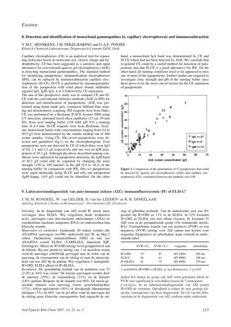
serum samples. Using CE, fifty-sev<strong>en</strong> paraproteins were detected<br />
and quantified (fig.1) on the electropherogram. Four<br />
paraproteins were not detected by CE of which three were IgG<br />
of 0.6, 1.1 and 2.2 g/l respectively and one was an IgM paraprotein<br />
of 20.3 g/l. Although the above described running conditions<br />
were optimized for paraprotein detection, the IgM band<br />
of 20.3 g/l could only be separated by changing the ionic<br />
str<strong>en</strong>gth (150 to 100 mmol/l) or the pH (9.9 to 10.2) of the<br />
running buffer. In comparison with IFE, fifty-six paraproteins<br />
were typed id<strong>en</strong>tically using IS-CE and only one paraprotein<br />
(IgM kappa, 14,9 g/l) could not be id<strong>en</strong>tified. On the other<br />
Ned Tijdschr Klin Chem 1997, vol. 22, no. 3<br />
hand, a monoclonal IgA band was demonstrated by CE and<br />
IS-CE which had not be<strong>en</strong> detected by AGE. We conclude that<br />
in g<strong>en</strong>eral CE could be a useful method for detection of paraproteins<br />
and that IS-CE is a good alternative for IFE. On the<br />
other hand, all running conditions tried so far appeared to miss<br />
one or more of the paraproteins. Further studies are required to<br />
investigate ionic str<strong>en</strong>gth and pH of the running buffer since<br />
these prove to be the most crucial factors for the CE separation<br />
of paraproteins.<br />
Figure 1. Comparison of the quantitation of 57 paraproteins that could<br />
be detected by agarose gel electrophoresis (AGE) and capillary electrophoresis<br />
(CE); correlation betwe<strong>en</strong> the methods was 0.96.<br />
9. Laboratoriumdiagnostiek van auto-immuun ziekt<strong>en</strong> (AIZ): immuunfluoresc<strong>en</strong>tie (IF) of ELISA?<br />
J. M. M. RONDEEL, W. van GELDER, H. van der LEEDEN 1 <strong>en</strong> R. B. DINKELAAR<br />
Afdeling <strong>Klinische</strong> Chemie <strong>en</strong> Reumatologie 1 , Drechtsted<strong>en</strong> ZH, Dordrecht<br />
Inleiding. In de diagnostiek van AIZ wordt IF steeds meer<br />
vervang<strong>en</strong> door ELISA. Wij vergelek<strong>en</strong> beide techniek<strong>en</strong><br />
m.b.t. aanvrag<strong>en</strong> voor anti-nucleaire antilicham<strong>en</strong> (ANA) <strong>en</strong><br />
extraheerbare nucleaire antig<strong>en</strong><strong>en</strong> (ENA) <strong>en</strong> onderzocht<strong>en</strong> hun<br />
klinische waarde.<br />
Material<strong>en</strong> <strong>en</strong> method<strong>en</strong>. Gedur<strong>en</strong>de 20 wek<strong>en</strong> werd<strong>en</strong> alle<br />
ANA/ENA aanvrag<strong>en</strong> (n=500) onderzocht met IF op Hep-2<br />
cell<strong>en</strong>, Ouchterlony immunodiffusie (OID) <strong>en</strong> met e<strong>en</strong><br />
ANA/ENA scre<strong>en</strong> ELISA (VARELISA; importeur IQP,<br />
Groning<strong>en</strong>). Alle<strong>en</strong> de IF+OID uitslag werd gerapporteerd aan<br />
de kliniek. Bij e<strong>en</strong> positieve uitslag van 1 of meerdere test<strong>en</strong><br />
werd de aanvrager schriftelijk gevraagd naar de red<strong>en</strong> van de<br />
aanvraag, de consequ<strong>en</strong>tie van de uitslag <strong>en</strong> naar de aanwezigheid<br />
van e<strong>en</strong> AIZ bij de patiënt. Wij vergelek<strong>en</strong> 3 strategieën:<br />
IF+OID, ELISA alle<strong>en</strong> <strong>en</strong> IF+ELISA.<br />
Resultat<strong>en</strong>. De gemiddelde leeftijd van de patiënt<strong>en</strong> was 53<br />
(2-92) jr; 64% was vrouw. De meeste aanvrag<strong>en</strong> werd<strong>en</strong> door<br />
de internist (29%), de reumatoloog (21%) <strong>en</strong> de huisarts<br />
(18%) gedaan. Respons op de <strong>en</strong>quête was 80%. De meest g<strong>en</strong>oemde<br />
red<strong>en</strong><strong>en</strong> voor aanvraag war<strong>en</strong>: gewrichtsklacht<strong>en</strong><br />
(37%), follow-up/controle (30%) of afwijk<strong>en</strong>de laboratorium<br />
uitslag<strong>en</strong> (7%). In 66% van de gevall<strong>en</strong> vond de aanvrager dat<br />
de uitslag ge<strong>en</strong> klinische consequ<strong>en</strong>ties had ongeacht de uit-<br />
slag of gebruikte techniek. Van de onderzochte sera was 8%<br />
positief bij IF+OID <strong>en</strong> 11% in de ELISA. In 12% kwam<strong>en</strong><br />
IF+OID <strong>en</strong> ELISA niet met elkaar overe<strong>en</strong>. Er kwam<strong>en</strong> 25<br />
AIZ voor in de geënquêteerde groep (18x reumatoide artritis,<br />
RA). Voorspell<strong>en</strong>de waarde van e<strong>en</strong> positieve (PVW) <strong>en</strong> e<strong>en</strong><br />
negatieve (NVW) uitslag voor AIZ sam<strong>en</strong> met kost<strong>en</strong> voor<br />
reag<strong>en</strong>tia (lijstprijz<strong>en</strong>) <strong>en</strong> arbeidsduur staan vermeld in onderstaande<br />
tabel.<br />
PVW (%) NVW (%) 2 reag<strong>en</strong>tia arbeidsduur<br />
IF+OID 53 1 83 ±Fl 4500,- 209 uur<br />
ELISA 26 61 ±Fl 9000,- 108 uur<br />
IF+ELISA 41 75 ±Fl 6000,- 230 uur<br />
1: p=0.04416 (IF+OID vs ELISA; χ 2 met Bonferroni); 2: p>0.05<br />
Indi<strong>en</strong> RA buit<strong>en</strong> de groep van AIZ werd gehoud<strong>en</strong> bleek de<br />
PVW niet significant te verschill<strong>en</strong> tuss<strong>en</strong> de 3 strategieën.<br />
Conclusies. In de laboratoriumdiagnostiek van AIZ g<strong>en</strong>iet<br />
IF+OID de voorkeur. Opvall<strong>en</strong>d is echter de zeer geringe klinische<br />
consequ<strong>en</strong>tie van deze diagnostiek. De rol van het laboratorium<br />
in de diagnostiek van AIZ verdi<strong>en</strong>t nader onderzoek.<br />
117