Karen Bedard and Karl-Heinz Krause
Karen Bedard and Karl-Heinz Krause
Karen Bedard and Karl-Heinz Krause

You also want an ePaper? Increase the reach of your titles
YUMPU automatically turns print PDFs into web optimized ePapers that Google loves.
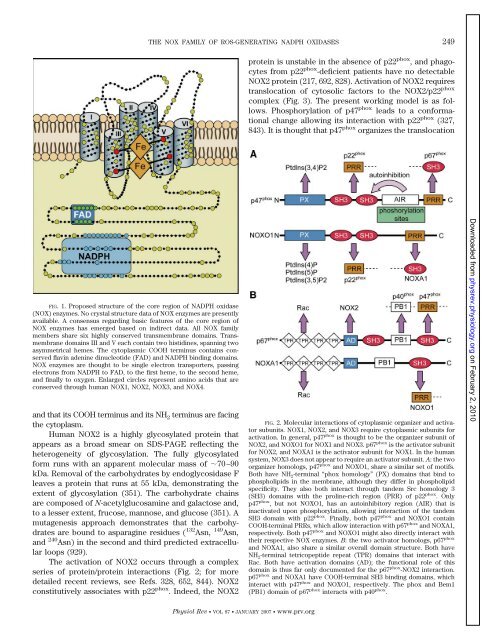
FIG. 1. Proposed structure of the core region of NADPH oxidase<br />
(NOX) enzymes. No crystal structure data of NOX enzymes are presently<br />
available. A consensus regarding basic features of the core region of<br />
NOX enzymes has emerged based on indirect data. All NOX family<br />
members share six highly conserved transmembrane domains. Transmembrane<br />
domains III <strong>and</strong> V each contain two histidines, spanning two<br />
asymmetrical hemes. The cytoplasmic COOH terminus contains conserved<br />
flavin adenine dinucleotide (FAD) <strong>and</strong> NADPH binding domains.<br />
NOX enzymes are thought to be single electron transporters, passing<br />
electrons from NADPH to FAD, to the first heme, to the second heme,<br />
<strong>and</strong> finally to oxygen. Enlarged circles represent amino acids that are<br />
conserved through human NOX1, NOX2, NOX3, <strong>and</strong> NOX4.<br />
<strong>and</strong> that its COOH terminus <strong>and</strong> its NH 2 terminus are facing<br />
the cytoplasm.<br />
Human NOX2 is a highly glycosylated protein that<br />
appears as a broad smear on SDS-PAGE reflecting the<br />
heterogeneity of glycosylation. The fully glycosylated<br />
form runs with an apparent molecular mass of �70–90<br />
kDa. Removal of the carbohydrates by endoglycosidase F<br />
leaves a protein that runs at 55 kDa, demonstrating the<br />
extent of glycosylation (351). The carbohydrate chains<br />
are composed of N-acetylglucosamine <strong>and</strong> galactose <strong>and</strong>,<br />
to a lesser extent, frucose, mannose, <strong>and</strong> glucose (351). A<br />
mutagenesis approach demonstrates that the carbohydrates<br />
are bound to asparagine residues ( 132 Asn, 149 Asn,<br />
<strong>and</strong> 240 Asn) in the second <strong>and</strong> third predicted extracellular<br />
loops (929).<br />
The activation of NOX2 occurs through a complex<br />
series of protein/protein interactions (Fig. 2; for more<br />
detailed recent reviews, see Refs. 328, 652, 844). NOX2<br />
constitutively associates with p22 phox . Indeed, the NOX2<br />
THE NOX FAMILY OF ROS-GENERATING NADPH OXIDASES 249<br />
Physiol Rev VOL 87 JANUARY 2007 www.prv.org<br />
protein is unstable in the absence of p22 phox , <strong>and</strong> phagocytes<br />
from p22 phox -deficient patients have no detectable<br />
NOX2 protein (217, 692, 828). Activation of NOX2 requires<br />
translocation of cytosolic factors to the NOX2/p22 phox<br />
complex (Fig. 3). The present working model is as follows.<br />
Phosphorylation of p47 phox leads to a conformational<br />
change allowing its interaction with p22 phox (327,<br />
843). It is thought that p47 phox organizes the translocation<br />
FIG. 2. Molecular interactions of cytoplasmic organizer <strong>and</strong> activator<br />
subunits. NOX1, NOX2, <strong>and</strong> NOX3 require cytoplasmic subunits for<br />
activation. In general, p47 phox is thought to be the organizer subunit of<br />
NOX2, <strong>and</strong> NOXO1 for NOX1 <strong>and</strong> NOX3. p67 phox is the activator subunit<br />
for NOX2, <strong>and</strong> NOXA1 is the activator subunit for NOX1. In the human<br />
system, NOX3 does not appear to require an activator subunit. A: the two<br />
organizer homologs, p47 phox <strong>and</strong> NOXO1, share a similar set of motifs.<br />
Both have NH 2-terminal “phox homology” (PX) domains that bind to<br />
phospholipids in the membrane, although they differ in phospholipid<br />
specificity. They also both interact through t<strong>and</strong>em Src homology 3<br />
(SH3) domains with the proline-rich region (PRR) of p22 phox . Only<br />
p47 phox , but not NOXO1, has an autoinhibitory region (AIR) that is<br />
inactivated upon phosphorylation, allowing interaction of the t<strong>and</strong>em<br />
SH3 domain with p22 phox . Finally, both p47 phox <strong>and</strong> NOXO1 contain<br />
COOH-terminal PRRs, which allow interaction with p67 phox <strong>and</strong> NOXA1,<br />
respectively. Both p47 phox <strong>and</strong> NOXO1 might also directly interact with<br />
their respective NOX enzymes. B: the two activator homologs, p67 phox<br />
<strong>and</strong> NOXA1, also share a similar overall domain structure. Both have<br />
NH 2-terminal tetricopeptide repeat (TPR) domains that interact with<br />
Rac. Both have activation domains (AD); the functional role of this<br />
domain is thus far only documented for the p67 phox -NOX2 interaction.<br />
p67 phox <strong>and</strong> NOXA1 have COOH-terminal SH3 binding domains, which<br />
interact with p47 phox <strong>and</strong> NOXO1, respectively. The phox <strong>and</strong> Bem1<br />
(PB1) domain of p67 phox interacts with p40 phox .<br />
Downloaded from<br />
physrev.physiology.org<br />
on February 2, 2010