Instituto de Ciencia de Materiales de Madrid - Materials Science ...
Instituto de Ciencia de Materiales de Madrid - Materials Science ...
Instituto de Ciencia de Materiales de Madrid - Materials Science ...

Create successful ePaper yourself
Turn your PDF publications into a flip-book with our unique Google optimized e-Paper software.
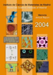
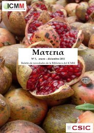
Portada: <strong>de</strong> arriba a abajo y <strong>de</strong> izquierda a <strong>de</strong>recha.<br />
Figura 1: Imagen <strong>de</strong> microscopía electrónica <strong>de</strong> transmisión<br />
que muestra la coalescencia <strong>de</strong> granos en una<br />
lámina <strong>de</strong>lgada ferroeléctrica <strong>de</strong> tantalato <strong>de</strong> estroncio<br />
y bismuto. (J. Ricote, M.L. Calzada y A. González,<br />
Departamento <strong>de</strong> <strong>Materiales</strong> Ferroeléctricos).<br />
Figura 2: Imagen <strong>de</strong> un microscopio <strong>de</strong> efecto túnel<br />
(STM) que muestra la superficie <strong>de</strong> un siliciuro <strong>de</strong> tierras<br />
raras crecido epitaxialmente sobre Si. Cada una <strong>de</strong><br />
las bolas representa un átomo <strong>de</strong> Si en la superficie. Se<br />
pue<strong>de</strong> distinguir que los átomos se disponen en dos<br />
alturas diferentes, formando una red hexagonal. El<br />
tamaño <strong>de</strong> la imagen es 26x23 Å2 (C. Rogero y J.A.<br />
Martín-Gago, Departamento <strong>de</strong> Física e Ingeniería <strong>de</strong><br />
Superficies)<br />
Figura 3: Nanocomposite liquen-epoxi como componente<br />
<strong>de</strong> sensores electroquímicos <strong>de</strong> metales pesados.<br />
(M. Dar<strong>de</strong>r, M. Colilla, N. Lara, E. Ruiz-Hitzky,<br />
Departamento <strong>de</strong> <strong>Materiales</strong> Porosos y Compuestos <strong>de</strong><br />
Intercalación)<br />
Figura 4: Topologia bidimensional <strong>de</strong> la Superficie <strong>de</strong><br />
Fermi <strong>de</strong> una lamina <strong>de</strong> Ag <strong>de</strong>positada en Si(111)-H<br />
<strong>de</strong>terminada por fotoemision. Distribución angular <strong>de</strong><br />
fotoelectrones medidos en el nivel <strong>de</strong> Fermi con una<br />
energía <strong>de</strong> foton <strong>de</strong> 32 eV en una pelicula <strong>de</strong> 6<br />
Monocapas <strong>de</strong> Ag <strong>de</strong>positada sobre H/Si(111)-(1x1) y<br />
recocida a 300° C durante 5 min. (M.C. Asensio y J.<br />
Avila, Departamento <strong>de</strong> Intercaras y Crecimineto)<br />
Figura 5: Micrografía <strong>de</strong> SEM <strong>de</strong> mezclas <strong>de</strong> 3SrO:2TiO 2<br />
mecanoactivadas, mostrando cristales <strong>de</strong> SrTiO 3<br />
. (T.<br />
Hungría, J.G. Lisoni y A. Castro. Departamento <strong>de</strong><br />
Sólidos Iónicos)<br />
Figura 6: Fotografía <strong>de</strong> un array <strong>de</strong> <strong>de</strong>tectores <strong>de</strong> infrarrojo<br />
piroeléctricos crecidos sobre MgO y sus microsoldaduras<br />
al zócalo. (P. Ramos, J. Mendiola, R. Jimenez,<br />
M.L. Calzada , A. Gonzalez y P. Tejedor, Departamento<br />
<strong>de</strong> <strong>Materiales</strong> Ferroeléctricos)<br />
Figura 7: Monocristal <strong>de</strong> la espinela LiMn 2<br />
O 4<br />
<strong>de</strong> hasta<br />
0.2mm como cátodo <strong>de</strong> baterías recargables <strong>de</strong> litio.<br />
(M.A. Monge, J.M. Amarilla, E. Gutiérrez-Puebla, J.A.<br />
Campa, I. Rasines, Departamento <strong>de</strong> Síntesis y<br />
Estructura <strong>de</strong> Óxidos y Departamento <strong>de</strong> Sólidos<br />
Iónicos)<br />
Cover: From top to bottom and left to right.<br />
Figure 1: Transmission electron microscopy image showing<br />
the coalescence of grains in a strontium bismuth<br />
tantalate ferroelectric thin film. (J. Ricote, M.L. Calzada<br />
and A. González, Ferroelectric <strong>Materials</strong> Department).<br />
Figure 2: STM image showing the surface termination<br />
of a rare-earth silici<strong>de</strong> epitaxially grown on Si. The<br />
bumps in the image correspond to Si atoms on the surface.<br />
Two different levels for the surface Si atoms, in a<br />
hexagonal arrangement can be distinguished in the<br />
image. The scanned area is 26x23 Å2. (C. Rogero y J.A.<br />
Martín-Gago, Department of Surface Physics and<br />
Engineering)<br />
Figure 3: Epoxy-lichen nanocomposite as component of<br />
heavy metal ions electrochemical sensors. (M. Dar<strong>de</strong>r,<br />
M. Colilla, N. Lara, E. Ruiz-Hitzky, Porous <strong>Materiales</strong> and<br />
Intercalation Compounds Department).<br />
Figure 4: Bidimensional Fermi Surface topology of the<br />
thick Ag film <strong>de</strong>posited onto Si(111)-H <strong>de</strong>termined<br />
using photoemission. Photoelectron angular distribution<br />
measured at the Ef with hv= 32 eV in a 6-ML Ag film<br />
<strong>de</strong>posited onto H/Si(111)-(1x1) and then annealing to<br />
300° C for 5 min. (M.C. Asensio y J. Avila, Department<br />
of Interfaces and Growth)<br />
Figure 5: SEM micrographs of 3SrO:2TiO 2<br />
mixture<br />
mechanoactivated, showing crystals of SrTiO 3<br />
. (T.<br />
Hungría, J.G. Lisoni and A. Castro. Ionic Solids<br />
Department).<br />
Figure 6: Array of pyroelectric infrarred <strong>de</strong>tector, <strong>de</strong>posited<br />
on MgO and its microboundings. (P. Ramos, J.<br />
Mendiola, R. Jimenez, M.L. Calzada , A. Gonzalez and P.<br />
Tejedor, Ferroelectric <strong>Materials</strong> Department).<br />
Figure 7: Spinel LiMn 2<br />
O 4<br />
crystals showing 0.2mm edg<strong>de</strong><br />
as catho<strong>de</strong> of rechargeable lithium batteries. (M.A.<br />
Monge, J.M. Amarilla, E. Gutiérrez-Puebla, J.A. Campa, I.<br />
Rasines, Synthesis and Structure of Oxi<strong>de</strong>s Dept. and<br />
Ionic Solids Department).<br />
Editores / Editors: Drs. F. Soria, E. Vila y D. J.I. Reguera<br />
Diseño / Design and Lay-out: J.I. Reguera (ICMM)<br />
Impresión / Printed by: P.G.M<br />
No. <strong>de</strong> ejemplares / Number of copies: 900<br />
Nuestro agra<strong>de</strong>cimiento a todo el personal <strong>de</strong>l <strong>Instituto</strong> que ha colaborado en la realización <strong>de</strong> esta Memoria.<br />
We <strong>de</strong>eply thank the Institute’s personnel for their cooperation