ferritina irma ct ref
ferritina irma ct ref
ferritina irma ct ref

You also want an ePaper? Increase the reach of your titles
YUMPU automatically turns print PDFs into web optimized ePapers that Google loves.
IMMUNORADIOMETRIC ASSAY FOR QUANTITATIVE DETERMINATION OF FERRITIN IN HUMAN<br />
SERUM.<br />
FOR IN VITRO DIAGNOSTIC USE ONLY<br />
CLINICAL APPLICATIONS<br />
The Ferritin molecule consists of 24 protein subunits, each of them with molecular weight of 20 kDa.<br />
Ferritin fun<strong>ct</strong>ions mainly as the intracellular site of iron storage (from which iron may be rapidly<br />
mobilized), thereby prote<strong>ct</strong>ing cells from the toxic effe<strong>ct</strong>s of unbound iron. Most Ferritin in the body is<br />
found in the liver cells as well as in the cells of the reticuloendothelial system of liver, spleen and bonemarrow.<br />
Small amounts are also found in the heart, pancreas and kidneys. Small but significant amounts<br />
of Ferritin are found in the human serum. The exa<strong>ct</strong> fun<strong>ct</strong>ion of serum Ferritin is unknown but there is a<br />
well established correlation between serum Ferritin and the body's iron stores. Serum Ferritin testing<br />
thus represents the simplest and less invasive method in monitoring any iron store change in the body.<br />
Ferritin serum levels are also affe<strong>ct</strong>ed by body changes due to age as well as sex. Average Ferritin<br />
levels in normal subje<strong>ct</strong>s (slightly higher at birth) tend to decrease during childhood, until the age of<br />
puberty is reached. From then on, a progressive increase of the body iron stores is observed in males,<br />
with a proportional rise in serum Ferritin. Females in the reprodu<strong>ct</strong>ive age instead tend to have lower<br />
amounts with more stable values. A Ferritin rise is only observed after menopause.<br />
Ferritin levels below normal ranges clearly indicate a lack of iron and allow to differentiate iron deficiency<br />
anemia from other forms of anemia. An iron overload, either due to an increase in iron uptake (as in<br />
idiopathic hemochromatosis) or else due to multiple transfusions, causes serum Ferritin to rise, often<br />
beyond normal ranges. High Ferritin levels are also observed for other clinical conditions such as liver<br />
disease, infe<strong>ct</strong>ious as well as inflammatory processes, leukemia, Hodgkin's disease and other forms of<br />
malignancy.<br />
PRINCIPLE OF THE ASSAY<br />
The present method employs two anti-Ferritin monoclonal antibodies which recognize two different<br />
epitopes of the molecule. One antibody is adsorbed in solid phase (coated tube), the other (labeled with<br />
iodine-125) is used as conjugate. The sample to be tested and the labeled antibody are incubated<br />
simultaneously in the coated tube. The amount of bound conjugate will thus be dire<strong>ct</strong>ly proportional to<br />
the antigen concentration in calibrators and samples. At the end of the incubation, the unbound material<br />
is removed by aspiration and washing. The radioa<strong>ct</strong>ivity in the tubes is measured in a gamma counter.<br />
REAGENTS PROVIDED WITH THE KIT: PREPARATION AND STABILITY<br />
− The reagents are sufficient for 100 tubes.<br />
− Store the kit at 2-8°C.<br />
− The expiry date of each reagent is shown on the vial label.<br />
1 - CT Coated Tubes: 100 tubes coated with mouse monoclonal anti-Ferritin antibody. Unused tubes<br />
should be stored at 2-8°C in the appropriate bag and accurately sealed.<br />
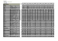
2 - CAL Calibrators: 7 vials (1 mL) of Ferritin (human liver) in phosphate buffer at the following<br />
concentrations: 0, 5, 20, 50, 250, 1000 and 2000 ng/mL. Preservative: NaN3 (