Cop ISPLAD - Salute per tutti
Cop ISPLAD - Salute per tutti
Cop ISPLAD - Salute per tutti

Create successful ePaper yourself
Turn your PDF publications into a flip-book with our unique Google optimized e-Paper software.
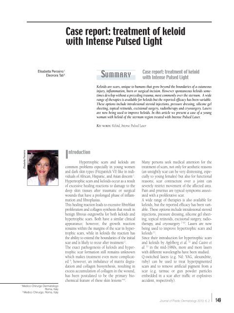
Elisabetta Perosino<br />
Elisabetta Perosino 1<br />
Eleonora Tati 2<br />
1 Medico Chirurgo Dermatologo<br />
Roma, Italy<br />
2 Medico Chirurgo, Roma, Italy<br />
Case report: treatment of keloid<br />
with Intense Pulsed Light<br />
Summary<br />
I ntroduction<br />
Hy<strong>per</strong>trophic scars and keloids are<br />
common problems especially in young women<br />
and dark skin types (Fitzpatrick VI) like in individuals<br />
of African, Hispanic, and Asian descent 1 .<br />
Hy<strong>per</strong>trophic scars and keloids occur as a result<br />
of excessive healing reactions to damage to the<br />
deep skin tissues after traumatic or surgical<br />
wounds that have a prolonged phase of inflammation<br />
and fibroplasias.<br />
This healing reaction leads to excessive fibroblast<br />
proliferation and collagen synthesis that result in<br />
benign fibrous outgrowths for both keloids and<br />
hy<strong>per</strong>trophic scars. Both have a similar clinical<br />
appearance; however, the growth reaction<br />
remains within the margins of the scar in hy<strong>per</strong>trophic<br />
scars, while in keloids the reaction has<br />
the ability to extend the boundaries of the initial<br />
scar and is likely to recur after treatment 2 .<br />
The exact pathogenesis of keloids and hy<strong>per</strong>trophic<br />
scar formation still remains unknown<br />
which makes treatment even more complicated<br />
3 ; however, an imbalance of matrix degradation<br />
and collagen biosynthesis, resulting in<br />
excess accumulation of collagen in the wound,<br />
has been postulated to be the primary biochemical<br />
feature of these skin lesions 4-6 .<br />
Case report: treatment of keloid<br />
with Intense Pulsed Light<br />
Keloids are scars, unique to humans that grow beyond the boundaries of a cutaneous<br />
injury, inflammation, burn or surgical incision. However spontaneous keloids sometimes<br />
develop without a preceding trauma, most commonly over the sternum. A wide<br />
range of therapies is available for keloids but the reported efficacy has been variable.<br />
These options include intralesional steroid injections, pressure dressing, silicone gel<br />
sheeting, topical retinoids, excisional surgery, radiotherapy and cryosurgery. Lasers<br />
are now being used to improve keloids. In this article we present a case of a young<br />
woman with keloid of the sternum region treated with Intense Pulsed Laser.<br />
KEY WORDS: Keloid, Intense Pulsed Laser<br />
Many <strong>per</strong>sons seek medical attention for the<br />
treatment of scars, not only for aesthetic reasons<br />
(an unsightly scar can be very distressing, especially<br />
to young females) but also for functional<br />
reasons; scar contracture over a joint can<br />
severely restrict movement of the affected area.<br />
Pain and pruritus are typical symptoms associated<br />
with a proliferative scar.<br />
A wide range of therapies is also available for<br />
keloids, but the reported efficacy has been variable.<br />
These options include intralesional steroid<br />
injections, pressure dressing, silicone gel sheeting,<br />
topical retinoids, excisional surgery, radiotherapy,<br />
and cryosurgery 7-10 . Lasers are now<br />
being used to improve hy<strong>per</strong>trophic scars and<br />
keloids 11 .<br />
Since their introduction for hy<strong>per</strong>trophic scars<br />
and keloids by Apfelberg et al. 12 and Castro et<br />
al. 13 in the mid-1980s, more and more lasers<br />
with different wavelengths have been studied.<br />
Q-switched lasers (e.g. Nd: YAG, alexandrite,<br />
ruby) can be used to treat hy<strong>per</strong>pigmented<br />
scars and to remove artificial pigment from a<br />
scar (e.g. tarmac or gun powder particles<br />
embedded in a scar after traffic or explosives<br />
accident, respectively).<br />
Journal of Plastic Dermatology 2010; 6, 2 149