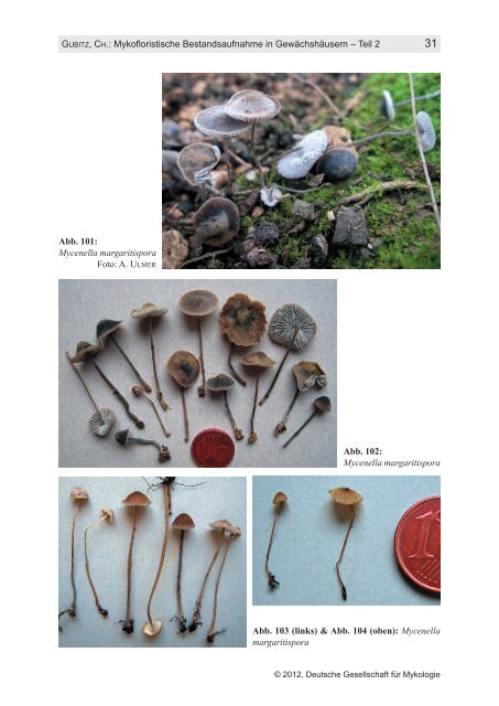
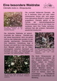
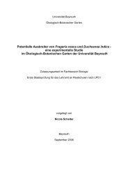
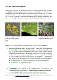
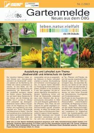
30 Z. MYKOL. 78/1, 2012 a 10 µm Abb. 100: Mycena cf. niveipes, a: Sporen, b: Basidien. Cheilo- und Pleurozystiden, c: Elemente der HDS, d: Caulozystiden Bemerkungen: Die Frage, ob Mycena oder Hydropus, konnte erst durch Hausknecht beantwortet werden. Seiner Untersuchung zu Folge gehört die Art in die Nähe von Mycena niveipes, die sich hauptsächlich durch viel längere Zystiden unterscheidet (ROBICH 2003). Eine striegelhaarige Stielbasis beschreibt BRESINSKY (1966) von einem Fund aus Lappland. Funde aus Warmhäusern sind nicht bekannt. Mycenella margaritispora (J. E. Lange) Singer Abb. 101-105 Geweihtragender Scheinhelmling Beschreibung nach 11 Kollektionen: Hut (5)10–20 mm Ø, stumpfkonisch, kegelig-glockig, gewölbt bis flach, zuweilen mit angedeutetem zentralem Buckel, nicht selten mit verbogenem und hochgeschlagenem Rand, älter mitunter (faltig) gerieft, stark weißlich bereift, darunter schwarz-, blei-, blaugrau, graubraun; trocken verblassend, mit meist dunkel bleibender Mitte. Lamellen angeheftet bis fast breit angewachsen, eng bis mäßig gedrängt, schmal bis etwas bauchig, nicht selten am Grunde anastomosierend, jung weiß, dann hellgrau, cremefarben, alt z. T. gelblich oder bräunlich; Schneiden gleich. Stiel 20–45 × 0,7–1 mm, zylindrisch, wie Hut bereift und gefärbt oder etwas heller, an Basis ± flockig-filzig bis striegelig. Geruch mehlig- oder spermatisch-tranig, Geschmack unauffällig. Sporen 5–8 × 5–7 µm (inkl. Ornament), rundlich, ± grobwarzig, höckerig, inamyolid, Apiculus groß; unreif nur mit schwacher Ornamentation, fast glatt, z. T. mit heterodiametrischem (an Entoloma-Sporen erinnernden) Umriss, mit großem Tropfen. Basidien 20–35 × 7 µm, (1,4)2sporig. Cheilozystiden 30–60(70) × 5–15 µm, zahlreich, vielgestaltig, flaschenförmig, spindelig-bauchig, lanzettlich, manchmal septiert; Spitze häufig mit hand-, geweihförmigen, koralloiden, bürstigen oder warzigen Auswüchsen, mitunter von harziger Substanz umgeben. © 2012, Deutsche Gesellschaft für Mykologie b c 10 µm d
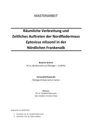
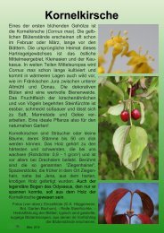
GUBITZ, CH.: Mykofloristische Bestandsaufnahme in Gewächshäusern – <strong>Teil</strong> 2 31 Abb. 101: Mycenella margaritispora Foto: A. ULMER Abb. 102: Mycenella margaritispora Abb. 103 (links) & Abb. 104 (oben): Mycenella margaritispora © 2012, Deutsche Gesellschaft für Mykologie
- Seite 1 und 2: ZEITSCHRIFT FÜR MYKOLOGIE, Band 78
- Seite 3 und 4: GUBITZ, CH.: Mykofloristische Besta
- Seite 5 und 6: GUBITZ, CH.: Mykofloristische Besta
- Seite 7 und 8: GUBITZ, CH.: Mykofloristische Besta
- Seite 9 und 10: GUBITZ, CH.: Mykofloristische Besta
- Seite 11 und 12: GUBITZ, CH.: Mykofloristische Besta
- Seite 13 und 14: GUBITZ, CH.: Mykofloristische Besta
- Seite 15 und 16: GUBITZ, CH.: Mykofloristische Besta
- Seite 17 und 18: GUBITZ, CH.: Mykofloristische Besta
- Seite 19 und 20: GUBITZ, CH.: Mykofloristische Besta
- Seite 21: GUBITZ, CH.: Mykofloristische Besta
- Seite 25 und 26: GUBITZ, CH.: Mykofloristische Besta
- Seite 27 und 28: GUBITZ, CH.: Mykofloristische Besta
- Seite 29 und 30: GUBITZ, CH.: Mykofloristische Besta
- Seite 31 und 32: GUBITZ, CH.: Mykofloristische Besta
- Seite 33 und 34: GUBITZ, CH.: Mykofloristische Besta
- Seite 35 und 36: GUBITZ, CH.: Mykofloristische Besta
- Seite 37 und 38: GUBITZ, CH.: Mykofloristische Besta
- Seite 39 und 40: GUBITZ, CH.: Mykofloristische Besta
- Seite 41 und 42: GUBITZ, CH.: Mykofloristische Besta
- Seite 43 und 44: GUBITZ, CH.: Mykofloristische Besta