Ch. 54 – Biliary System
Ch. 54 – Biliary System
Ch. 54 – Biliary System
You also want an ePaper? Increase the reach of your titles
YUMPU automatically turns print PDFs into web optimized ePapers that Google loves.
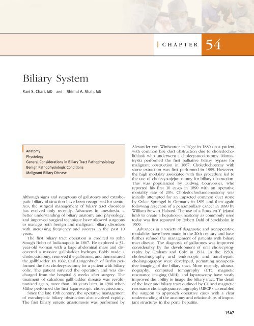
<strong>Biliary</strong> <strong>System</strong><br />
Ravi S. <strong>Ch</strong>ari, MD and Shimul A. Shah, MD<br />
Anatomy<br />
Physiology<br />
General Considerations in <strong>Biliary</strong> Tract Pathophysiology<br />
Benign Pathophysiologic Conditions<br />
Malignant <strong>Biliary</strong> Disease<br />
Although signs and symptoms of gallstones and extrahepatic<br />
biliary obstruction have been recognized for centuries,<br />
the surgical management of biliary tract disorders<br />
has evolved only recently. Advances in anesthesia, a<br />
better understanding of biliary anatomy and physiology,<br />
and improved surgical technique have allowed surgeons<br />
to manage both benign and malignant biliary disorders<br />
with increasing frequency and success in the past 10<br />
years.<br />
The fi rst biliary tract operation is credited to John<br />
Stough Bobb of Indianapolis in 1867. He explored a 32year-old<br />
woman with a large abdominal mass and discovered<br />
a massive gallbladder hydrops. Bobb made a<br />
cholecystotomy, removed the gallstones, and then sutured<br />
the gallbladder. In 1882, Carl Langenbuch of Berlin performed<br />
the fi rst cholecystectomy for a patient with biliary<br />
colic. The patient survived the operation and was discharged<br />
from the hospital 8 weeks after surgery. The<br />
treatment of calculous gallbladder disease was revolutionized<br />
again, more than 100 years later, in 1986 when<br />
Mühe performed the fi rst laparoscopic cholecystectomy.<br />
Since the late 19th century, the operative management<br />
of extrahepatic biliary obstruction also evolved rapidly.<br />
The fi rst biliary enteric anastomosis was performed by<br />
CHAPTER <strong>54</strong><br />
Alexander von Winiwarter in Liège in 1880 on a patient<br />
with common bile duct obstruction due to choledocholithiasis<br />
who underwent a cholecystocolostomy. Monastryski<br />
performed the fi rst palliative biliary bypass for<br />
malignant obstruction in 1887. <strong>Ch</strong>oledochotomy with<br />
stone extraction was fi rst performed in 1889. However,<br />
the high mortality associated with this procedure led to<br />
the use of cholecystojejunostomy for biliary obstruction.<br />
This was popularized by Ludwig Courvoisier, who<br />
reported his fi rst 10 cases in 1890 with an operative<br />
mortality rate of 20%. <strong>Ch</strong>oledochoduodenostomy was<br />
initially attempted for an impacted common duct stone<br />
by Oskar Sprengel in Germany in 1891 and then again<br />
following resection of a periampullary cancer in 1898 by<br />
William Stewart Halsted. The use of a Roux-en-Y jejunal<br />
limb to create a hepaticojejunostomy as commonly used<br />
today was fi rst reported by Robert Dahl of Stockholm in<br />
1909.<br />
Advances in a variety of diagnostic and nonoperative<br />
modalities have been made in the 20th century and have<br />
further refi ned the management of patients with biliary<br />
tract disease. The diagnosis of gallstones was improved<br />
considerably by the development of oral cholecysto graphy<br />
by Graham and Cole in 1924. In the 1950s,<br />
cholescintography and endoscopic and transhepatic<br />
cholangiography were developed, permitting nonoperative<br />
imaging of the biliary tract. More recently, ultrasonography,<br />
computed tomography (CT), magnetic<br />
resonance imaging (MRI), and laparoscopy have vastly<br />
improved the ability to image the biliary tract. The detail<br />
of the liver and biliary tract outlined by CT and magnetic<br />
resonance cholangiopancreatography (MRCP) has enabled<br />
the surgeon to approach operative cases with a clear<br />
understanding of the anatomy and relationships of important<br />
structures in the porta hepatitis.<br />
1<strong>54</strong>7
1<strong>54</strong>8 Section X Abdomen<br />
ANATOMY<br />
Cystic artery<br />
Common<br />
bile duct<br />
Right hepatic duct<br />
Left hepatic duct<br />
Pancreatic<br />
duct<br />
Extrahepatic <strong>Biliary</strong> Tract<br />
The extrahepatic biliary tract consists of the bifurcation<br />
of the left and right hepatic ducts, the common hepatic<br />
duct, the common bile duct, and the cystic duct and<br />
gallbladder (Fig. <strong>54</strong>-1). The left hepatic duct is formed<br />
by the ducts draining segments II, III, and IV of the liver,<br />
courses horizontally along the base of segment IV, and<br />
has an extrahepatic length of about 2 cm. The right<br />
hepatic duct is formed by the right posterior (segments<br />
IV and VII) and right anterior (segments V and VIII)<br />
hepatic ducts and has a shorter extrahepatic length. The<br />
hepatic duct bifurcation is usually extrahepatic and anterior<br />
to the portal vein bifurcation. The biliary confl uence<br />
is separated from the posterior aspect of the caudate lobe<br />
(segment I) of the liver by the hilar plate, which consists<br />
of a fusion of connective tissue enclosing the biliary and<br />
vascular structures within the Glisson capsule (Fig. <strong>54</strong>-2).<br />
The common hepatic duct lays anterolateral to the hepatic<br />
artery and portal vein in the hepatoduodenal ligament<br />
and joins the cystic duct to form the common bile duct.<br />
The common bile duct extends from the cystic duct<strong>–</strong><br />
common hepatic duct junction inferiorly to the papilla of<br />
Fundus<br />
Right hepatic artery<br />
Corpus<br />
Portal vein<br />
Gastroduodenal artery<br />
Common hepatic artery<br />
Neck<br />
Figure <strong>54</strong>-1 Anatomy of the biliary system and its relationship to surrounding structures.<br />
A<br />
B<br />
VI<br />
VII<br />
V<br />
VIII<br />
Papilla<br />
C<br />
IV<br />
Common<br />
hepatic duct<br />
Cystic duct<br />
Hartmann’s<br />
pouch<br />
Common<br />
bile duct<br />
Figure <strong>54</strong>-2 Anatomy of the hilar plate. Note the cystic plate (A)<br />
above the gallbladder, the hilar plate (B) above the biliary confl uence,<br />
and the umbilical plate (C) above the umbilical portion of<br />
the portal vein. Large, curving arrows indicate plane of dissection<br />
of the cystic plate during cholecystectomy and of the hilar<br />
plate at the base of segment IV during approaches to the left<br />
hepatic duct. (From Blumgart LH, Hann LE: Surgical and radiological<br />
anatomy of the liver and biliary tract. In Blumgart LH,<br />
Fong Y [ed]: Surgery of the Liver and <strong>Biliary</strong> Tract. New York,<br />
WB Saunders, 2000, pp 13-14.)<br />
II<br />
III
Vater, where it empties into the duodenum. The common<br />
bile duct varies in length from 5 to 9 cm depending on<br />
its junction with the cystic duct and is divided into three<br />
segments: supraduodenal, retroduodenal, and intrapancreatic.<br />
The distal common bile duct and pancreatic duct<br />
may join outside the duodenal wall to form a long<br />
common channel, within the duodenal wall to form a<br />
short common channel, or they may enter the duodenum<br />
through two distinct ostia.<br />
The gallbladder is a pear-shaped reservoir in continuity<br />
with the common hepatic and common bile ducts<br />
through the cystic duct. It is usually 7 to 10 cm in length,<br />
is 3 to 5 cm in diameter, and has a capacity of 30 to<br />
60 mL. The gallbladder lies on the inferior surface of the<br />
liver partially enveloped in a layer of peritoneum. The<br />
gallbladder is anatomically divided into the fundus, body,<br />
infundibulum, and neck, which empties into the cystic<br />
duct. Both the gallbladder neck and the cystic duct<br />
contain spirally oriented mucosal folds known as the<br />
valves of Heister. The valves prevent the passage of gallstones<br />
and excessive distention or collapse of the cystic<br />
duct, despite variations in ductal pressure. The cystic duct<br />
varies in length from 1 to 5 cm and in diameter from 3<br />
to 7 mm; it usually joins the common hepatic duct at an<br />
acute angle. Small veins and lymphatics course between<br />
the gallbladder fossa and the gallbladder wall, connecting<br />
the lymphatic and venous drainage of the liver and gallbladder.<br />
These connections are the cause of the direct<br />
infl ammatory and carcinomatous spread from the gallbladder<br />
into the liver.<br />
Anatomic variations in the cystic duct and hepatic<br />
ducts are common. Frequent variations in the hepatic<br />
ductal anatomy are shown in Figure <strong>54</strong>-3. Drainage to<br />
the caudate lobe (segment I) is not shown but can arise<br />
directly from common bile duct, right hepatic duct, or<br />
left hepatic duct. Variations of the left hepatic duct are<br />
much less common than those of the right hepatic duct.<br />
The cystic duct usually enters the common bile duct at<br />
an acute angle, but may run parallel to the common<br />
hepatic duct for a variable distance before joining it, or<br />
may join the right hepatic duct or a segmental right<br />
hepatic duct. An accessory hepatic duct or cholecystohepatic<br />
duct (duct of Luschka) may also enter the gallbladder<br />
through the gallbladder fossa and, if encountered<br />
during a cholecystectomy, should be ligated to prevent<br />
a biliary fi stula.<br />
Anomalies of the gallbladder are much less frequent<br />
than variations in ductal anatomy. Agenesis of the gallbladder<br />
has been reported (∼200 cases), and duplication<br />
of the gallbladder (two separate gallbladders, each with<br />
its own cystic duct) occurs in 1 of 4000 births.<br />
Vascular Anatomy<br />
The blood supply to the extrahepatic biliary tree originates<br />
(1) distally from the gastroduodenal, retroduodenal,<br />
and posterosuperior pancreatoduodenal arteries and (2)<br />
proximally from the right hepatic and cystic arteries.<br />
These arteries supply the common bile and common<br />
hepatic ducts through branches running parallel to the<br />
duct in the 3- and 9-o’clock positions. The extrahepatic<br />
ra<br />
rp lh<br />
A57% B12%<br />
ra<br />
rp lh<br />
<strong>Ch</strong>apter <strong>54</strong> <strong>Biliary</strong> <strong>System</strong> 1<strong>54</strong>9<br />
rp<br />
C20% 16% 4%<br />
C1 C2<br />
rp<br />
D6%<br />
ra<br />
rp<br />
E3%<br />
ra<br />
D1<br />
F2%<br />
5%<br />
lh<br />
2% 1%<br />
E1 E2<br />
rp<br />
I<br />
IV<br />
II<br />
ra<br />
biliary tree is vulnerable to ischemic injury. To avoid<br />
disrupting the fragile inconstant blood supply to the duct,<br />
it is important not to strip the investing areolar tissue<br />
around it during dissection and isolation. Ischemia of<br />
the bile duct will not be readily evident at time of<br />
dissection but can result in biliary stricture or leak<br />
postoperatively.<br />
rp<br />
rp<br />
ra<br />
ra<br />
rp<br />
ra<br />
ra<br />
lh<br />
1%<br />
D2<br />
Figure <strong>54</strong>-3 Variations in the confl uence of the left and right<br />
hepatic ducts. A, Typical anatomy of the confl uence. B, Trifurcation<br />
of left, right anterior, and right posterior hepatic ducts.<br />
C, Aberrant drainage of a right anterior (C1) or posterior (C2)<br />
sectoral hepatic duct into the common hepatic duct. D-F, Less<br />
common variations in hepatic ductal anatomy. (From Smadja C,<br />
Blumgart L: The biliary tract and the anatomy of biliary exposure.<br />
In Blumgart L [ed]: Surgery of the Liver and <strong>Biliary</strong> Tract.<br />
New York, <strong>Ch</strong>urchill Livingstone, 1994, pp 11-24.)<br />
III<br />
IV<br />
I<br />
lh<br />
lh<br />
lh<br />
III<br />
II
1550 Section X Abdomen<br />
Cystic artery<br />
Cystic duct<br />
Common duct<br />
The gallbladder is supplied by a single cystic artery,<br />
but in 12% of cases, a double cystic artery (anterior and<br />
posterior) may exist. The origin and course of the cystic<br />
artery is highly variable and is one of the most variable<br />
in the body. The cystic artery may originate from the left<br />
hepatic, common hepatic, gastroduodenal, or superior<br />
mesenteric arteries. The cystic artery divides into superfi<br />
cial and deep branches before entering the gallbladder.<br />
The cystic artery usually lies superior to the cystic duct<br />
and passes posterior to the common hepatic duct, but its<br />
course varies with its origin. The common hepatic duct,<br />
the liver, and the cystic duct defi ne the boundaries of<br />
Calot’s triangle (Fig. <strong>54</strong>-4). Located within this triangle<br />
are important structures: the cystic artery, the right hepatic<br />
artery, and the cystic duct lymph node. The Calot node<br />
is the main route of lymphatic drainage of the gallbladder<br />
and is therefore commonly involved in infl ammatory or<br />
neoplastic diseases of the gallbladder.<br />
PHYSIOLOGY<br />
X<br />
Hepatic duct<br />
Portal vein<br />
Hepatic artery<br />
Figure <strong>54</strong>-4 The triangle of Calot is bounded by the cystic duct,<br />
the common hepatic duct, and the inferior border of the liver.<br />
(From Gilchrist BF, Trunkey DD, <strong>Biliary</strong> Tract trauma. In<br />
Zuidema GD [ed]: Shackelford’s surgery of the alimentary tract,<br />
3rd ed. WB Saunders, Philadelphia, 1991, pp 257.)<br />
Bile Ducts<br />
The bile ducts, gallbladder, and sphincter of Oddi modify,<br />
store, and regulate the fl ow of bile. The liver produces<br />
500 to 1000 mL of bile per day and excretes it into the<br />
bile canaliculi. During its passage through the bile ductules<br />
and hepatic duct, canalicular bile is modifi ed by the<br />
absorption and secretion of electrolytes and water. The<br />
secretion of bile is responsive to neurogenic, humoral,<br />
and chemical stimuli. Vagal stimulation increases bile<br />
secretion, whereas splanchnic nerve stimulation results<br />
in decreased bile fl ow. The gastrointestinal hormone,<br />
secretin, stimulates bile fl ow primarily by increasing the<br />
active secretion of chloride-rich fl uid by the bile ducts<br />
and ductules. Secretin release is stimulated by hydrochloric<br />
acid, proteins, and fatty acids in the duodenum. Bile<br />
ductular secretion is also stimulated by cholecystokinin<br />
(CCK), gastrin, and other hormones. The bile duct epi-<br />
thelium is also capable of water and electrolyte absorption,<br />
which may be of primary importance in the storage<br />
of bile during fasting in patients who have previously<br />
undergone cholecystectomy.<br />
Bile is composed of water, electrolytes, bile salts, proteins,<br />
lipids, and bile pigments. Sodium, potassium,<br />
calcium, and chlorine have the same concentration in bile<br />
as in plasma or extracellular fl uid. The primary bile salts,<br />
cholate and chenodeoxycholate, are synthesized in the<br />
liver by cholesterol. They are conjugated there with<br />
taurine and glycine, and act within the bile as anions<br />
(bile acids) that are balanced by sodium. Bile salts are<br />
excreted into the bile by the hepatocyte and aid in the<br />
digestion and absorption of fats in the intestines. About<br />
95% of the bile acid pool is reabsorbed and returned<br />
through the portal venous system to the liver, also known<br />
as the enterohepatic circulation (Fig. <strong>54</strong>-5). The remaining<br />
5% is excreted in the stool.<br />
<strong>Ch</strong>olesterol and phospholipids synthesized in the liver<br />
are the principal lipids found in bile. The synthesis of<br />
phospholipid and cholesterol by the liver is regulated in<br />
part by bile acids. The color of bile is due to the presence<br />
of the pigment bilirubin diglucuronide, which is the<br />
metabolic product from the breakdown of hemoglobin,<br />
and is present in bile in concentrations 100 times greater<br />
than plasma. Once in the intestine, bacteria convert it<br />
into urobilinogen, a small fraction of which is absorbed<br />
and secreted into the bile.<br />
Gallbladder<br />
The gallbladder concentrates and stores hepatic bile<br />
during the fasting state and delivers bile into the duodenum<br />
in response to a meal. Since the usual capacity of<br />
the gallbladder is only about 30 to 60 mL, the remarkable<br />
absorptive capacity of the gallbladder accounts for its<br />
ability to store much of the 600 mL of bile produced each<br />
day. The gallbladder mucosa has the greatest absorptive<br />
capacity per unit area of any structure in the body. Bile<br />
is usually concentrated 5- to 10-fold by the absorption of<br />
water and electrolytes leading to a marked change in bile<br />
composition.<br />
Active NaCl transport by the gallbladder epithelium is<br />
the driving force for the concentration of bile. Water is<br />
passively absorbed in response to the osmotic force generated<br />
by solute absorption. The concentration of bile<br />
may affect the solubility of two important components of<br />
gallstones: calcium and cholesterol. Although the gallbladder<br />
mucosa absorbs calcium, this process is not<br />
nearly as effi cient as for sodium or water, leading to<br />
greater relative increase in calcium concentration. As the<br />
gallbladder bile becomes concentrated, several changes<br />
occur in the capacity of bile to solubilize cholesterol. The<br />
solubility in the micellar fraction is increased, but the<br />
stability of phospholipid-cholesterol vesicles is greatly<br />
decreased. Because cholesterol crystal precipitation<br />
occurs preferentially by vesicular rather than micellar<br />
mechanisms, the net effect of concentrating bile is an<br />
increased tendency for cholesterol nucleation.<br />
The gallbladder epithelial cell secretes at least two<br />
important products into the gallbladder lumen: glycopro-
Urinary<br />
excretion<br />
(95% of biliary secretion)<br />
Synthesis<br />
(0.2<strong>–</strong>0.6 g/d)<br />
teins and hydrogen ions. Secretion of mucus glycoprotein<br />
occurs primarily from the glands of the gallbladder<br />
neck and cystic duct. The resultant mucin gel is believed<br />
to constitute an important part of the unstirred layer<br />
(diffusion-resistant barrier) that separates the gallbladder<br />
cell membrane from the luminal bile. This mucus barrier<br />
may be very important in protecting the gallbladder epithelium<br />
from the strong detergent effect of the highly<br />
concentrated bile salts found in the gallbladder. However,<br />
considerable evidence also suggests that mucin glycoproteins<br />
play a role as a pronucleating agent for cholesterol<br />
crystallization. The transport of hydrogen ions by the<br />
gallbladder epithelium leads to a decrease in gallbladder<br />
bile pH through a sodium-exchange mechanism. Acidifi -<br />
cation of bile promotes calcium solubility, thereby preventing<br />
its precipitation as calcium salts. The gallbladder’s<br />
normal acidifi cation process lowers the pH of entering<br />
hepatic bile from 7.5 to 7.8 down to 7.1 to 7.3.<br />
The gallbladder fi lls from the continuous production<br />
of bile by the liver against the force of a contracted<br />
sphincter of Oddi. As the pressure within the common<br />
bile duct exceeds that within the gallbladder lumen,<br />
hepatic bile enters the gallbladder by retrograde fl ow<br />
through the cystic duct, wherein it is rapidly concentrated.<br />
Periods of fi lling are punctuated by brief episodes<br />
of partial emptying (∼10%-15% of its volume) of concentrated<br />
gallbladder bile that are coordinated through the<br />
duodenum of phase III of the migrating myoelectric<br />
complex (MMC).<br />
Fecal excretion<br />
(0.2<strong>–</strong>0.6 g/d)<br />
<strong>Biliary</strong> secretion = pool x cycles<br />
(12<strong>–</strong>36 g/d) (~3 g) x (4<strong>–</strong>12/d)<br />
<strong>Ch</strong>apter <strong>54</strong> <strong>Biliary</strong> <strong>System</strong> 1551<br />
Figure <strong>54</strong>-5 Enterohepatic circulation. (From Arias IM, Popper H, Jakoby WB, et al: The liver: Biology and<br />
pathobiology. Philadelphia: Raven Press, 1988, p 576.)<br />
Following a meal, the gallbladder contracts in response<br />
to both a vagally mediated cephalic phase of activity and<br />
the release of CCK, the major regulator of gallbladder<br />
function. In the next 60 to 120 minutes, about 50% to<br />
70% of gallbladder bile is steadily emptied into the intestinal<br />
tract. CCK is localized to the proximal small intestine,<br />
especially the duodenal epithelial cells, where its<br />
release is stimulated by intraluminal fat, amino acids, and<br />
gastric acid and inhibited by bile. In addition to stimulating<br />
gallbladder contractions, CCK also acts to functionally<br />
inhibit the normal phasic motor activity of the sphincter<br />
of Oddi. Gallbladder refi lling then occurs gradually over<br />
the next 60 to 90 minutes.<br />
Sphincter of Oddi<br />
The sphincter of Oddi is a complex structure that is<br />
functionally independent from the duodenal musculature.<br />
It creates a high-pressure zone between the bile<br />
duct and the duodenum. The sphincter regulates the fl ow<br />
of bile and pancreatic juice into the duodenum, prevents<br />
the regurgitation of duodenal contents into the biliary<br />
tract, and also diverts bile into the gallbladder. The<br />
sphincter of Oddi also has very high-pressure phasic<br />
contractions, which play a role in preventing the regurgitation<br />
of duodenal contents into the biliary tract.<br />
Both neural and hormonal factors infl uence the sphincter<br />
of Oddi. In response to CCK, both sphincter of Oddi<br />
pressure and phasic wave activity diminish. After a meal,
1552 Section X Abdomen<br />
Suspect common<br />
bile duct stones<br />
Lap cholecystectomy<br />
with IOC and lap CDBE<br />
or<br />
ERCP with stone extraction,<br />
papillotomy; lap cholecystectomy<br />
sphincter pressure relaxes in coordination with gallbladder<br />
contraction, thereby allowing the passive fl ow of bile<br />
into the duodenum. During fasting, high-pressure phasic<br />
contractions of the sphincter of Oddi persist through all<br />
phases of the MMC. Sphincter of Oddi activity appears<br />
to be coordinated with the partial gallbladder emptying<br />
and increases in the bile fl ow that occur during phase III<br />
of the MMC. This activity may be a preventive mechanism<br />
against the accumulation of biliary crystals during<br />
fasting.<br />
GENERAL CONSIDERATIONS IN BILIARY<br />
TRACT PATHOPHYSIOLOGY<br />
Suspect extrahepatic<br />
biliary obstruction<br />
Ultrasound<br />
Symptoms<br />
Symptoms attributable to biliary tract pathology are<br />
usually the result of obstruction, infection, or both.<br />
Obstruction can be extramural (e.g., pancreatic cancer),<br />
intramural (cholangiocarcinoma), or intraluminal (cho-<br />
Jaundice<br />
History and physical<br />
Dilated ducts Normal ducts<br />
Proximal obstruction<br />
Palliative<br />
PTC<br />
Suspect malignant<br />
obstruction<br />
3-phase<br />
CT scan<br />
with venous delay<br />
Operation<br />
Suspect intrahepatic<br />
disease<br />
CT scan to rule out liver mass<br />
Hepatitis screen<br />
Possible liver biopsy<br />
Distal obstruction<br />
Palliative<br />
ERCP<br />
Figure <strong>54</strong>-6 Diagnostic algorithm for patients presenting with jaundice. CBDE, common bile duct exploration; CT,<br />
computed tomography; ERCP, endoscopic retrograde cholangiopancreatography; IOC, intraoperative cholangiogram;<br />
lap, laparoscopic; PTC, percutaneous transhepatic cholangiography.<br />
ledocholithiasis). Similar to infections in other parts of<br />
the body, biliary infections are usually due to three<br />
factors: a susceptible host, suffi cient inoculum, and stasis.<br />
The most common symptoms related to biliary tract<br />
disease are abdominal pain, jaundice, fever, and nausea<br />
and vomiting.<br />
Abdominal Pain<br />
Gallstones and infl ammation of the gallbladder are the<br />
most frequent causes of abdominal pain from biliary tract<br />
disease. Acute obstruction of the gallbladder by calculi<br />
results in biliary colic, a common misnomer because the<br />
pain is not colicky in the epigastrium or right upper<br />
quadrant. <strong>Biliary</strong> colic is a constant pain that builds in<br />
intensity, and can radiate to the back, interscapular<br />
region, or right shoulder. The pain is described as a<br />
bandlike tightness of the upper abdomen that may be<br />
associated with nausea and vomiting. This is due to a<br />
normal gallbladder contracting against a luminal obstruction,<br />
such as a gallstone impacted in the neck of the<br />
gallbladder, the cystic duct, or the common bile duct.
The pain is most commonly triggered by fatty foods, but<br />
it can also be initiated by other types of food or even<br />
occur spontaneously. An association with meals is present<br />
in only 50% of patients, and in these patients, the pain<br />
often develops more than 1 hour after eating.<br />
The pain of biliary colic is distinct from that associated<br />
with acute cholecystitis. Although biliary colic can also<br />
be localized to the right upper quadrant, the pain of acute<br />
cholecystitis is exacerbated by touch, is somatic in nature,<br />
and is often associated with fever and leukocytosis. Irritation<br />
of the visceral and parietal peritoneum due to transmural<br />
infl ammation from cholecystitis results in a positive<br />
Murphy’s sign. This physical exam fi nding (in a patient<br />
abruptly arresting his or her inspiratory effort because of<br />
pain as the examiner palpates under the right costal<br />
margin) is indicative of acute cholecystitis.<br />
Jaundice<br />
When the serum concentration of bilirubin exceeds about<br />
2.5 mg/dL, a yellowish discoloration of the sclera becomes<br />
evident (scleral icterus). Jaundice represents a similar<br />
discoloration of the skin, with serum bilirubin levels in<br />
excess of 5 mg/dL. The changes in color represent deposition<br />
of bile pigments in the affected tissues. The presence<br />
of conjugated bilirubin in the urine is one of the<br />
fi rst changes noted by patients.<br />
Workup and diagnosis of the jaundiced patient require<br />
an algorithm similar that in to Figure <strong>54</strong>-6. Disorders<br />
resulting in jaundice can be divided into those causing<br />
“medical” jaundice, such as increased production,<br />
decreased hepatocyte transport or conjugation, or<br />
impaired excretion of bilirubin, and those causing “surgical”<br />
jaundice through impaired delivery of bilirubin into<br />
the intestine. Common causes of increased bilirubin production<br />
include the hemolytic anemias and acquired<br />
causes of hemolysis, including sepsis, burns, transfusion<br />
reactions, and medications. Impaired excretion of bilirubin<br />
leads to intrahepatic cholestasis and conjugated<br />
hyperbilirubinemia and can be due to conditions like<br />
viral or alcoholic hepatitis, cirrhosis, and drug-induced<br />
cholestasis.<br />
<strong>Ch</strong>apter <strong>54</strong> <strong>Biliary</strong> <strong>System</strong> 1553<br />
Table <strong>54</strong>-1 Accuracy of Preferred Imaging Modalities for Different <strong>Biliary</strong> Tract Diagnoses Causing Right Upper Quadrant Pain<br />
SUSPECTED DIAGNOSIS IMAGING MODALITY SENSITIVITY (%) SPECIFICITY (%)<br />
<strong>Ch</strong>olelithiasis Ultrasound 95 99<br />
Acute calculous cholecystitis Ultrasound 88 80<br />
HIDA 95 95<br />
Acute acalculous cholecystitis Ultrasound 36-93 17-89<br />
HIDA 70-80 90-100<br />
<strong>Ch</strong>oledocholithiasis ERCP 95 89<br />
MRC 95 98<br />
I/OP cholangiogram 78 97<br />
Lap ultrasound 80 99<br />
<strong>Biliary</strong> dyskinesia HIDA 94 80<br />
ERCP, endoscopic retrograde cholangiopancreatography; HIDA, cholecystokinin hepatobiliary 2,6-dimethyl-iminodiacetic acid scan; I/OP, intraoperative;<br />
Lap, laparoscopic; MRC, magnetic resonance cholangiography.<br />
From Trowbridge RL, Rutkowski NK, Shojania KG: Does this patient have acute cholecystitis? JAMA 289:80-86, 2003.<br />
Fever<br />
Signifi cant elevations in body temperature (≥38.0°C) represent<br />
a systemic manifestation of an initially localized<br />
infl ammatory process. Bacterial contamination of the<br />
biliary system is a common feature of acute cholecystitis<br />
or choledocholithiasis with obstruction, and can be<br />
expected following percutaneous or endoscopic cholangiography.<br />
The combination of right upper quadrant<br />
abdominal pain, jaundice, and fever, known as <strong>Ch</strong>arcot’s<br />
triad, signifi es an active infection of the biliary system<br />
termed acute cholangitis. The addition of an altered<br />
mental status and hypotension to the above fi ndings<br />
represents severe cholangitis and is termed pentad of<br />
Reynolds.<br />
Laboratory Tests<br />
<strong>Biliary</strong> colic, in the absence of gallbladder wall pathology<br />
or common bile duct obstruction, does not produce<br />
abnormal laboratory test values. On the other hand,<br />
obstructive choledocholithiasis is commonly associated<br />
with both liver dysfunction and acute cellular injury with<br />
resultant elevations in liver function tests. Hepatocellular<br />
injury results in increased levels of unconjugated or indirect<br />
reacting bilirubin due to an increase in bilirubin<br />
production or a decrease in hepatocyte uptake with conjugation.<br />
Conjugated or direct hyperbilirubinemia is due<br />
to defects in bilirubin excretion (intrahepatic cholestasis)<br />
or extrahepatic biliary obstruction. In addition to hyperbilirubinemia,<br />
an increased alkaline phosphatase level<br />
is virtually pathognomonic of bile duct obstruction. In<br />
patients with a high clinical suspicion of cholecystitis, but<br />
with associated elevations of bilirubin, alkaline phosphatase,<br />
and aminotransferase, cholangitis should be suspected.<br />
Serum transaminase (aspartate and alanine) levels<br />
can also be mildly elevated in biliary system disease,<br />
either because of direct injury of the liver adjacent to an<br />
infl amed gallbladder or from the effect of biliary sepsis<br />
on hepatocellular membrane integrity. Leukocytosis,<br />
composed primarily of neutrophils, is often present with<br />
acute cholecystitis or cholangitis, but is a nonspecifi c
15<strong>54</strong> Section X Abdomen<br />
Figure <strong>54</strong>-7 Ultrasound of liver identifying mass at hepatic duct<br />
bifurcation (long arrow), pressing on confl uence of right and<br />
left hepatic ducts (short arrow).<br />
fi nding that does not distinguish them from other infectious<br />
or infl ammatory causes.<br />
Studies<br />
Plain Radiographs<br />
Although frequently obtained during the initial evaluation<br />
of abdominal pain, plain radiographs of the abdomen in<br />
patients with complaints localized to the right upper<br />
quadrant are rarely helpful. Only about 15% of gallstones<br />
contain enough calcium to render them radiopaque and<br />
therefore visible on plain abdominal fi lms. Plain fi lms are<br />
important to exclude other potential diagnoses, such as<br />
perforated ulcer with free intraperitoneal air, bowel<br />
obstruction with dilated loops of bowel, or right lower<br />
lobe pneumonia on chest x-ray, that may mimic biliary<br />
tract disease.<br />
Ultrasonography<br />
Ultrasound of the abdomen is an extremely useful and<br />
accurate method for identifying gallstones and pathologic<br />
changes in the gallbladder consistent with acute cholecystitis.<br />
Abdominal ultrasound, if performed by an experienced<br />
operator, should be part of the routine evaluation<br />
of patients suspected of having gallstone disease, given<br />
the high specifi city (>98%) and sensitivity (>95%) of this<br />
test for the diagnosis of cholelithiasis 1 (Table <strong>54</strong>-1). In<br />
addition to identifying gallstones, ultrasound can also<br />
detail signs of cholecystitis such as thickening of the<br />
gallbladder wall, pericholecystic fl uid, and impacted<br />
stone in the neck of the gallbladder. It is often the initial<br />
Figure <strong>54</strong>-8 CT cholangiogram shows enhanced imaging of the<br />
biliary system comparable to MRC. Intrahepatic and extrahepatic<br />
biliary ducts are clearly seen in this patient for evaluation for<br />
living donor right hepatectomy.<br />
screening test for patients with suspected extrahepatic<br />
biliary obstruction (Fig. <strong>54</strong>-7). Dilation of the extrahepatic<br />
(>10 mm) or intrahepatic (>4 mm) bile ducts suggests<br />
biliary obstruction. Intraoperative ultrasound is now used<br />
frequently to further evaluate intrahepatic lesions, assess<br />
resectability, and determine involvement of vascular<br />
structures. 2<br />
Oral <strong>Ch</strong>olecystography<br />
Once considered the diagnostic test of choice for gallstones,<br />
oral cholecystography has been replaced by ultrasonography.<br />
It identifi es fi lling defects in a visualized,<br />
opacifi ed gallbladder after oral administration of a radiopaque<br />
compound that passes into the gallbladder. Oral<br />
cholecystography is of no value in patients with vomiting,<br />
biliary obstruction, jaundice, or hepatic failure.<br />
Computed Tomography<br />
Although abdominal CT scanning is probably the most<br />
informative single radiographic tool for examining intraabdominal<br />
pathology, its overall value for the diagnosis<br />
of biliary tract disease pales in comparison to ultrasonography.<br />
The disadvantage is largely because gallstones and<br />
bile appear nearly isodense on CT; that is, it is diffi cult<br />
to distinguish gallstones from bile, unless the stones are<br />
heavily calcifi ed. CT identifi es gallstones within the biliary<br />
tree and gallbladder with a sensitivity of only about 55%<br />
to 65%. Conversely, CT is more accurate at identifying<br />
the site and cause of extrahepatic biliary obstruction.<br />
Abdominal CT is a powerful tool for evaluating biliary<br />
tract disease when the differential diagnosis includes a<br />
question of hepatobiliary or pancreatic neoplasm, liver<br />
abscess, or hepatic parenchymal disease (e.g., biliary cirrhosis,<br />
organ atrophy). Use of CT cholangiogram provides<br />
improved defi nition of the biliary tract comparable<br />
to magnetic resonance cholangiography (MRC; Fig. <strong>54</strong>-8).<br />
Angiograms have now essentially been replaced by triplephase<br />
liver CT angiogram.
<strong>Ch</strong>olangiography<br />
<strong>Ch</strong>olangiography functionally involves the installation of<br />
contrast directly into the biliary tree and is the most<br />
accurate and sensitive method available to anatomically<br />
delineate the intrahepatic and extrahepatic biliary tree. It<br />
is most useful when the precise location or cause of<br />
biliary pathology needs to be ascertained. MRC is noninvasive<br />
and provides excellent anatomic detail. No contrast<br />
is administered because bile/water density is<br />
phase-contrasted. CT cholangiography requires the<br />
administration of intravenous (IV) contrast that is excreted<br />
in the biliary system. Neither of these is considered<br />
invasive. Both endoscopic retrograde cholangiopancreatography<br />
(ERCP) and percutaneous transhepatic cholangiography<br />
(PTC) are invasive procedures with a 2% to<br />
5% risk of complications but offer the opportunity for a<br />
therapeutic intervention. ERCP is most useful in imaging<br />
patients with hepatobiliary malignancies and choledocholithiasis.<br />
It illustrates distal common bile duct or<br />
ampullary obstruction, can provide tissue samples for<br />
pathologic diagnosis, and can palliate patients with complete<br />
biliary obstruction using prosthetic stents. However,<br />
it gives no information regarding tumor size, local invasion,<br />
or distant spread, and is of limited use in staging. 2<br />
Transhepatic cholangiography is the preferred technique<br />
in patients with proximal biliary obstruction or in patients<br />
in whom ERCP is not technically possible. Percutaneous<br />
transhepatic cholangiography can be followed by placement<br />
of transhepatic catheters, which can decompress<br />
the biliary system, function as anatomical landmarks<br />
during surgical reconstruction, or provide access for nonoperative<br />
dilation of strictures.<br />
Scintigraphy<br />
<strong>Biliary</strong> scintigraphy is useful to visualize the biliary tree,<br />
assess liver and gallbladder function, and diagnose several<br />
common disorders including cholecystitis. Although it is<br />
an excellent test to decide whether the common bile and<br />
cystic ducts are patent, biliary scintigraphy does not<br />
identify gallstones or give any detailed anatomic information.<br />
Nonvisualization of the gallbladder at 2 hours after<br />
injection is reliable evidence of cystic duct obstruction.<br />
<strong>Biliary</strong> scintigraphy followed by CCK administration is<br />
helpful for documenting biliary dyskinesia when gallbladder<br />
contraction accompanies biliary tract pain in<br />
patients without evidence of stones (CCK hepatobiliary<br />
2,6-dimethyl-iminodiacetic acid [HIDA]). These agents are<br />
iminodiacetic acid (IDA)-based compounds and are processed<br />
in the liver and excreted (H originally stood for<br />
hydroxy, but today stands for hepatobiliary because other<br />
IDA derivatives, such as proisopropyl-IDA [PIPIDA], are<br />
more commonly used, but are still referred to as HIDA<br />
scans).<br />
Laparoscopy<br />
Advancement in laparoscopic skill has coincided with the<br />
increased use of laparoscopy for diagnosis and treatment<br />
of biliary tract disorders. It is most effective when used<br />
in conjunction with laparoscopic ultrasound in the staging<br />
and operative management of biliary malignancies. Intra-<br />
<strong>Ch</strong>apter <strong>54</strong> <strong>Biliary</strong> <strong>System</strong> 1555<br />
operative ultrasound is now used frequently to further<br />
evaluate intrahepatic lesions, assess resectability, and<br />
determine involvement of vascular structures. 2,3 Although<br />
the need for laparoscopy may have diminished as a result<br />
of advancements in radiologic techniques like CT, laparoscopy<br />
still best identifi es micrometastases much beyond<br />
the discrimination of the CT scan; in addition, biopsy<br />
of micrometastases can be undertaken with the<br />
laparoscope.<br />
FDG-PET Scanning<br />
Fluorodeoxyglucose positron emission tomography<br />
(FDG-PET) is a whole-body technique that allows detection<br />
of unsuspected metastases that may lead to major<br />
changes in the surgical management of these patients.<br />
PET imaging with the fl uorinated glucose analogue,<br />
18 FDG, can be used to exploit the metabolic differences<br />
between benign and malignant cells for imaging purposes.<br />
Therefore, 18 FDG-PET imaging has become well<br />
established for differentiation of benign from malignant<br />
lesions, staging malignant lesions, detection of malignancy<br />
recurrence, and monitoring therapy for various<br />
malignancies (Fig. <strong>54</strong>-9). Recent studies have shown that<br />
18 FDG-PET is accurate in predicting the presence of<br />
nodular cholangiocarcinoma (mass >1 cm) and gallbladder<br />
carcinoma (sensitivity, 78%). 4 18 FDG-PET is not useful<br />
for detection of carcinomatosis, and infl ammatory changes<br />
related to biliary stents may cause interpretation<br />
diffi culties.<br />
Bacteriology<br />
Local recurrence<br />
Metastasis<br />
Figure <strong>54</strong>-9 Fluorodeoxyglucose positron emission tomography<br />
(FDG-PET) imaging in a patient undergoing surveillance after<br />
treatment for cholangiocarcinoma. The FDG-PET images demonstrate<br />
FDG uptake corresponding to the hilum on the respective<br />
CT image, indicating local recurrence and metastatic spread.<br />
Bile in the gallbladder or bile ducts in the absence of<br />
gallstones or any other biliary tract disease is normally<br />
sterile. In the presence of gallstones or biliary obstruction,<br />
the prevalence of bactibilia increases. The percentage<br />
of positive gallbladder bile cultures among patients<br />
with symptomatic gallstones and chronic cholecystitis<br />
ranges from 11% to 30%. The prevalence of positive<br />
gallbladder bile cultures is higher in patients with acute
1556 Section X Abdomen<br />
Box <strong>54</strong>-1 Common Bacterium Species<br />
Found in <strong>Biliary</strong> Tract Infections*<br />
Enterobacteriaceae (68% incidence)<br />
Escherichia coli<br />
Klebsiella species<br />
Enterobacter species<br />
Enterococcus species (14% incidence)<br />
Anaerobes (10% incidence)<br />
Bacteroides species<br />
Clostridium species (7% incidence)<br />
Streptococcus species (rare)<br />
Pseudomonas species (rare)<br />
Candida species (rare)<br />
*<strong>Ch</strong>olecystitis, cholangitis, biliary sepsis, or common duct obstruction.<br />
From Thompson JE Jr, Pitt HA, Doty JE, et al: Broad spectrum penicillin<br />
as an adequate therapy for acute cholangitis. Surg Gynecol Obstet<br />
1990;171:275-282.<br />
cholecystitis than chronic cholecystitis (46% versus 22%)<br />
and increases further in the presence of common bile<br />
duct stones. Positive bile cultures are signifi cantly more<br />
common in elderly (>60 years) patients with symptomatic<br />
gallstones than in younger patients (45% versus 16%). 5,6<br />
Gram-negative aerobes are the organisms most frequently<br />
isolated from bile in patients with symptomatic gallstones,<br />
acute cholecystitis, or cholangitis (Box <strong>54</strong>-1).<br />
Escherichia coli and Klebsiella species are the most<br />
common gram-negative bacteria isolated. However, Pseudomonas<br />
and Enterobacter species are being seen with<br />
increased frequency, particularly in patients with malignant<br />
biliary obstruction. 5 Other common isolates include<br />
the gram-positive aerobes, Enterococcus species, and<br />
Streptococcus viridans. Anaerobic bacteria, such as Bacteroides<br />
and Clostridium species, are infrequent but<br />
remain signifi cant pathogens in biliary infections. Candida<br />
species are also being increasingly recognized as a signifi<br />
cant biliary pathogen particularly in critically ill<br />
patients.<br />
The source of bacteria in patients with biliary tract<br />
infections is controversial. Most theories favor an ascending<br />
route through the duodenum as the main source of<br />
biliary bacteria. The bacterial fl ora in the small intestine<br />
is similar to that detected in the biliary tract.<br />
Antibiotic Selection<br />
Antibiotics should be used prophylactically in most<br />
patients undergoing elective biliary tract surgery or other<br />
biliary tract manipulations such as ERCP or PTC. In lowrisk<br />
patients undergoing laparoscopic cholecystectomy<br />
for biliary colic or chronic cholecystitis, there is no benefi t<br />
of prophylactic antibiotics. In high-risk patients, such as<br />
elderly patients, patients with recent acute cholecystitis,<br />
and those with high risk for conversion to open cholecystectomy,<br />
a single dose of the fi rst-generation cephalosporin,<br />
cefazolin, provides good coverage against the<br />
gram-negative aerobes commonly isolated from bile and<br />
skin fl ora.<br />
Box <strong>54</strong>-2 Risk Factors for Gallstones<br />
Obesity*<br />
Rapid weight loss<br />
<strong>Ch</strong>ildbearing<br />
Multiparity<br />
Female sex<br />
First-degree relatives<br />
Drugs: ceftriaxone, postmenopausal estrogens, total parenteral<br />
nutrition<br />
Ethnicity: Native American (Pima Indian), Scandinavian<br />
Ileal disease, resection or bypass<br />
Increasing age<br />
*Obesity is defi ned as body mass index greater than 30 kg/m2 .<br />
Adapted from Bellows CF, Berger DH, Crass RA: Management of<br />
gallstones. Am Fam Physician 72:637-642, 2005.<br />
Therapeutic antibiotics should be used in patients with<br />
acute cholecystitis and cholangitis and should cover<br />
gram-negative aerobes, gram-positive coverage, and<br />
anaerobes.<br />
BENIGN PATHOPHYSIOLOGIC CONDITIONS<br />
Calculous <strong>Biliary</strong> Disease<br />
Epidemiology<br />
Gallstones are among the most common gastrointestinal<br />
illness requiring hospitalization and frequently occur in<br />
young, otherwise healthy people with a prevalence of<br />
11% to 36% in autopsy reports. Female sex, obesity,<br />
pregnancy, fatty foods, Crohn’s disease, terminal ileal<br />
resection, gastric surgery, hereditary spherocytosis, sickle<br />
cell disease, and thalassemia are all associated with an<br />
increased risk for developing gallstones 7 (Box <strong>54</strong>-2). Only<br />
fi rst-degree relatives of patients with gallstones and<br />
obesity (defi ned as body mass index >30 kg/m 2 ) have<br />
been identifi ed as strong risk factors for development of<br />
symptomatic gallstone disease. 8<br />
Gallstone Pathogenesis<br />
Gallstones represent an inability to maintain certain<br />
biliary solutes, primarily cholesterol and calcium salts, in<br />
a solubilized state. Gallstones are classifi ed by their cholesterol<br />
content as either cholesterol or pigment stones.<br />
Pigment stones are further classifi ed as either black or<br />
brown. Pure cholesterol gallstones are uncommon (10%),<br />
with most cholesterol stones containing calcium salts in<br />
their center, or nidus. In the United States, 70% to 80%<br />
of gallstones are cholesterol, and black pigment stones<br />
account for most of the remaining 20% to 30%.<br />
<strong>Biliary</strong> sludge refers to a mixture of cholesterol crystals,<br />
calcium bilirubinate granules, and a mucin gel matrix.<br />
It is most commonly found in prolonged fasting states or<br />
with the use of parental nutrition. The fi nding of macromolecular<br />
complexes of mucin and bilirubin suggests<br />
that sludge may serve as the nidus for gallstone<br />
pathogenesis.
<strong>Ch</strong>olesterol Gallstones<br />
The pathogenesis of cholesterol gallstones involves three<br />
stages:<br />
1. <strong>Ch</strong>olesterol supersaturation in bile<br />
2. Crystal nucleation<br />
3. Stone growth<br />
Gallbladder mucosal and motor function plays a key<br />
role in gallstone formation. The key to maintaining<br />
cholesterol in solution is the formation of micelles, a bile<br />
salt<strong>–</strong>phospholipid-cholesterol complex, and cholesterolphospholipid<br />
vesicles. In states of excess cholesterol<br />
production, these large vesicles may also exceed their<br />
capability to transport cholesterol, and crystal precipitation<br />
may occur. One third of biliary cholesterol is transported<br />
in micelles, but the cholesterol-phospholipid<br />
vesicles carry the majority of biliary cholesterol. By plotting<br />
the percentages of each component on triangular<br />
coordinates, the micellar zone in which cholesterol is<br />
completely soluble can be demonstrated (Fig. <strong>54</strong>-10). In<br />
the area above the curve, bile is supersaturated with<br />
cholesterol, and precipitation of cholesterol crystals can<br />
occur.<br />
Pigment Gallstones<br />
Pigment stones contain less than 20% cholesterol and are<br />
dark owing to the presence of calcium bilirubinate. Otherwise,<br />
black and brown pigment stones have little in<br />
common and should be considered as separate entities.<br />
Black pigment stones are small and tarry, and are frequently<br />
associated with hemolytic conditions such as<br />
hereditary spherocytosis and sickle cell disease or cir-<br />
60<br />
40<br />
2<br />
phases<br />
Percent cholesterol<br />
20<br />
80<br />
1 phase<br />
100<br />
3<br />
phases<br />
20<br />
<strong>Ch</strong> crystals<br />
Micelles<br />
Lamelar<br />
2<br />
phases<br />
40<br />
Percent lecithin<br />
60<br />
1<br />
phase<br />
100 80 60 40 20<br />
Percent Na taurocholate<br />
80<br />
100<br />
<strong>Ch</strong>apter <strong>54</strong> <strong>Biliary</strong> <strong>System</strong> 1557<br />
Figure <strong>54</strong>-10 Triangular-phase diagram with axes plotted in percent cholesterol, lecithin (phospholipid), and the<br />
bile salt sodium taurocholate. Below the solid line, cholesterol is maintained in solution in micelles. Above the<br />
solid line, bile is supersaturated with cholesterol and precipitation of cholesterol crystals can occur. <strong>Ch</strong>, cholesterol.<br />
(From Donovan JM, Carey MC: Separation and quantitation of cholesterol “carriers” in bile. Hepatology<br />
12:94S, 1990.)<br />
rhosis. In hemolytic states, the bilirubin load and concentration<br />
of unconjugated bilirubin increases. Cirrhosis<br />
may lead to increased secretion of unconjugated bilirubin.<br />
These stones are usually not associated with infected<br />
bile and are located almost exclusively in the gallbladder.<br />
Black stones account for a high percentage of gallstones<br />
in Asian countries such as Japan compared with the<br />
Western hemisphere.<br />
Brown pigment stones are soft and earthy in texture<br />
and are typically found in bile ducts, especially in Asian<br />
populations. Brown stones often contain more cholesterol<br />
and calcium palmitate and occur as primary common<br />
duct stones in Western patients with disorders of biliary<br />
motility and associated bacterial infection. Bacteria producing<br />
slime such as E. coli secrete β-glucuronidase that<br />
causes enzymatic hydrolysis of soluble conjugated bilirubin<br />
glucuronide to produce insoluble free bilirubin,<br />
which then precipitates with calcium. 9<br />
Natural History<br />
Most patients remain asymptomatic from their gallstones.<br />
Although the mechanism is unclear, some patients<br />
develop symptomatic gallstones, with biliary colic caused<br />
by a stone obstructing the cystic duct. Additional complications<br />
related to gallstones include acute cholecystitis,<br />
choledocholithiasis with or without cholangitis, gallstone<br />
pancreatitis, gallstone ileus, and gallbladder carcinoma.<br />
Gallstones are commonly found incidentally at laparotomy<br />
or on imaging either by ultrasonography or CT<br />
scan. Only 1% to 2% of asymptomatic individuals with<br />
gallstones develop serious symptoms or complications<br />
related to their gallstones per year; therefore, only about<br />
1% require a cholecystectomy. Once symptomatic,
1558 Section X Abdomen<br />
patients tend to have recurring symptoms, usually<br />
repeated episodes of biliary colic. 10 Over a 20-year period,<br />
two thirds of asymptomatic patients with gallstones<br />
remain symptom-free.<br />
<strong>Ch</strong>ronic Calculous <strong>Ch</strong>olecystitis<br />
Ongoing infl ammation with recurrent episodes of biliary<br />
colic or pain from cystic duct obstruction is referred to<br />
as chronic cholecystitis. About two thirds of patients with<br />
gallstone disease present with these repeated attacks.<br />
Although the pathologic changes in the gallbladder can<br />
vary, repeated attacks, scarring, and a nonfunctioning<br />
gallbladder are the rule. Histologically, chronic cholecystitis<br />
is characterized by an increase in subepithelial and<br />
subserosal fi brosis and a mononuclear cell infi ltrate.<br />
Clinical Presentation<br />
The primary symptom of chronic cholecystitis or symptomatic<br />
cholelithiasis is pain, often referred to as biliary<br />
colic (see earlier section, Abdominal Pain). The pain is<br />
constant and usually lasts 1 to 5 hours. The attacks<br />
usually last for more than 1 hour but subsides by 24<br />
hours; if pain persists longer than 1 day, acute cholecystitis<br />
is likely the underlying etiology. The attacks are discrete<br />
and severe enough that patients can accurately<br />
recall and number them. Other symptoms such as nausea<br />
and vomiting often accompany each episode, and bloating<br />
and belching may also be present in 50% of cases.<br />
Fever and jaundice are rare with simple biliary colic.<br />
Patients without symptoms, about two thirds of patients<br />
with gallstones, develop symptoms infrequently and<br />
complications at an even lower rate. In most cases, treatment<br />
is not necessary in these asymptomatic patients.<br />
Patients with gallstones but an atypical presentation<br />
should have other causes of right upper quadrant pain<br />
ruled out such as peptic ulcer disease, pneumonia, renal<br />
calculi, liver disease, hernia, refl ux, or angina.<br />
The physical examination and liver function tests are<br />
usually completely normal in patients with chronic cholecystitis,<br />
particularly if they are pain-free. During an<br />
episode of biliary colic, mild right upper quadrant tenderness<br />
may also be present.<br />
Diagnosis<br />
The diagnosis of symptomatic gallstones or chronic calculous<br />
cholecystitis relies on the clinical presentation and<br />
evidence of gallstones on diagnostic imaging. The presence<br />
of symptoms, typically biliary colic, attributable to<br />
the gallbladder is necessary to consider any treatment for<br />
gallstones. An abdominal ultrasound is the standard diagnostic<br />
exam for gallstones (Fig. <strong>54</strong>-11). Ultrasonography<br />
also provides important anatomic information for the<br />
surgeon—presence of polyps, common bile duct diameter,<br />
or any hepatic parenchymal abnormalities. Occasionally,<br />
patients with typical attacks of biliary pain have no<br />
evidence of stones on ultrasonography, or only sludge is<br />
present. If the patient has recurrent attacks of typical<br />
biliary colic and sludge is detected on two or more occasions,<br />
cholecystectomy is indicated. In addition to sludge<br />
and stones, cholesterolosis and adenomyomatosis of the<br />
gallbladder may cause typical biliary symptoms and may<br />
be detected on ultrasonography. <strong>Ch</strong>olesterolosis is caused<br />
by the accumulation of cholesterol in macrophages in the<br />
gallbladder mucosa, either locally or as polyps. It produces<br />
the classic macroscopic appearance of a “strawberry<br />
gallbladder.” Granulomatous polyps develop in the<br />
lumen at the fundus; the gallbladder wall is thickened,<br />
and septa or strictures may be seen in the gallbladder.<br />
In patients with these symptoms, cholecystectomy is the<br />
treatment of choice.<br />
Management<br />
The optimal treatment for patients with symptomatic<br />
cholelithiasis is elective laparoscopic cholecystectomy<br />
(Box <strong>54</strong>-3). Patients should be advised to avoid dietary<br />
fats and large meals while awaiting surgery. Diabetic<br />
patients should have a cholecystectomy promptly because<br />
they are at higher risk for acute cholecystitis or even<br />
gangrenous cholecystitis. Pregnant women with symp-<br />
Figure <strong>54</strong>-11 Gallbladder ultrasound in patient with biliary colic<br />
demonstrating multiple dependent echogenic foci with posterior<br />
acoustic shadowing consistent with gallstones.<br />
Box <strong>54</strong>-3 Indications for <strong>Ch</strong>olecystectomy<br />
Urgent*<br />
Acute cholecystitis<br />
Emphysematous cholecystitis<br />
Empyema of the gallbladder<br />
Perforation of the gallbladder<br />
Previous choledocholithiasis with endoscopic duct clearance<br />
Elective<br />
<strong>Biliary</strong> dyskinesia<br />
<strong>Ch</strong>ronic cholecystitis<br />
Symptomatic cholelithiasis<br />
*Within 24 to 72 hours.
tomatic gallstones who fail expectant management with<br />
dietary modifi cation can safely undergo surgery during<br />
the second trimester. <strong>Ch</strong>olecystectomy offers excellent<br />
long-term results for patients with symptomatic gallstones.<br />
About 90% of patients are rendered symptom-free<br />
after cholecystectomy. For patients with atypical symptoms<br />
or painless dyspepsia (fatty food intolerance, fl atulence,<br />
belching, or bloating), the percentage of patients<br />
experiencing relief of symptoms falls.<br />
Acute Calculous <strong>Ch</strong>olecystitis<br />
Pathophysiology<br />
Acute cholecystitis is related to gallstones in 90% to 95%<br />
of cases. Obstruction of the cystic duct leading to biliary<br />
colic is the initial event in acute cholecystitis. If the cystic<br />
duct remains obstructed, the gallbladder distends, and<br />
the gallbladder wall then becomes infl amed and edematous.<br />
Initially, acute cholecystitis is an infl ammatory<br />
process with a thickened and reddish wall with subserosal<br />
hemorrhage. The mucosa may show hyperemia and<br />
patchy areas of necrosis. In the most common scenario,<br />
the gallstone dislodges, and the infl ammation will gradually<br />
resolve. In the most severe cases, this process can<br />
lead to ischemia and necrosis of the gallbladder wall<br />
(5%-10%). Acute gangrenous cholecystitis results in formation<br />
of an abscess or empyema within the gallbladder.<br />
When gas-forming organisms are part of the secondary<br />
bacterial infection, gas may be seen in the gallbladder<br />
lumen and in the wall of the gallbladder on imaging<br />
resulting in emphysematous cholecystitis.<br />
Clinical Presentation<br />
Right upper quadrant pain, similar in severity to but much<br />
longer in duration than pain from previous episodes of<br />
biliary colic, is the most common symptom of acute<br />
cholecystitis. Other common symptoms include fever,<br />
nausea, and vomiting. On physical exam, right upper<br />
quadrant tenderness and guarding are usually present<br />
inferior to the right costal margin, distinguishing the<br />
episode from simple biliary colic. When infl ammation<br />
spreads to the peritoneum, patients develop more diffuse<br />
tenderness, guarding and rigidity. A mass, the gallbladder<br />
and adherent omentum, is occasionally palpable, and<br />
Murphy’s sign, inspiratory arrest with deep palpation in<br />
the right upper quadrant, may also be present. A mild<br />
leukocytosis is usually present (12,000-14,000 cells/mm 3 ).<br />
In addition, mild elevations in serum bilirubin (>4 mg/<br />
dL), alkaline phosphatase, the transaminases, and amylase<br />
may be present. Severe jaundice is suggestive of common<br />
bile duct stones or obstruction of the bile ducts by severe<br />
pericholecystic infl ammation secondary to impaction of<br />
a stone in the infundibulum of the gallbladder that<br />
mechanically obstructs the bile duct, known as Mirizzi’s<br />
syndrome.<br />
Diagnosis<br />
Ultrasound is the most useful radiographic test for diagnosing<br />
acute cholecystitis, with sensitivity and specifi city<br />
of 85% and 95%, respectively. It is sensitive for identifying<br />
the presence of gallstones. Ultrasound also shows the<br />
<strong>Ch</strong>apter <strong>54</strong> <strong>Biliary</strong> <strong>System</strong> 1559<br />
presence of thickening of the gallbladder wall (>4 mm),<br />
pericholecystic fl uid, gallbladder distention, impacted<br />
stone, and a sonographic Murphy’s sign (focal tenderness<br />
directly over the gallbladder).<br />
<strong>Biliary</strong> radionuclide scanning is used less frequently<br />
today but may be helpful in atypical cases. No fi lling of<br />
the gallbladder with the radiotracer ( 99m Tc-HIDA) after 4<br />
hours indicates an obstructed cystic duct with a sensitivity<br />
and specifi city for acute cholecystitis of 95%. A normal<br />
HIDA scan excludes acute cholecystitis. However, when<br />
the patient is fasting for more than 5 days, HIDA scan is<br />
much less helpful, with a 40% false-positive rate. CT scan,<br />
although performed frequently in patients with abdominal<br />
pain, may identify some of the fi ndings mentioned<br />
previously, similar to ultrasonography, but is less sensitive<br />
than ultrasonography for acute cholecystitis.<br />
Management<br />
After the diagnosis of acute cholecystitis is made, IV<br />
fl uids, antibiotics, and analgesia should be initiated. Antibiotics<br />
should cover gram-negative aerobes as well as<br />
anaerobes (see Box <strong>54</strong>-1). More than half of patients with<br />
acute cholecystitis have positive cultures from the gallbladder<br />
bile. Because it is diffi cult to know who is secondarily<br />
infected, IV antibiotics are an appropriate part<br />
of the management.<br />
<strong>Ch</strong>olecystectomy is the defi nitive treatment for patients<br />
with acute cholecystitis. Early cholecystectomy performed<br />
within 2 to 3 days of presentation is preferred over interval<br />
or delayed cholecystectomy that is performed 6 to 10<br />
weeks after initial medical therapy. 11,12 About 20% of<br />
patients fail initial medical therapy and require surgery<br />
during the initial admission or before the end of the<br />
planned cooling-off period.<br />
Laparoscopic cholecystectomy is the preferred<br />
approach to patients with acute cholecystitis. Conversion<br />
to an open procedure should be made if the infl ammation<br />
prevents adequate visualization of important structures.<br />
The conversion rate to an open cholecystectomy<br />
is higher (4%-35%) in the setting of acute cholecystitis<br />
than with chronic cholecystitis. Numerous studies have<br />
shown the morbidity rate, hospital stay, and time to<br />
return to work are lower in patients undergoing laparoscopic<br />
cholecystectomy than open cholecystectomy. 13-15<br />
Early laparoscopic cholecystectomy, due to a reduced<br />
length of hospital stay and readmissions, is a more costeffective<br />
approach than open cholecystectomy for acute<br />
cholecystitis. 16 Patients who are operated on early in the<br />
course of their illness (within 48 hours) are more likely<br />
to have their procedure completed laparoscopically (4%<br />
versus 23%) than patients with a longer duration of symptoms.<br />
17 Additional factors predicting the need to convert<br />
to an open cholecystectomy include increased patient<br />
age, male gender, elevated American Society of Anesthesiologists<br />
class, obesity, and thickened gallbladder wall<br />
(>4 mm).<br />
Acute cholecystitis may progress to empyema of the<br />
gallbladder, emphysematous cholecystitis, or perforation<br />
of the gallbladder despite antibiotic therapy. In each case,<br />
emergency cholecystectomy is indicated, if the patient
1560 Section X Abdomen<br />
can safely withstand an anesthetic. In most patients,<br />
cholecystectomy can be performed and is the best treatment<br />
of complicated acute cholecystitis. Occasionally, the<br />
infl ammatory process obscures the structures in the triangle<br />
of Calot, precluding safe dissection and ligation of<br />
the cystic duct. In these patients, partial cholecystectomy,<br />
cauterization of the remaining gallbladder mucosa, and<br />
drainage avoid injury to the common bile duct. In patients<br />
considered too unstable to tolerate a laparotomy, percutaneous<br />
transhepatic cholecystostomy under local anesthesia<br />
can be performed to drain the gallbladder. This<br />
procedure leaves the gallbladder in place, which may be<br />
a source of ongoing sepsis. Drainage and IV antibiotics,<br />
followed by interval laparoscopic cholecystectomy,<br />
can then be performed after 3 to 4 months to allow<br />
the patient to recover and the acute infl ammation to<br />
resolve.<br />
<strong>Ch</strong>oledocholithiasis<br />
Common bile duct stones are classifi ed by their point of<br />
origin and are found in 6% to 12% of patients with stones<br />
in the gallbladder. Most common bile duct stones in<br />
Western countries form initially in the gallbladder and<br />
migrate through the cystic duct into the common bile<br />
duct. These stones are identifi ed as secondary calculi to<br />
distinguish them from primary common bile duct stones,<br />
which form within the biliary tract. Common duct stones<br />
are also defi ned as retained if they are discovered within<br />
2 years of cholecystectomy, or recurrent if they are<br />
detected more than 2 years after cholecystectomy. The<br />
secondary stones are usually of the brown pigment type.<br />
Identifi cation of brown stones in the common bile duct<br />
should alert the surgeon to the high likelihood of recurrent<br />
stones and need for a biliary-enteric drainage procedure.<br />
The primary stones are associated with biliary<br />
stasis and infection and are more commonly seen in<br />
Asian populations. The causes of biliary stasis that lead<br />
to the development of primary stones include biliary<br />
stricture, papillary stenosis, tumors, or other (secondary)<br />
stones.<br />
Presentation<br />
Common bile duct stones may be silent and are often<br />
discovered incidentally. In these patients, biliary obstruction<br />
is transient, and laboratory tests may be normal.<br />
About 1% to 2% of patients with normal liver function<br />
tests managed with laparoscopic cholecystectomy without<br />
a routine cholangiogram for gallstones present with a<br />
retained stone after cholecystectomy.<br />
Clinical features suspicious for biliary obstruction due<br />
to common bile duct stones include biliary colic, jaundice,<br />
lightening of the stools, and darkening of the urine.<br />
In addition, fever and chills may be present in patients<br />
with choledocholithiasis and cholangitis. Serum bilirubin<br />
(>3.0 mg/dL), serum aminotransferases, and alkaline<br />
phosphatase all are commonly elevated in patients with<br />
biliary obstruction but are neither sensitive nor specifi c<br />
for the presence of common duct stones. Of these, serum<br />
bilirubin has the highest positive predictive value (28%-<br />
50%) for the presence of choledocholithiasis. However,<br />
laboratory values may be normal in as many as one third<br />
of patients with choledocholithiasis.<br />
Diagnosis<br />
Ultrasonography, commonly the fi rst test, can document<br />
stones in the gallbladder and estimate the diameter of<br />
the common bile duct. A dilated bile duct (>8 mm in<br />
diameter) on ultrasonography in a patient with gallstones,<br />
jaundice and biliary pain is highly suggestive of choledocholithiasis.<br />
As stones in the distal bile duct slowly move<br />
down, bowel gas can preclude their visibility on ultrasound;<br />
echogenic shadows consistent with calculi in the<br />
common bile duct are visible in only 60% to 70% of<br />
patients with choledocholithiasis. Among patients with<br />
gallstones, the prevalence of choledocholithiasis is signifi<br />
cantly higher in the setting of a dilated common bile<br />
duct (diameter >5 mm) than in patients with a nondilated<br />
duct (58% versus 1%). 18 MRC provides excellent anatomic<br />
detail, with sensitivity and specifi city of 95% and 98%,<br />
respectively, for common bile duct stones, avoids the<br />
need for invasive ERCP in more than 50% of patients and<br />
can be used as a screening test for patients at low or<br />
moderate risk for having common duct stones before<br />
ERCP.<br />
ERCP is the diagnostic and potentially therapeutic test<br />
of choice for patients with suspected common bile duct<br />
stones (Fig. <strong>54</strong>-12). Cannulation of the ampulla of Vater<br />
and diagnostic cholangiography are achieved in more<br />
than 90% of cases. Minimal morbidity rates of less than<br />
5% are now achieved in experienced hands and mainly<br />
consist of cholangitis and pancreatitis. Endoscopic ultrasound<br />
(EUS) can also be used to identify bile duct stones<br />
without cannulation of the ampulla and its associated<br />
risks, but it is less sensitive than ERCP.<br />
Management<br />
Endoscopic <strong>Ch</strong>olangiography<br />
The use of endoscopic cholangiography in patients with<br />
suspected common bile duct stones not only confi rms<br />
the diagnosis but also provides ductal clearance of the<br />
stones and sphincterotomy before subsequent laparoscopic<br />
cholecystectomy. Endoscopic clearance of stones<br />
from the common bile duct can avoid the need for an<br />
open operation if expertise in laparoscopic common bile<br />
duct exploration is not available. Patients with worsening<br />
cholangitis, ampullary stone impaction, biliary pancreatitis,<br />
multiple comorbidities, and cirrhosis are considered<br />
good candidates for preoperative endoscopic therapy.<br />
When endoscopic evaluation is carried out routinely in<br />
all patients, 86% are normal. If clearance is not possible<br />
because of multiple stones, intrahepatic stones, impacted<br />
stones, diffi culty with cannulation, duodenal diverticula,<br />
or biliary stricture, this information is known before<br />
surgery. Endoscopic sphincterotomy with stone extraction<br />
is well tolerated in most patients, with a 5% to 8%<br />
complication rate. Complete clearance of all common<br />
duct stones is achieved endoscopically in 71% to 75% of<br />
patients at the fi rst procedure and in 84% to 95% of<br />
patients after multiple endoscopic procedures. 19
A B<br />
After endoscopic sphincterotomy and stone extraction,<br />
patients with gallstones still remain at high risk for developing<br />
future biliary complications. A signifi cantly greater<br />
incidence of recurrent biliary symptoms is expected<br />
among patients managed with a wait-and-see approach<br />
versus laparoscopic cholecystectomy (47% versus 2%;<br />
P > .0001) after endoscopic stone extraction. Thirty-seven<br />
percent of patients managed expectantly later required<br />
cholecystectomy. 20 Therefore, prompt cholecystectomy<br />
after endoscopic clearance of the common bile duct<br />
should be performed during the hospital admission if the<br />
patient is fi t for surgery. On the other hand, patients older<br />
than 70 years should have their ductal stones cleared<br />
endoscopically as their sole therapy; only about 15%<br />
become symptomatic from their gallbladder stones in<br />
their remaining lifetime, which can then be treated as<br />
symptoms arise.<br />
Laparoscopic Common Bile Duct Exploration<br />
An intraoperative cholangiogram at the time of cholecystectomy<br />
will also document the presence of common bile<br />
duct stones (Fig. <strong>54</strong>-13). Laparoscopic common bile duct<br />
exploration through the cystic duct or with formal choledochotomy<br />
allows the stones to be retrieved during the<br />
<strong>Ch</strong>apter <strong>54</strong> <strong>Biliary</strong> <strong>System</strong> 1561<br />
Figure <strong>54</strong>-12 A, MRCP showing multiple, small stones in a nondilated common bile duct. B, Corresponding endoscopic<br />
retrograde cholangiopancreatography image. (From Moon JH, <strong>Ch</strong>o YD, <strong>Ch</strong>a SW, et al: The detection of<br />
bile duct stones in suspected biliary pancreatitis: Comparison of MRCP, ERCP, and intraductal US. Am J Gastroenterol<br />
100:1051-1057, 2005.)<br />
same procedure. If the expertise and instrumentation for<br />
laparoscopic common bile duct exploration are not available,<br />
a drain should be placed and left adjacent next to<br />
the cystic duct and an endoscopic cholangiogram performed<br />
the following day. An open common bile duct<br />
exploration should be performed if endoscopic intervention<br />
is not available or not feasible because of anatomic<br />
restrictions or expertise. If a choledochotomy is performed,<br />
a T tube is left in place. The purpose of the T<br />
tube is to provide access to the biliary system for postoperative<br />
radiologic stone extraction. The size of the tube<br />
is therefore of importance, in that tubes smaller than 16<br />
French do not allow for postoperative radiologic instrumentation<br />
without dilation of the tract; a minimum of 4<br />
to 6 weeks should pass for the tract to mature before<br />
instrumentation.<br />
Open Common Bile Duct Exploration<br />
With the increased use of endoscopic, percutaneous, and<br />
laparoscopic techniques, open common bile duct exploration<br />
is rarely performed today. It should be performed<br />
when a concomitant biliary drainage procedure is indicated.<br />
Open common bile duct exploration is associated<br />
with low operative mortality (1%-2%) and operative mor-
1562 Section X Abdomen<br />
Figure <strong>54</strong>-13 Intraoperative cholangiogram confi rming the presence<br />
of common bile duct stones. Calculi are indicated with<br />
arrows. (Courtesy of Michael D. Holzman, MD.)<br />
bidity (8%-16%). The rate of retained common bile stones<br />
using intraoperative choledochoscopy is less than 5%.<br />
Stones impacted in the ampulla may be diffi cult for both<br />
endoscopic ductal clearance and common bile duct<br />
exploration. In these cases, transduodenal sphincteroplasty<br />
and stone extraction should be performed;<br />
alternatively, if this is not successful, a choledochoduodenostomy<br />
or a Roux-en-Y choledochojejunostomy<br />
should be performed.<br />
Gallstone Pancreatitis<br />
Blockage of the pancreatic duct by an impacted stone or<br />
temporary obstruction by a stone passing through the<br />
ampulla may lead to pancreatitis by an unknown mechanism.<br />
An ultrasound in patients with acute pancreatitis of<br />
uncertain cause is critical to assess for gallstones and<br />
choledocholithiasis. An ERCP with sphincterotomy and<br />
stone extraction is the initial treatment and may relieve<br />
the pancreatitis. Once the pancreatitis has subsided, the<br />
gallbladder should be removed during the same admission.<br />
If the pancreatitis is self-limited, the stone has likely<br />
passed. For these patients, cholecystectomy and an intraoperative<br />
cholangiogram are indicated.<br />
<strong>Biliary</strong> Dyskinesia<br />
Patients who present with typical symptoms of biliary<br />
colic but have no evidence of gallstones on ultrasound<br />
exam should be investigated for biliary dyskinesia. Once<br />
other possible diagnoses have been excluded with CT<br />
scan, upper endoscopy, or ERCP, a CCK-Tc-HIDA scan<br />
should be performed to rule out biliary dyskinesia. CCK<br />
is infused IV after the gallbladder has been fi lled with<br />
R. subcostal<br />
Lateral<br />
Medial<br />
Subxiphoid<br />
Supraumbilical<br />
Figure <strong>54</strong>-14 Trocar placement for laparoscopic cholecystectomy.<br />
The laparoscope is placed through a 10-mm port just<br />
above the umbilicus. Additional ports are placed in the epigastrium<br />
and subcostally in the midclavicular and near the anterior<br />
axillary lines. (From Cameron J: Atlas of Surgery, vol 2. Philadelphia,<br />
BC Decker, 1994.)<br />
the 99m Tc-labeled radionuclide. Twenty minutes after the<br />
administration of CCK, a gallbladder ejection fraction is<br />
determined. An ejection fraction less than 35% at 20<br />
minutes is considered abnormal.<br />
Patients with symptoms of biliary colic and an abnormal<br />
gallbladder ejection fraction should be managed<br />
with a laparoscopic cholecystectomy. Between 85% and<br />
94% of patients with a low gallbladder ejection fraction<br />
and symptoms of biliary colic will be asymptomatic or<br />
improved by cholecystectomy. Commonly, there is histopathologic<br />
evidence of chronic cholecystitis.<br />
Sphincter of Oddi Dysfunction<br />
Pain similar to biliary colic with normal liver function<br />
tests and episodes of acute pancreatitis have been attributed<br />
to a poorly defi ned syndrome known as dysfunction<br />
of the sphincter of Oddi. The pathogenesis is unclear, but<br />
theories that have been postulated include gallstone<br />
migration inducing fi brosis of the sphincter, trauma, pancreatitis,<br />
and congenital anomalies. About 1% of patients<br />
undergoing cholecystectomy have sphincter of Oddi dysfunction.<br />
A dilated common bile duct (>12 mm diameter)<br />
or increase in common bile duct diameter in response to<br />
CCK is a typical ultrasound fi nding. Delayed emptying of<br />
contrast medium from the common bile duct after ERCP<br />
is also indicative of abnormal sphincter function. Ampullary<br />
manometry indicating an elevated basal sphincter<br />
pressure (>40 mm Hg) has been correlated with good<br />
results after treatment that consists of endoscopic or<br />
operative sphincterotomy. 21
Surgery for Calculous <strong>Biliary</strong> Disease<br />
Laparoscopic <strong>Ch</strong>olecystectomy<br />
Since the introduction of laparoscopic cholecystectomy,<br />
the number of cholecystectomies performed in the United<br />
States has increased from about 500,000 per year to<br />
700,000 per year. Contraindications to laparoscopic cholecystectomy<br />
include coagulopathy, severe chronic<br />
obstructive pulmonary disease, end-stage liver disease,<br />
and congestive heart failure. Currently, the major contraindication<br />
to completing a laparoscopic cholecystectomy<br />
is inability to clearly identify all of the anatomic structures.<br />
The conversion rate for elective laparoscopic<br />
cholecystectomy should be around 5%, whereas the conversion<br />
rate in the setting of acute cholecystitis may be<br />
as high as 30%. Conversion to an open procedure is not<br />
a failure, and the possibility should be discussed with the<br />
patient preoperatively.<br />
Patients undergoing laparoscopic cholecystectomy<br />
should be prepared and draped in a similar fashion to<br />
open cholecystectomy. The patient is supine on the operating<br />
table with the surgeon standing on the patient’s left.<br />
The pneumoperitoneum is created with carbon dioxide<br />
gas, either with an open technique or by closed-needle<br />
technique. With the open technique, a small incision is<br />
made either above or below the umbilicus into the peritoneal<br />
cavity. A special blunt-tipped cannula (Hasson)<br />
with a gas-tight sleeve is inserted into the peritoneal<br />
cavity and anchored to the fascia. This technique is often<br />
used following previous abdominal surgery and should<br />
Right<br />
subcostal<br />
lateral<br />
and medial<br />
instrument<br />
positions<br />
Fundus of<br />
gallbladder<br />
<strong>Ch</strong>apter <strong>54</strong> <strong>Biliary</strong> <strong>System</strong> 1563<br />
avoid infrequent but life-threatening trocar injuries. In the<br />
closed technique, a special hollow insuffl ation needle<br />
(Veress) with a retractable cutting sheath is inserted into<br />
the peritoneal cavity through a periumbilical incision and<br />
used for insuffl ation. There is no difference in inadvertent<br />
bowel or tissue injury between the two techniques.<br />
The laparoscope with the attached video camera is<br />
then inserted into the umbilical port and the abdomen<br />
inspected. The additional ports are inserted under direct<br />
vision (Fig. <strong>54</strong>-14). The medial 5-mm cannula is used<br />
to grasp the gallbladder infundibulum (Fig. <strong>54</strong>-15) and<br />
retract it laterally with the pull toward the right pelvis, to<br />
expose the triangle of Calot; it is important to widely<br />
expose the triangle of Calot with this direction of retraction<br />
to fully enable identifi cation of possible aberrant<br />
biliary anatomy. This maneuver may require taking down<br />
the adhesions between the omentum or duodenum and<br />
the gallbladder. Most of the dissection can be performed<br />
using a dissector, hook, or scissors. The junction of the<br />
gallbladder and cystic duct is identifi ed and dissection<br />
continued until the cystic artery and duct is clearly seen<br />
entering the gallbladder (Fig. <strong>54</strong>-16). A helpful anatomic<br />
landmark is the cystic lymph node. Careful extended<br />
dissection of the base of the gallbladder off the liver bed<br />
is essential to defi ne the duct and artery. The outdated<br />
infundibular technique of cystic duct dissection and identifi<br />
cation does not fully expose the triangle of Calot and<br />
leads to misidentifi cation and is a setup for bile duct<br />
injury. Partial dissection of the base of the gallbladder off<br />
the liver bed before dividing either the artery or cystic<br />
Liver<br />
Cystic duct<br />
and<br />
artery<br />
Subxiphoid<br />
instrument<br />
position<br />
Figure <strong>54</strong>-15 The gallbladder is retracted cephalad using the grasper on the gallbladder fundus and at the infundibulum,<br />
with the direction of pull toward the right pelvis. The peritoneum overlying the gallbladder infundibulum<br />
and neck and the cystic duct is divided bluntly, exposing the cystic duct. (From Cameron J: Atlas of Surgery, vol<br />
2. Philadelphia, BC Becker, 1994.)
1564 Section X Abdomen<br />
Gallbladder<br />
Cystic artery<br />
Cystic duct<br />
Figure <strong>54</strong>-16 View obtained after dissection within the triangle of Calot demonstrating the cystic duct and cystic<br />
artery clearly entering the gallbladder. At this point, it is safe to ligate and divide the cystic duct. Visualization of<br />
the common bile duct is not necessary. (From Strasberg SM, Hertl M, Soper NJ: An analysis of the problem of<br />
biliary injury during laparoscopic cholecystectomy. J Am Coll Surg 180:101-125, 1995.)<br />
duct enables identifi cation of all the anatomy and minimizes<br />
risk for bile duct injury.<br />
The next step is ligation of the cystic artery. The artery<br />
is usually encountered running parallel to and behind the<br />
cystic duct. Clips are placed proximally and distally on<br />
the artery, which is then divided. If indicated, an intraoperative<br />
cholangiogram may now be performed by<br />
placing a hemoclip proximally on the cystic duct, incising<br />
the anterior surface of the duct, and passing a cholangiogram<br />
catheter into the cystic duct. Once the cholangiogram<br />
is completed, two clips are placed distally on<br />
the cystic duct, which is then divided. A large cystic duct<br />
may require placement of a pretied loop ligature or a<br />
standard suture tied laparoscopically for secure closure.<br />
Finally, the gallbladder is dissected out of the gallbladder<br />
fossa with electrocautery. Just before removing the gallbladder<br />
from the liver, the operative fi eld is carefully<br />
searched for hemostasis. The gallbladder is then dissected<br />
off the liver and removed through the umbilical<br />
port. If the gallbladder is acutely infl amed, gangrenous,<br />
or entered during the dissection, a plastic specimen<br />
retrieval bag should be used for removal from the abdominal<br />
cavity. Any bile or blood that has accumulated should<br />
be irrigated and sucked away, and if stones were spilled,<br />
they should be retrieved. Any concern about bile accumulation<br />
or leak should prompt placement of a closedsuction<br />
drain through one of the 5-mm ports and left<br />
underneath the right lobe of the liver close to the gallbladder<br />
fossa.<br />
Elective laparoscopic cholecystectomy can be safely<br />
performed as an outpatient procedure, and most centers<br />
in the United States have demonstrated a strong trend<br />
toward ambulatory cholecystectomy. 22 Among patients<br />
selected for outpatient management, 77% to 97% of<br />
patients can be successfully discharged without hospital<br />
admission. 23<br />
Intraoperative <strong>Ch</strong>olangiogram or Ultrasound<br />
Use of routine cholangiogram or ultrasound to identify<br />
so-called silent common bile duct stones is controversial.<br />
Routine intraoperative cholangiography will detect stones<br />
in about 7% of patients, outline the anatomy, and identify<br />
potential biliary injuries, 24 although it does not prevent<br />
them. A selective intraoperative cholangiogram should be<br />
performed when the patient has a history of abnormal<br />
liver function tests, a large duct and small stones, or a<br />
dilated common bile duct, or if preoperative endoscopic<br />
cholangiography was not performed in a patient with<br />
suspected choledocholithiasis (Box <strong>54</strong>-4). Laparoscopic<br />
ultrasonography is as accurate as intraoperative cholangiography<br />
in detecting common bile duct stones but is<br />
operator dependent and requires expertise with ultrasound<br />
and interpretation of results. 25<br />
Open <strong>Ch</strong>olecystectomy<br />
Open cholecystectomy has become an uncommon procedure.<br />
It is now usually performed either as a conversion<br />
from an attempted laparoscopic cholecystectomy or<br />
as a second procedure in patients who require laparotomy<br />
for another reason. It should be performed in any<br />
patient who cannot tolerate pneumoperitoneum because<br />
of poor pulmonary or cardiac reserve (Box <strong>54</strong>-5). An<br />
important consideration for open cholecystectomy is in<br />
patients in whom gallbladder cancer is suspected<br />
preoperatively.<br />
From a technical standpoint, open cholecystectomy<br />
can be performed similarly to the laparoscopic approach.<br />
After the cystic artery and duct have been identifi ed, the
Box <strong>54</strong>-4 Indications for Intraoperative<br />
<strong>Ch</strong>olangiogram<br />
Elevated preoperative liver enzymes (AST, ALT, ALP, bilirubin)<br />
Unclear anatomy during laparoscopic dissection<br />
Suspicion of intraoperative injury to biliary tract<br />
Dilated common bile duct on preoperative imaging<br />
Gallstone pancreatitis without endoscopic clearance of common<br />
bile duct<br />
Jaundice<br />
Large common bile duct and small stones<br />
Unsuccessful preoperative endoscopic retrograde<br />
cholangiopancreatography for choledocholithiasis<br />
gallbladder is dissected from the liver bed, starting with<br />
the fundus. Alternatively, the retrograde technique can<br />
be used where the dissection is initiated with the fundus<br />
and the artery and duct identifi ed, ligated, and divided<br />
as a fi nal step. It is important to keep the dissection as<br />
close to the gallbladder as possible, to avoid dissection<br />
into the liver and subsequent bleeding. The dissection is<br />
carried proximally toward the cystic artery and the cystic<br />
duct, which are then ligated and divided.<br />
Common Bile Duct Exploration<br />
In patients with large and multiple stones or dilated<br />
ducts, and when endoscopic therapy fails, laparoscopic<br />
common bile duct exploration is indicated. If unsuccessful,<br />
conversion to open surgery is necessary, whereas<br />
postoperative ERCP should be used as the last resource.<br />
After intraoperative cholangiogram indicates the presence<br />
of choledocholithiasis, the surgeon is faced with many<br />
options. A wide array of techniques and tools are at a<br />
surgeon’s disposal. The initial step is to determine the<br />
important factors regarding which modality of treatment<br />
will best serve the patient. Factors such as diameter and<br />
anatomy of the cystic and common bile ducts, number<br />
and size of CBD stones, clinical status of the patient, and<br />
most importantly, technical skill of the surgeon should<br />
all be considered.<br />
Laparoscopic Approach<br />
If the stones are small, use of saline irrigation through<br />
the cholangiogram catheter to fl ush the stones into the<br />
duodenum is all that is necessary. Relaxation of the<br />
sphincter of Oddi with glucagon may be helpful. If irrigation<br />
is unsuccessful, a balloon catheter may be passed<br />
through the cystic duct and down the common bile duct,<br />
where it is infl ated and withdrawn to retrieve the stones.<br />
The next attempt should be made with a wire basket<br />
passed under fl uoroscopic guidance to catch the stones.<br />
If needed, a fl exible choledochoscope is inserted directly<br />
into the cystic duct (Fig. <strong>54</strong>-17). The cystic duct may have<br />
to be dilated to allow its passage. Although most authors<br />
advocate fi rst attempting a transcystic access, there are<br />
clear indications for a direct common bile duct exploration<br />
fi rst. The most common scenarios are the presence<br />
of more than fi ve common bile duct stones, stones larger<br />
than 9 mm, and common hepatic duct stones. Once in<br />
the common bile duct, the stones may be caught into<br />
<strong>Ch</strong>apter <strong>54</strong> <strong>Biliary</strong> <strong>System</strong> 1565<br />
Box <strong>54</strong>-5 Indications for Open <strong>Ch</strong>olecystectomy<br />
Poor pulmonary or cardiac reserve<br />
Suspected or known gallbladder cancer<br />
Cirrhosis and portal hypertension<br />
Third-trimester pregnancy<br />
Combined procedure<br />
a wire basket under direct vision or pushed into the<br />
duodenum.<br />
When the duct has been cleared, the cystic duct is<br />
ligated and cut and the cholecystectomy is completed. If<br />
the cystic duct cannot be dilated, a longitudinal choledochotomy<br />
should be made on the anterior wall. After<br />
adequate dissection, anterior choledochotomy can be<br />
used. A longitudinal incision is favored because of the<br />
pattern of terminal arterioles supplying the common bile<br />
duct. Postoperative biliary drainage with a T tube may<br />
then be necessary. Clearance of all common bile duct<br />
stones is achieved in 75% to 95% of cases with laparoscopic<br />
common bile duct exploration. 19 Many studies<br />
have shown similar effi cacy of laparoscopic common bile<br />
duct exploration as endoscopic sphincterotomy and laparoscopic<br />
cholecystectomy, while demonstrating signifi -<br />
cant cost analysis in favor of common bile duct exploration<br />
at one setting.<br />
Open Approach<br />
With the increased use of endoscopic, percutaneous, and<br />
laparoscopic techniques to treat common bile duct stones,<br />
the open common bile duct exploration is rarely performed<br />
today. It is now commonly used as a last resort<br />
if the above therapies fail. The techniques and operative<br />
manipulations are similar to the laparoscopic approach.<br />
In patients with a nondilated bile duct (>4 mm), a transduodenal<br />
sphincterotomy should be performed and the<br />
duct explored through the sphincteroplasty. This avoids<br />
the need for a postoperative T tube and the potential for<br />
a late bile duct stricture. With the use of intraoperative<br />
choledochoscopy, the rate of retained common bile<br />
stones is less than 5%.<br />
Drainage Procedures<br />
If the stones cannot be cleared or when the duct is very<br />
dilated (>1.5 cm in diameter), a choledochal drainage<br />
procedure should be performed. Options for biliary<br />
drainage include a side-to-side choledochoduodenostomy<br />
or a choledochojejunostomy with a Roux-en-Y limb<br />
of jejunum. Surgeon preference dictates which approach<br />
is used. Use of the duodenum is preferred if the duodenum<br />
can be fully mobilized (Kocher maneuver) and there<br />
is minimal scarring. Any concerns about tension should<br />
prompt use of the jejunum either as an end-to-side or<br />
side-to-side confi guration.<br />
Transduodenal Sphincterotomy<br />
In the current era, endoscopic sphincterotomy has<br />
replaced open transduodenal sphincterotomy. If an open<br />
procedure for common bile duct stones is being done in<br />
which the stones are impacted, recurrent, or multiple, the
1566 Section X Abdomen<br />
A<br />
B<br />
Cystic<br />
duct<br />
Dilators of<br />
increasing<br />
diameter<br />
Eyepiece Stone basket<br />
introduced into<br />
choledochoscope<br />
5 mm<br />
<strong>Ch</strong>oledochoscope Common<br />
bile duct<br />
Papilla<br />
of Vater<br />
Stone basket<br />
engages stone<br />
Stone<br />
Ampulla<br />
of Vater<br />
Withdrawal<br />
of basket and<br />
stone including<br />
choledochoscope<br />
Figure <strong>54</strong>-17 A, Laparoscopic common bile duct exploration. After dilation of the cystic duct, the fl exible choledochoscope<br />
is inserted into the abdomen through the small trocar and maneuvered into the distal common bile duct.<br />
B, A stone basket is passed through the working channel of the choledochoscope and is used to snare a common<br />
duct stone. The stone basket and choledochoscope are withdrawn together. (From Curet M, Zucker K: Laparoscopic<br />
surgery of the biliary tract and liver. In Zuidema G [ed]: Shackelford’s Surgery of the Alimentary Tract,<br />
3rd ed. Philadelphia, WB Saunders, 1996, pp 257-278.)
transduodenal approach may be feasible. The duodenum<br />
is incised transversely. The sphincter then is incised at<br />
the 11-o’clock position to avoid injury to the pancreatic<br />
duct (Fig. <strong>54</strong>-18). The impacted stones are removed, as<br />
are large stones, from the duct. There is no need to fully<br />
clear the duct of stones because they can now pass<br />
spontaneously through the cut sphincter.<br />
Postcholecystectomy Syndromes<br />
Because of the frequency of laparoscopic cholecystectomy<br />
being performed today, the surgeon should be<br />
aware of an entire spectrum of early problems inherent<br />
to this procedure. Identifi cation and prompt treatment<br />
are critical for good outcomes (Fig. <strong>54</strong>-19).<br />
Bile Duct Injury and Ligation<br />
Most benign strictures follow iatrogenic bile duct injury,<br />
most commonly during laparoscopic cholecystectomy.<br />
Most injuries are recognized intraoperatively or during<br />
the early postoperative period, and with appropriate<br />
management, the long-term results are acceptable.<br />
Long-term sequelae of unrecognized or inappropriately<br />
managed biliary strictures may lead to recurrent cholangitis,<br />
secondary biliary cirrhosis, and portal<br />
hypertension.<br />
Pathogenesis<br />
The incidence of biliary injuries rose sharply in the 1990s<br />
with the widespread use of laparoscopic cholecystectomy.<br />
Although the incidence of bile duct injuries during<br />
open cholecystectomy is only 0.1% to 0.2%, estimates of<br />
bile duct injuries and leaks after laparoscopic cholecystectomy<br />
are reported around 0.85% in several large<br />
studies.<br />
A number of factors may be involved in the occurrence<br />
of bile duct injuries during laparoscopic cholecystectomy.<br />
These include acute or chronic infl ammation,<br />
obesity, anatomic variations, and bleeding. Surgical technique<br />
with inadequate exposure and failure to identify<br />
structures before ligating or dividing them are the most<br />
common cause of signifi cant biliary injury. The bile duct<br />
injury rate is increased in patients with complications<br />
of gallstones, including acute cholecystitis, pancreatitis,<br />
cholangitis, and obstructive jaundice. Surgeon training<br />
and experience were recognized as factors in early reports<br />
of laparoscopic bile duct injuries. As surgeon experience<br />
increases beyond 20 cases, the bile duct injury rate<br />
decreases. Recent reports have indicated that errors<br />
leading to laparoscopic bile duct injuries stem from<br />
misperception, not errors of skill, knowledge, or judgment.<br />
The primary cause of error in 97% of cases was a<br />
visual perceptual illusion, whereas only 3% of injuries<br />
were due to faults of technical skill. 26<br />
Aberrant biliary anatomy is often cited as a factor in<br />
biliary injuries. The bile duct may be narrow and can be<br />
mistaken for the cystic duct. The cystic duct may travel<br />
parallel to the common bile duct before joining it, misleading<br />
the surgeon to the wrong place. Also, the cystic<br />
duct may enter the right hepatic duct, and the right<br />
hepatic duct may run aberrantly, coursing through the<br />
<strong>Ch</strong>apter <strong>54</strong> <strong>Biliary</strong> <strong>System</strong> 1567<br />
Figure <strong>54</strong>-18 A transduodenal sphincterotomy can be used to<br />
remove distal bile duct stones that may be affected. An incision<br />
is made enlarging the papilla along the long axis of the duct, and<br />
the calculus is either expressed or removed using stone forceps.<br />
(From Zollinger RM Jr, Zollinger RM: Atlas of Surgical Operations,<br />
7th ed. New York, McGraw Hill, 1993.)<br />
triangle of Calot and entering the common hepatic duct.<br />
A number of other technical factors have been implicated<br />
in biliary injuries. The classic injury occurs when excessive<br />
cephalad retraction of the gallbladder may align the<br />
cystic duct with the common bile duct, allowing the latter<br />
to be mistaken for the cystic duct (Fig. <strong>54</strong>-20). Careless<br />
use of electrocautery may lead to thermal injury. Dissection<br />
deep into the liver parenchyma may cause injury to<br />
intrahepatic ducts, and poor clip placement close to the<br />
hilar area or to structures not well visualized can result<br />
in a clip across the bile duct.<br />
The routine use of intraoperative cholangiography to<br />
prevent bile duct injury is controversial. 24 It may limit the<br />
extent of injury, but does not seem to prevent it. However,<br />
if a bile duct injury is suspected during cholecystectomy,<br />
a cholangiogram must be obtained to identify the<br />
anatomy.<br />
Presentation<br />
Patients with bile duct injuries can present intraoperatively,<br />
in the early postoperative period, or months or<br />
years after the initial injury. About 25% of major ductal<br />
injuries are recognized intraoperatively because of bile<br />
leakage, an abnormal cholangiogram, or late recognition<br />
of the anatomy. The most common presentation of a<br />
complete occlusion of the common hepatic or bile duct
1568 Section X Abdomen<br />
Postcholecystectomy<br />
pain<br />
No biloma<br />
Pain after laparoscopic cholecystectomy<br />
No jaundice Jaundice<br />
CT/Ultrasound<br />
LFTs<br />
*<br />
Significant<br />
postcholecystectomy<br />
pain<br />
Biloma<br />
<strong>Biliary</strong> leak<br />
(cystic duct stump leak,<br />
duct of Luschka leak,<br />
major ductal injury)<br />
CT/Ultrasound<br />
LFTs<br />
Common bile duct injury<br />
or stricture<br />
ERCP<br />
No biloma<br />
Retained stone<br />
Figure <strong>54</strong>-19 Workup and diagnostic algorithm of patients with right upper quadrant pain after laparoscopic cholecystectomy.<br />
CT, computed tomography; ERCP, endoscopic retrograde cholangiopancreatography; LFT, liver<br />
function test.<br />
*Signifi cant and persistent pain should by itself necessitate evaluation of the biliary system even if no biloma is<br />
found.<br />
Cystic duct<br />
Common<br />
bile duct<br />
Right hepatic<br />
artery<br />
Common<br />
hepatic duct<br />
Figure <strong>54</strong>-20 The classic laparoscopic cholecystectomy bile duct injury. The cystic duct and common bile duct<br />
are aligned by traction on the gallbladder. A segment of the common bile and hepatic ducts is resected. (From<br />
Branum G, Schmitt C, Baillie J, et al: Management of major biliary complications after laparoscopic cholecystectomy.<br />
Ann Surg 217:532-<strong>54</strong>1, 1993.)
is jaundice with or without abdominal pain. Patients<br />
may also present months or years after prior surgery<br />
with cholangitis or cirrhosis secondary to a biliary tract<br />
injury.<br />
Diagnosis and Management<br />
The management of bile duct injury is dependent on the<br />
timing of diagnosis and extent and level of injury. Inappropriate<br />
management of biliary strictures may result in<br />
signifi cant morbidity and mortality secondary to complications<br />
such as biliary cirrhosis or cholangitis. In a 12year<br />
review of 130 patients with postoperative biliary<br />
strictures, the causes of mortality were all related to the<br />
presence of liver parenchymal disease with portal hypertension.<br />
Twenty-three of these patients had evidence of<br />
portal hypertension at the time of referral. 27<br />
Management of the Bile Duct Injury Recognized at the Time<br />
of <strong>Ch</strong>olecystectomy Isolated, small, non<strong>–</strong>cautery-based<br />
partial lateral bile duct injury recognized at time of cholecystectomy<br />
can be managed with placement of a T<br />
tube. The T tube can be placed at the site of the injury<br />
if this is similar in size to a choledochotomy. However,<br />
if the biliary injury is more extensive, or if there is signifi<br />
cant thermal damage owing to cautery-based trauma,<br />
or if the injury involves more than 50% of the circumference<br />
of the bile duct wall, an end-to-side choledochojejunostomy<br />
with a Roux-en-Y loop of jejunum should be<br />
performed. Similarly, major bile duct injuries, including<br />
transections of the common bile or common hepatic<br />
duct, can be repaired if recognized at the time of cholecystectomy.<br />
Isolated hepatic ducts smaller than 3 mm or<br />
those draining a single hepatic segment can be safely<br />
ligated. Ducts larger than 3 mm are more likely to drain<br />
several segments or an entire lobe and need to be reimplanted.<br />
It cannot be overstated that signifi cant experience<br />
and judgment are critical to the decision to conduct<br />
a repair at the time of injury. If one is uncertain or underexperienced,<br />
and no colleague with suffi cient expertise<br />
is immediately available, placing a drain followed by<br />
referral to an experienced center is the most appropriate<br />
course of action.<br />
Management of the Bile Duct Injury Recognized After <strong>Ch</strong>olecystectomy<br />
Most large series report the incidence of ductal<br />
injury after laparoscopic cholecystectomy to be 0.3% to<br />
0.85%. Historically, after open cholecystectomy, 10% of<br />
patients presented within the fi rst week, 70% within 6<br />
months, and 80% within 1 year. In a recent study of 156<br />
patients referred for management of biliary strictures<br />
resulting from bile duct injuries, 9.3% of injuries were<br />
recognized during laparoscopic versus 0% during open<br />
cholecystectomy. 28 In this series of 156 patients with<br />
postoperative biliary strictures, 49 patients (31.4%) presented<br />
with leaks, 42 (26.9%) presented with jaundice,<br />
and 50 (32.1%) presented with cholangitis. 28 In general,<br />
patients with a bile leak will present early, whereas<br />
patients with postoperative biliary strictures alone often<br />
present with jaundice or cholangitis months to years after<br />
the initial injury.<br />
Diagnosis Abdominal imaging with ultrasonography or<br />
CT should be performed in patients with signs of abdominal<br />
pain or peritonitis, sepsis, or any other clinical sus-<br />
<strong>Ch</strong>apter <strong>54</strong> <strong>Biliary</strong> <strong>System</strong> 1569<br />
Figure <strong>54</strong>-21 Transhepatic cholangiography confi rming ligation<br />
of the common hepatic duct just distal to the bifurcation<br />
(arrow).<br />
picion of biloma. Such patients must be stabilized with<br />
immediate parenteral antibiotics and image-guided percutaneous<br />
drainage of any fl uid collections. Patients with<br />
signs and symptoms of cholangitis should undergo urgent<br />
cholangiogram with bile duct drainage. <strong>Ch</strong>olangiography<br />
should be performed to establish the presence of ductal<br />
stricture, identify the level of the stricture, and identify<br />
the nature of the injury when necessary. In one study of<br />
88 patients with bile duct injuries from laparoscopic cholecystectomy,<br />
attempts at repair were unsuccessful in 27<br />
of 28 (96%) when preoperative cholangiograms were not<br />
performed, and 69% unsuccessful when data from cholangiograms<br />
were incomplete. 29 It is important that the<br />
method of cholangiography should provide detail of the<br />
intrahepatic ductal system and the bile duct confl uence.<br />
Although PTC is the imaging method of choice for most<br />
postoperative biliary strictures, expertise with this is not<br />
available at all centers (Fig. <strong>54</strong>-21). ERCP may be easier<br />
to obtain in a patient with a biliary stricture and cholangitis<br />
who requires urgent cholangiography and biliary<br />
decompression. However, this is only useful in patients<br />
with bile duct continuity. Cystic duct leaks or small tangential<br />
injuries can be treated with endoscopic stenting.<br />
In situations in which the biliary stricture is too tight to<br />
pass with ERCP, PTC may be performed for proximal<br />
biliary decompression.<br />
CT arteriography should be considered in the preoperative<br />
evaluation of patients with benign biliary
1570 Section X Abdomen<br />
Partial injury<br />
(30%) to CBD<br />
Cautery injury<br />
Intraoperative<br />
cholangiogram<br />
Roux-en-Y<br />
choledochojejunostomy<br />
strictures. Unrecognized injury to the hepatic artery or a<br />
portal vein branch occurs with a frequency of 12% to<br />
47% concomitant with a bile duct injury. Certainly, if<br />
signifi cant bleeding required urgent control at the time<br />
of the original operation, a vascular injury should<br />
be considered. However, the clinical consequences of<br />
hepatic artery injury are not fully known, but at least one<br />
study suggested that the presence of a right hepatic artery<br />
disruption should not affect the surgical repair of a bile<br />
duct injury. 30,31 In patients presenting with late strictures<br />
with evidence of liver dysfunction, a CT arteriogram<br />
should be performed to evaluate for evidence of portal<br />
hypertension.<br />
Intraoperative Considerations<br />
The management of postoperative biliary strictures following<br />
ductal injury depends on the degree of injury, the<br />
presence of stricture-induced complications, and the<br />
operative risk of the patient. After recognition of a bile<br />
duct injury or stricture, a multidisciplinary team consisting<br />
of experienced interventional radiologists, endoscopists,<br />
and surgeons, coordinated by an experienced hepatobiliary<br />
surgeon, should plan the following specifi c goals:<br />
1. Control the infection (abscess or cholangitis)<br />
2. Drain the biloma<br />
3. Complete the cholangiography<br />
4. Provide defi nitive therapy with controlled reconstruction<br />
or stenting (Fig. <strong>54</strong>-22)<br />
These goals do not mandate elaborate workup and<br />
delayed repair in all cases. Initial experience suggested<br />
that immediate repair of bile duct injury from cholecystectomy<br />
can give good results with low morbidity when<br />
performed properly; more recent data, however, suggest<br />
Complete transection<br />
of CBD<br />
>3 mm<br />
in size<br />
Injury to isolated<br />
hepatic duct<br />
Reimplantation or<br />
reconstruct with Roux-en-Y<br />
hepaticojejunostomy<br />
An adequate incision permitting full visualization is<br />
necessary for a good biliary-enteric anastomosis. A right<br />
subcostal incision extended to either the midline (hockey<br />
stick) or the left (chevron incision) is usually necessary.<br />
The liver should be completely freed from the diaphragm,<br />
and adhesions from previous operations should be taken<br />
down to facilitate creation of a Roux-en-Y jejunal limb if<br />
necessary (Fig. <strong>54</strong>-23). The Hepp-Couinaud approach to<br />
bile duct reconstruction is the best option in most circumstances.<br />
This technique requires dissection of the<br />
hilar plate to expose the left hepatic duct and allow for<br />
a side-to-side anastomosis of the left hepatic duct to the<br />
Roux-en-Y jejunal limb. The use of a Roux-en-Y jejunal<br />
limb is favored over a direct choledochoduodenostomy<br />
or choledochojejunostomy because it also allows for the<br />
creation of an “access loop” of the proximal portion of<br />
the Roux-en-Y limb for future interventional radiologic<br />
access.<br />
Interventional Radiologic and Endoscopic Techniques<br />
Interventional radiologic techniques are useful in patients<br />
with bile duct injuries, leaks, or postoperative strictures.<br />
These techniques allow percutaneous drainage of abdominal<br />
fl uid collections, preoperative identifi cation of the<br />
ductal anatomy through percutaneous transhepatic cholangiography,<br />
and stricture dilation with or without placement<br />
of palliative stents for bile drainage in the patient<br />
whose overall physiologic status precludes a major operation.<br />
Percutaneous transhepatic dilation can be employed<br />
in patients with intrahepatic ductal disease and in patients<br />
in whom ERCP is not possible. It is often used as an<br />
adjunct to operative repair in order to assist with identifi<br />
cation of the proximal biliary tree for reconstruction and<br />
for the dilation of anastomotic strictures.<br />
The success rate of percutaneous transhepatic dilation<br />
is reported between 50% and 70%. Patients with anastomotic<br />
strictures (including biliary-enteric anastomotic<br />
strictures) have the highest success rates. A study of 89<br />
patients treated for major bile duct injuries following<br />
laparoscopic cholecystectomy showed that by a mean<br />
follow-up of 27 months, percutaneous dilation yielded<br />
only a 64% success rate in ischemic strictures versus 92%<br />
at 33.4 months in patients with biliary-enteric anastomotic<br />
strictures. 33 In addition, although mortality following percutaneous<br />
dilation is low, complication rates are reported<br />
as high as 35%, and complications consist mainly of<br />
hemobilia, cholangitis, and bile leaks. These procedures<br />
often require multiple sessions of dilation to achieve<br />
long-term success rates.<br />
When reported from large-volume centers, endoscopic<br />
and percutaneous methods of dilation have equivalent<br />
effi cacy. Endoscopic dilation of benign extrahepatic<br />
biliary strictures is also a useful adjunctive option in<br />
patents with a dominant extrahepatic stricture causing<br />
clinical symptoms. In general, treatment of biliary strictures<br />
with this technique, as with interventional radiologic<br />
methods, requires multiple sessions of dilations, and<br />
nonischemic strictures (anastomotic strictures) respond<br />
best. Endoscopic dilation also has a low mortality rate,<br />
but it has a signifi cant morbidity rate. The more common<br />
complications following endoscopic biliary interventions<br />
<strong>Ch</strong>apter <strong>54</strong> <strong>Biliary</strong> <strong>System</strong> 1571<br />
Figure <strong>54</strong>-23 End-to-side biliary reconstruction with Roux-en-Y<br />
hepaticojejunostomy. Note than anterior row of PDS sutures is<br />
placed fi rst to elevate anterior wall of bile duct and facilitate<br />
exposure.<br />
include hemobilia, bile leak, pancreatitis, and cholangitis.<br />
In a study of 101 patients with benign biliary strictures,<br />
66 patients were treated endoscopically, with a reported<br />
re-stricture rate of 18% at 3 months and a complication<br />
rate of 35%. 34 Although endoscopic and interventional<br />
radiologic procedures are not ideal long-term treatments<br />
for unresponsive biliary strictures, endoscopic stenting<br />
and drainage is a successful treatment option for cystic<br />
duct leak or small common bile duct leaks following<br />
laparoscopic cholecystectomy.<br />
Careful long-term follow-up of patients with biliary<br />
strictures treated with percutaneous or endoscopic dilation<br />
methods is required because ischemic biliary strictures<br />
will not respond permanently to dilation. Early<br />
retreatment (through repeat dilation or biliary-enteric reconstruction)<br />
of postdilation recurrent strictures is essential<br />
to prevent secondary biliary cirrhosis. The risk for additive<br />
morbidity from the required repeat sessions and the<br />
risk for late stricture recurrence should be considered and<br />
discussed with patients when treatment options for<br />
benign biliary strictures are being considered.<br />
Outcomes<br />
Acceptable long-term results can be achieved in most<br />
patients undergoing operative repair of bile duct injuries.<br />
More than 90% of patients are free of jaundice and cholangitis<br />
after operative repair of a laparoscopic bile duct<br />
injury. 28 The best results are obtained when the injury is<br />
recognized during the cholecystectomy and repaired by<br />
an experienced biliary surgeon. Postoperative injuries<br />
identifi ed in the presence of concomitant biliary leak<br />
should be repaired once the biliary leak has subsided<br />
and tissue planes are less infl amed, usually 6 weeks after<br />
initial laparoscopic cholecystectomy. Complications of
1572 Section X Abdomen<br />
biliary reconstruction can be managed nonoperatively,<br />
and mortality rates have been less than 1%. 32 Common<br />
complications include recurrent cholangitis, external<br />
biliary fi stula, bile leak, and hemobilia. Restenosis of<br />
a biliary-enteric anastomosis occurs in about 10% of<br />
patients, and may manifest up to 20 years later. About<br />
two thirds of recurrent strictures become symptomatic<br />
within 2 years after repair. The more proximal strictures<br />
are associated with a lower success rate than are distal<br />
ones. Percutaneous balloon dilation with stenting has a<br />
signifi cantly lower success rate (64%) than operative<br />
repair. Although most patients are free of jaundice and<br />
cholangitis after operative repair of a bile duct injury,<br />
there appears to be a signifi cant impact of the injury on<br />
the quality of life. 35<br />
Postcholecystectomy Pain<br />
Abdominal pain or other symptoms originally attributed<br />
to the gallbladder may persist or recur months or years<br />
after cholecystectomy. Improvements in imaging have<br />
decreased the incidence of this pain, which was once<br />
reported to be as high as 20%. Patients presenting with<br />
right upper quadrant pain, jaundice, and chills shortly<br />
after cholecystectomy should be evaluated for retained<br />
stones or biliary leak. Other causes of abdominal pain in<br />
patients with normal liver function tests should also be<br />
investigated. Another possibility in a small number of<br />
patients with persistent pain following cholecystectomy<br />
is abnormalities in the sphincter of Oddi such as stenosing<br />
papillitis or sphincter dysfunction.<br />
Retained <strong>Biliary</strong> Stones<br />
Retained or recurrent stones following cholecystectomy<br />
present soon after (
A<br />
Recurrent Pyogenic <strong>Ch</strong>olangitis<br />
<strong>Ch</strong>olangiohepatitis or intrahepatic stones are endemic in<br />
East Asia. It is uncommon in North America except in<br />
the <strong>Ch</strong>inese population. It is more common in people<br />
with poor economic status and living standards. The<br />
infectious aspect of this disease is caused by bacterial<br />
contamination, usually biliary pathogens, and biliary parasites,<br />
such as Clonorchis sinensis, Opisthorchis viverrini,<br />
and Ascaris lumbricoides. <strong>Biliary</strong> sludge and dead bacterial<br />
cell bodies form brown pigment stones formed<br />
throughout the biliary tree, which cause partial obstruction.<br />
<strong>Biliary</strong> strictures and repeated episodes of cholangitis<br />
are the common clinical course and may lead to<br />
hepatic abscesses and cirrhosis. Patients are at risk for<br />
cholangiocarcinoma due to persistent biliary infection<br />
and irritation from stones and sludge.<br />
Presentation<br />
Patients with recurrent pyogenic cholangitis (RPC) commonly<br />
present with frequent episodes of pain, fever, and<br />
jaundice. MRC and PTC are the primary imaging modalities<br />
for monitoring of disease progression (Fig. <strong>54</strong>-25).<br />
They are useful for identifying location and severity of<br />
<strong>Ch</strong>apter <strong>54</strong> <strong>Biliary</strong> <strong>System</strong> 1573<br />
Figure <strong>54</strong>-24 A posteroanterior radiograph obtained during a barium examination of the small bowel shows an<br />
irregular collection of barium in the right upper quadrant (A, arrowheads), representing partial fi lling of the cystic<br />
duct. Both jejunum and ileum are markedly dilated, with dilution of the barium in a pattern consistent with small<br />
bowel obstruction. There is abrupt termination of the barium column at the site of an oval intraluminal fi lling<br />
defect (A, arrow). A view of the end of the barium column shows luminal obstruction by a smooth intraluminal<br />
mass (B, arrows) with faint calcifi cation of the peripheral rim. Exploratory laparotomy revealed a foreign body in<br />
the terminal ileum that was 4 cm by 4 cm and felt hard. (From Kaiser AM, Molmenti EP: Gallstone ileus. N Engl<br />
J Med 335:942, 1996.)<br />
B<br />
stones and strictures and allow decompression of the<br />
biliary tree in a septic patient.<br />
Management<br />
Patients with RPC should be treated with a multidisciplinary<br />
approach including endoscopists, interventional<br />
radiologists, and surgeons because of the frequency and<br />
inaccessibility of strictures and stones. The long-term goal<br />
of therapy is to extract stones, remove debris, and relieve<br />
strictures. Because clearance of all stones at any one<br />
operation is diffi cult, Roux-en-Y hepaticojejunostomy<br />
with a subcutaneous afferent limb (Hudson loop) is a<br />
safe and effective way to provide access to the biliary<br />
tree for stone extractions. 36 <strong>Ch</strong>olangiocarcinoma has been<br />
reported in 1% to 10% of patients with RPC; therefore,<br />
patients with adequate hepatic reserve and a dominant<br />
lobe with stones should undergo extended hepatectomy<br />
of the involved side, Roux-en-Y hepaticojejunostomy,<br />
and Hudson loop. This would remove the stones and<br />
reduce the future risk for cholangiocarcinoma. About 50%<br />
of patients require further percutaneous choledochoscopy<br />
or balloon dilation to clear any remaining stones or<br />
manage persistent strictures.
1574 Section X Abdomen<br />
Figure <strong>54</strong>-25 Endoscopic retrograde cholangiopancreatography<br />
of patient with recurrent pyogenic cholangitis. Note the presence<br />
of intrahepatic stone and stricturing of the bile ducts.<br />
Noncalculous <strong>Biliary</strong> Disease<br />
Acute Acalculous <strong>Ch</strong>olecystitis<br />
Acute infl ammation of the gallbladder can occur without<br />
gallstones. Acute acalculous cholecystitis accounts for 5%<br />
to 10% of all patients with acute cholecystitis and is the<br />
diagnosis in about 1% to 2% of patients undergoing cholecystectomy.<br />
It has a more fulminant course than acute<br />
calculous cholecystitis and more commonly progresses<br />
to gangrene, empyema, or perforation. Acute acalculous<br />
cholecystitis occurs most frequently in elderly and critically<br />
ill patients after trauma, burns, long-term parenteral<br />
nutrition, and major operations such as abdominal aneurysm<br />
repair and cardiopulmonary bypass. Although the<br />
exact etiology is unclear, gallbladder stasis and ischemia<br />
have been implicated as causative factors. Stasis is<br />
common in critically ill patients not being fed enterally<br />
and may lead to colonization of the gallbladder with<br />
bacteria. Visceral ischemia is also common in patients<br />
with acute acalculous cholecystitis and may explain the<br />
high incidence of gallbladder gangrene.<br />
The signs and symptoms of acute acalculous cholecystitis<br />
parallel acute calculous cholecystitis. Patients may<br />
also present with only unexplained fever, leukocytosis,<br />
and hyperamylasemia without right upper quadrant tenderness.<br />
If untreated, rapid progression to gangrene and<br />
perforation may occur. 37 Radiologic fi ndings are also<br />
similar except for the absence of gallstones. Ultrasonography<br />
is the diagnostic test of choice, especially because<br />
it can be done at the bedside. <strong>Ch</strong>olescintigraphy demonstrates<br />
absent gallbladder fi lling, but the false-positive<br />
rate may be as high as 40%.<br />
Emergency cholecystectomy is the appropriate treatment<br />
for acute acalculous cholecystitis for patients who<br />
are stable enough to tolerate the anesthetic and procedure.<br />
Because of the high incidence of gangrene, perforation,<br />
and empyema, open cholecystectomy is often the<br />
preferred approach. Because most patients are critically<br />
ill, the morbidity rate of acute acalculous cholecystitis in<br />
recent series is 40%. If patients are unfi t for surgery,<br />
percutaneous, ultrasound-guided, or CT-guided cholecystostomy<br />
is the treatment of choice. If the diagnosis is<br />
uncertain, percutaneous cholecystostomy can be both<br />
diagnostic and therapeutic. About 90% of patients will<br />
improve with percutaneous cholecystostomy.<br />
Acute <strong>Ch</strong>olangitis<br />
<strong>Ch</strong>olangitis is an ascending bacterial infection of the<br />
biliary ductal system with obstruction most commonly<br />
due to choledochal stones. Although cultures of the gallbladder<br />
and bile ducts are usually sterile, in the presence<br />
of common bile duct stones or other obstructing pathology,<br />
the incidence of positive bile cultures increases. The<br />
most common organisms present in the bile in patients<br />
with cholangitis include E. coli, Klebsiella pneumoniae,<br />
Streptococcus faecalis, and Bacteroides fragilis. Even in<br />
the presence of high bacteria counts, clinical cholangitis<br />
and bacteremia do not develop unless obstruction causes<br />
elevated intraductal pressures. Although stones are the<br />
most common cause of obstruction in cholangitis, other<br />
etiologies include benign and malignant strictures, anastomotic<br />
strictures, cholangiocarcinoma, and periampullary<br />
cancer.<br />
Diagnosis<br />
<strong>Ch</strong>olangitis may be either self-limited or toxic with severe<br />
illness, including jaundice, fever, abdominal pain, mental<br />
status changes, and hypotension (Reynold’s pentad).<br />
Fever and chills are the most common presentation and<br />
are due to cholangiovenous and cholangiolymphatic<br />
refl ux. Normal biliary pressures range from 7 to 14 cm<br />
H 2O; in the presence of bactibilia and normal biliary<br />
pressures, hepatic vein blood and perihepatic lymph are<br />
sterile. However, with partial or complete biliary obstruction,<br />
intrabiliary pressures rise to 18 to 20 cm H 2O, and<br />
organisms rapidly appear in both the blood and lymph.<br />
The most common causes of biliary obstruction are<br />
choledocholithiasis, benign strictures, biliary-enteric anastomotic<br />
strictures, and cholangiocarcinoma and periampullary<br />
cancer. Before 1980, choledocholithiasis was the<br />
cause of about 80% of the reported cases of cholangitis,<br />
but in recent years, malignant strictures have become a<br />
more frequent cause in tertiary referral centers. Leukocytosis,<br />
hyperbilirubinemia, and elevations of alkaline phosphatase<br />
and transaminases all are common in patients with<br />
cholangitis. Although ultrasound, CT, and MRI may be<br />
helpful in identifying the cause of obstruction, cholangiography<br />
is mandatory as a diagnostic and potentially therapeutic<br />
intervention. If ERCP is not available, PTC should<br />
be performed. <strong>Ch</strong>olangiography will identify the level of<br />
and reason for obstruction, allow for culture and possible<br />
biopsy if a mass is present, and provide drainage of the<br />
bile ducts with stents, catheters, or dilation.
Management<br />
IV antibiotics and aggressive hydration are the initial<br />
treatment in patients with acute cholangitis. Identifying<br />
severity of ascending cholangitis is crucial to the diagnostic<br />
and therapeutic management of the patient (Fig.<br />
<strong>54</strong>-26). Patients in septic shock with toxic cholangitis may<br />
require intensive care unit monitoring and vasopressors<br />
to support blood pressure. Most patients will respond to<br />
these measures alone. However, in 15% of cases, urgent<br />
biliary decompression will be necessary. <strong>Biliary</strong> decompression<br />
may be performed endoscopically or by a percutaneous<br />
transhepatic route based on the level of the<br />
obstruction. Patients with a proximal hilar obstruction or<br />
a biliary-enteric anastomotic stricture should be drained<br />
transhepatically. <strong>Ch</strong>oledocholithiasis or suspected ampullary<br />
tumors are best accessed endoscopically. If ERCP or<br />
PTC is not possible, an emergent operation and decompression<br />
of the common bile duct with a T tube should<br />
be performed. Defi nitive operative therapy should be<br />
deferred until the cholangitis has been treated, the patient<br />
stabilized, and the diagnosis confi rmed.<br />
Overall, the mortality rate associated with an episode<br />
of gallstone cholangitis is about 2%, but is much higher<br />
in patients with toxic cholangitis (5%). Renal failure,<br />
hepatic abscess, and malignancy are all associated with<br />
Ascending cholangitis<br />
Acute, but not toxic Toxic<br />
IV antibiotic and<br />
IV hydration<br />
Ultrasound<br />
Stone disease Ductal dilation,<br />
suspect mass<br />
ERCP or<br />
LAP<br />
<strong>Ch</strong>olecystectomy<br />
with IOC<br />
CT/MRCP<br />
Resuscitation and stabilization<br />
IV antibiotic and IV hydration<br />
Urgent decompression<br />
Proximal obstruction Distal obstruction<br />
PTC Unsuccessful ERCP<br />
Unsuccessful<br />
decompression<br />
Operative<br />
decompression<br />
<strong>Ch</strong>apter <strong>54</strong> <strong>Biliary</strong> <strong>System</strong> 1575<br />
Successful<br />
decompression<br />
Figure <strong>54</strong>-26 Diagnostic and therapeutic algorithm of patients with ascending cholangitis. CT, computed tomography;<br />
ERCP, endoscopic retrograde cholangiopancreatography; IOC, intraoperative cholangiogram; LAP, laparoscopic;<br />
MRCP; magnetic resonance cholangiopancreatography; PTC, percutaneous transhepatic cholangiography.<br />
higher morbidity and mortality. Hepatic abscess is frequently<br />
observed in patients with biliary pathology and<br />
should be considered in patients who do not respond to<br />
therapy.<br />
Primary Sclerosing <strong>Ch</strong>olangitis<br />
Primary sclerosing cholangitis (PSC) is a cholestatic liver<br />
disease characterized by fi brotic strictures involving the<br />
intrahepatic and extrahepatic biliary tree in the absence<br />
of a known precipitating cause (Fig. <strong>54</strong>-27). Some patients<br />
remain asymptomatic for years, whereas others rapidly<br />
progress to bile duct loss, cirrhosis, and liver failure.<br />
Genetic and immunologic factors appear to have a role<br />
in the pathogenesis of this disease. It is more common<br />
in certain HLA haplotypes such as B8/DR3.<br />
The diseases with the strongest association with PSC<br />
are infl ammatory bowel disease and primarily ulcerative<br />
colitis. The incidence of ulcerative colitis in patients with<br />
PSC ranges from 60% to 72%. Patients with PSC are at<br />
increased risk for developing cholangiocarcinoma. The<br />
risk for developing cholangiocarcinoma is 1% per year<br />
in patients with PSC. Between 10% and 15% of patients<br />
undergoing liver transplantation as treatment of PSC will<br />
have an unsuspected cholangiocarcinoma on explant<br />
analysis.
1576 Section X Abdomen<br />
Figure <strong>54</strong>-27 MRC documenting diffuse biliary strictures characteristic<br />
of primary sclerosing cholangitis.<br />
Clinical Presentation<br />
There are no pathognomonic signs of PSC, and the<br />
natural history is variable. Although patients can be<br />
asymptomatic for up to 15 years, prolonged disease ultimately<br />
leads to progressive hepatic failure. Mean age at<br />
presentation is 40 to 45 years, and most patients are<br />
male. 38 About 75% of patients are symptomatic at presentation<br />
with evidence of cholestatic liver disease such as<br />
jaundice, pruritus, and fatigue. Symptoms of bacterial<br />
cholangitis are uncommon. The condition is characterized<br />
by relapses and remissions, with quiescent periods.<br />
The median survival for patients with PSC from the time<br />
of diagnosis ranges from 10 to 12 years.<br />
Diagnosis<br />
<strong>Ch</strong>olangiography confi rms the diagnosis of PSC with<br />
evidence of diffuse multifocal strictures commonly found<br />
in both intrahepatic and extrahepatic bile ducts. Involvement<br />
of the extrahepatic ducts alone without intrahepatic<br />
duct involvement occurs in 5% to 10% of patients with<br />
PSC. Despite the presence of diffuse disease in most<br />
patients with PSC, the hepatic duct bifurcation is often<br />
the most severely strictured segment of the biliary tree.<br />
A liver biopsy to determine the degree of hepatic fi brosis<br />
or the presence of cirrhosis is also critical in selecting<br />
therapy.<br />
Management<br />
Primary sclerosing cholangitis is a progressive disease<br />
that eventually results in biliary cirrhosis. Patients with<br />
persistent biliary sepsis, biliary cirrhosis, and manifestations<br />
of end-stage liver disease (ascites, variceal bleeding)<br />
are best treated with liver transplantation. Five-year graft<br />
and patient survival after transplantation are excellent<br />
Figure <strong>54</strong>-28 MRC demonstrating anastomotic stricture (arrow)<br />
following right lobe living donor liver transplantation. Note the<br />
slight dilation of the biliary tree proximal to the stricture and<br />
signifi cant dilation distal.<br />
at 72% and 85%, respectively. Recurrent PSC has been<br />
reported in up to 10% of patients and may require<br />
retransplantation.<br />
An important consideration in the management of PSC<br />
is the risk for superimposed cholangiocarcinoma, which<br />
occurs in 10% to 20% of cases. 39 Rapid onset of jaundice,<br />
pruritus, and weight loss may be clues to the diagnosis<br />
of cholangiocarcinoma but are not specifi c. Cancer<br />
antigen (CA) 19-9 may be helpful if levels are greater<br />
than 100 U/mL but is not predictive. Repeated brushings<br />
for cytology by endoscopic or percutaneous approaches<br />
should be employed to distinguish benign from malignant<br />
dominant strictures. Long-term survival in patients<br />
with a small incidental cholangiocarcinoma (
Box <strong>54</strong>-6 Todani Modifi cation of Alonso-Lej<br />
Classifi cation of <strong>Ch</strong>oledochal Cysts<br />
Type I: Dilation of the extrahepatic biliary tree. Type I dilations<br />
are further classifi ed according to the shape of the affected<br />
segment into type Ia: cystic dilation; type Ib: focal segmental<br />
dilation; type Ic, fusiform dilation.<br />
Type II: Diverticular dilation of the extrahepatic biliary tree<br />
Type III: Cystic dilation of the intraduodenal portion of the<br />
common bile duct (choledochocele)<br />
Type IVa: Dilation of the extrahepatic and intrahepatic biliary<br />
tree<br />
Type IVb: Dilation of multiple sections of the extrahepatic bile<br />
ducts<br />
Type V: Dilation confi ned to the intrahepatic bile ducts (Caroli’s<br />
disease)<br />
Data from Todani T, Watanabe Y, Narusue M, et al: Congenital bile duct<br />
cysts: Classifi cation, operative procedures, and review of thirty-seven cases<br />
including cancer arising from choledochal cyst. Am J Surg 134:263-269,<br />
1977.<br />
surgery is limited. These patients are not common,<br />
however, because bilateral involvement is the usual presentation.<br />
Resection of the extrahepatic biliary tree with<br />
bilateral hepaticojejunostomies may have a role in patients<br />
without cirrhosis or signifi cant fi brosis on biopsy, but<br />
timing, aggressiveness, and selection remain unclear.<br />
Limited data have shown that overall survival has been<br />
signifi cantly longer in noncirrhotic patients treated with<br />
resection than in patients managed nonoperatively. 38<br />
Liver transplantation has produced excellent results in<br />
patients with PSC and end-stage liver disease, with a 5year<br />
actuarial patient survival rate of 72% and a 5-year<br />
graft survival rate of 72% reported.<br />
<strong>Biliary</strong> Strictures<br />
Benign bile duct strictures have numerous causes, including<br />
chronic pancreatitis, common bile duct stones, acute<br />
cholangitis, biliary obstruction due to cholecystolithiasis<br />
(Mirizzi’s syndrome), PSC, RPC, post-transplantation strictures,<br />
and biliary-enteric anastomosis. Patients most commonly<br />
present with episodes of cholangitis or jaundice.<br />
<strong>Ch</strong>olangiography provides diagnostic and therapeutic<br />
information regarding location, severity, and causes of<br />
biliary strictures. Percutaneous or endoscopic dilation<br />
and stent placement give good results in more than half<br />
of patients. Surgery with Roux-en-Y choledochojejunostomy<br />
or hepaticojejunostomy is the standard of care with<br />
excellent results in 80% to 90% of cases. 28<br />
Anastomotic biliary strictures after liver transplantation<br />
or biliary-enteric bypass can usually be managed nonoperatively<br />
(Fig. <strong>54</strong>-28). Percutaneous transhepatic stents<br />
are required for patients with Roux-en-Y reconstruction<br />
because of diffi culty accessing the papilla. This provides<br />
symptomatic relief but may not provide long-term success.<br />
<strong>Biliary</strong> strictures after duct-to-duct reconstruction are best<br />
treated with endoscopic stenting initially, and if unsuccessful,<br />
then Roux-en-Y biliary-enteric reconstruction.<br />
I<br />
<strong>Ch</strong>apter <strong>54</strong> <strong>Biliary</strong> <strong>System</strong> 1577<br />
II III<br />
IVa IVb V<br />
Figure <strong>54</strong>-29 Todani modifi cation of Alonso-Lej classifi cation of<br />
choledochal cysts. (From <strong>Ch</strong>ijiiwa K, Koga A: Surgical management<br />
and long-term follow-up of patients with choledochal<br />
cysts. Am J Surg 165:238-242, 1993.)<br />
<strong>Biliary</strong> Cysts<br />
Cystic dilation of the biliary ducts, also known as a choledochal<br />
cyst, is an uncommon but serious condition that<br />
requires surgical treatment. Although choledochal cysts<br />
frequently present in infancy and childhood, the disease<br />
is more commonly diagnosed in adults. The incidence of<br />
choledochal cysts is only between 1 in 100,000 and 1 in<br />
150,000 people in Western countries but is much more<br />
common in Japan. They occur three to eight times more<br />
commonly in women. This congenital disorder involves<br />
isolated or combined dilation of the extrahepatic or intrahepatic<br />
biliary tree.<br />
Pathogenesis<br />
The most commonly accepted theory is an anomalous<br />
pancreatic duct<strong>–</strong>biliary duct junction (APBDJ). A high<br />
common bile duct<strong>–</strong>pancreatic duct junction creates a long<br />
common channel. This results in refl ux of pancreatic fl uid<br />
into the distal common hepatic duct and results in<br />
mucosal injury, chronic infl ammation, and weakening of<br />
the bile duct wall. This proposed mechanism is supported<br />
by elevated levels of amylase in choledochal<br />
cysts.<br />
In 1977, Todani modifi ed the Alonso-Lej classifi cation<br />
(Box <strong>54</strong>-6 and Fig. <strong>54</strong>-29) and combined the extrahepatic<br />
and intrahepatic types into a classifi cation that is currently<br />
used by most surgeons. Type I cysts are the most common<br />
and make up 50% of choledochal cysts.<br />
Clinical Presentation<br />
The classic clinical triad associated with choledochal cysts<br />
includes right upper quadrant pain, jaundice, and an
1578 Section X Abdomen<br />
abdominal mass. Only 10% of patients present with this<br />
triad. Adults have a slightly different presentation (abdominal<br />
pain and jaundice) than children because of a higher<br />
incidence of bile calculi or sludge and pancreatic-biliary<br />
ductal malformation.<br />
Severe hepatobiliary complications may result from<br />
long-standing biliary cysts if left untreated. Portal hypertension<br />
may develop as a result of portal vein compression<br />
by the adjacent cyst or cirrhosis secondary to<br />
long-term biliary obstruction. Some patients may present<br />
with variceal hemorrhage as an initial manifestation. Very<br />
rarely, patients may present with bilious ascites and peritonitis<br />
as a result of rupture of a choledochal cyst, and<br />
pseudocysts may appear surrounding the common bile<br />
duct. Those patients also have a high incidence of developing<br />
sludge, cholelithiasis, or choledocholithiasis and<br />
have commonly had a prior cholecystectomy.<br />
The incidence of carcinoma (bile duct, hepatic, or<br />
gallbladder in origin) in the choledochal cyst ranges from<br />
2.5% to 26%, which is well above the rate of less than<br />
1% for the general population. Many patients have biliary<br />
cancer at the time of initial presentation. <strong>Ch</strong>ronic infl ammation<br />
caused by bile stagnation has been suggested as<br />
a possible mechanism for the development of cancer by<br />
causing metaplasia of the epithelium of the cystic wall.<br />
Laboratory evaluation may demonstrate liver dysfunction<br />
in 60% of adult patients but is not specifi c. The<br />
diagnosis can be established with ultrasound or CT scan<br />
(Fig. <strong>54</strong>-30A). <strong>Ch</strong>olangiography is required to determine<br />
the type of choledochal cyst and plan operative treatment<br />
(see Fig. <strong>54</strong>-30B). ERCP is more useful in defi ning the<br />
distal ductal anatomy and the presence of APBDJ, whereas<br />
PTC is useful in defi ning the proximal ductal anatomy<br />
and the presence of intrahepatic disease.<br />
Management<br />
The goals of management are to relieve symptoms and<br />
prevent long-term complications of biliary cysts such as<br />
cholangitis, portal hypertension, cirrhosis, and potential<br />
carcinoma. <strong>Ch</strong>olecystectomy, resection of the extrahepatic<br />
biliary tract including the choledochal cyst, with<br />
Roux-en-Y hepaticojejunostomy is the appropriate treatment<br />
for type I and II choledochal cysts. The goal of cyst<br />
excision is to completely remove the lining mucosa of<br />
the bile duct cysts (Fig. <strong>54</strong>-31), with oversewing of the<br />
distal duct. Simple enteric drainage (choledochocystojejunosotomy)<br />
of the bile duct cyst is associated with recurrent<br />
biliary stasis and infection 24 and does not prevent<br />
development of biliary malignancy. Todani and colleagues<br />
have reported a 10-year mean interval between<br />
internal drainage and detection of carcinoma. 41 <strong>Ch</strong>olangiocarcinoma<br />
is uncommon in children with choledochal<br />
cysts, but the risk in adults may be as high as 30%. Resection<br />
of the extrahepatic cyst is also recommended for<br />
type IV cysts; if the intrahepatic cysts are confi ned to one<br />
side of the liver, hepatic resection of the involved liver<br />
is recommended. With long-standing Caroli’s disease<br />
(Fig. <strong>54</strong>-32), hepatic parenchymal damage may ensue<br />
from diffuse biliary dilation and periportal fi brosis and<br />
eventually result in cirrhosis and liver failure. Liver trans-<br />
A<br />
B<br />
Figure <strong>54</strong>-30 A, Computed tomography scan demonstrating type<br />
I choledochal cyst. B, Endoscopic retrograde cholangiopancreatography<br />
visualizing anatomy and relationship to nearby structures<br />
of choledochal cyst is important in operative planning.<br />
(From Shah SA, Alsoufi B, Callery MP: Bile duct cysts. Available<br />
online: http://www.unboundsurgery.com.)<br />
plantation offers a potential cure without long-term complications<br />
of recurrences of Caroli’s disease.<br />
Polyploid Lesions of the Gallbladder<br />
Benign masses classifi ed as polyploid lesions of the gallbladder<br />
include benign pseudotumors, such as cholesterol<br />
polyps and adenomyomatosis, and adenomas, and<br />
appear in 3% to 7% of normal subjects undergoing<br />
abdominal ultrasound and in 2% to 12% of cholecystectomy<br />
specimens. <strong>Ch</strong>olesterol polyps are the most common
Figure <strong>54</strong>-31 Operative photography demonstrating neck of<br />
biliary cyst during excision of a type I choledochal cyst.<br />
Figure <strong>54</strong>-32 Computed tomography demonstrating Caroli’s<br />
disease.<br />
benign masses of the gallbladder and are usually smaller<br />
than 10 mm, have a characteristic echogenic pedunculated<br />
appearance on ultrasound, and are often multiple<br />
(30% of cases). Adenomyomatosis appears as a sessile<br />
polyp with characteristic microcysts on ultrasound and is<br />
often larger than 10 mm. Adenoma may be diffi cult to<br />
distinguish from adenocarcinoma of the gallbladder; the<br />
main differentiating feature is a lack of transmural invasion<br />
on ultrasound, which is sometimes diffi cult to accurately<br />
assess. Risk factors associated with malignancy are<br />
age older than 60 years, coexistence of gallstones, a<br />
documented increase in size, and size larger than 10 mm.<br />
All patients with symptomatic polyploid lesions of the<br />
<strong>Ch</strong>apter <strong>54</strong> <strong>Biliary</strong> <strong>System</strong> 1579<br />
gallbladder should undergo laparoscopic cholecystectomy.<br />
Any patient with risk factors or a suspicion of in<br />
situ or frank adenocarcinoma should undergo open cholecystectomy.<br />
Lesions smaller than 10 mm that are asymptomatic<br />
and without ultrasound features of neoplasia may<br />
be safely observed with follow-up imaging.<br />
Benign <strong>Biliary</strong> Masses<br />
The most common benign tumor of the extrahepatic<br />
biliary tree is the papilloma or adenoma, which arises<br />
from the glandular epithelium lining the ducts. Most of<br />
these tumors are found in or close to the ampulla (47%).<br />
Jaundice is the presenting symptom in most patients but<br />
can also be accompanied by right upper quadrant pain.<br />
A benign adenomatous tumor should be included in the<br />
differential in all secondary operations performed for<br />
obstruction of the biliary system. It is a soft lesion, is<br />
diffi cult to palpate, and presents very little resistance to<br />
ductal probes. Treatment should consist of total resection<br />
of symptomatic lesions with some duct wall as well. High<br />
recurrence rates have been reported after simple curettage<br />
of the polyps. Lesions situated at the ampulla can<br />
usually be managed by transduodenal papillotomy or<br />
wide local excision.<br />
Benign infl ammatory tumors of the bile duct, known<br />
as pseudotumors and benign fi brosing disease can mimic<br />
biliary tract tumors. Their histology consists of only<br />
infl ammatory cells and fi brosis. Pseudotumors appear to<br />
occur most frequently above the biliary bifurcation but<br />
are still usually extrahepatic. About 10% of resected cholangiocarcinomas<br />
are pseudotumors in origin.<br />
MALIGNANT BILIARY DISEASE<br />
Gallbladder Cancer<br />
Cancer of the gallbladder is an aggressive malignancy<br />
that occurs predominantly in elderly people. Besides the<br />
exceptional cases detected incidentally at the time of<br />
cholecystectomy for gallstone disease, which are usually<br />
early stage, the prognosis for most patients is poor. Series<br />
from the Western hemisphere have reported 5-year survival<br />
rates of only 5% to 38%. 42-44 Unfortunately, many of<br />
these tumors are unresectable at presentation, and most<br />
must be managed nonoperatively. Recently, an aggressive<br />
surgical approach for patients with localized gallbladder<br />
cancer has produced encouraging results with an<br />
acceptable morbidity.<br />
Incidence<br />
Gallbladder cancer is the fi fth most common gastrointestinal<br />
malignancy, with about 5000 new cases diagnosed<br />
annually in the United States. Cancer of the gallbladder<br />
is two to three times more common in women than men,<br />
in part because of the higher incidence of gallstones in<br />
women. More than 75% of patients with this malignancy<br />
are older than 65 years. The incidence of gallbladder<br />
cancer varies considerably with both ethnic background<br />
and geographic location. In the United States, gallbladder<br />
cancer is more common in Native Americans. Similarly,
1580 Section X Abdomen<br />
Box <strong>54</strong>-7 Risk Factors for Gallbladder Carcinoma<br />
Gallstones<br />
Porcelain gallbladder<br />
Anomalous pancreatobiliary junction<br />
<strong>Ch</strong>oledochal cysts<br />
Adenomatous gallbladder polyps<br />
Primary sclerosing cholangitis<br />
Obesity<br />
Salmonella typhi infection<br />
in <strong>Ch</strong>ile, the incidence of gallbladder cancer is particularly<br />
high.<br />
Etiology<br />
Several factors have been associated with an increased<br />
risk for developing gallbladder cancer (Box <strong>54</strong>-7).<br />
Although not entirely clear, the pathogenesis is likely<br />
related to chronic infl ammation. Among these factors,<br />
gallstones are the most common because of the high<br />
prevalence in the general population. The association<br />
between an APBDJ, a porcelain gallbladder, and other<br />
biliary disorders such as choledochal cysts and primary<br />
sclerosing cholangitis and gallbladder cancer has been<br />
recognized more recently.<br />
A strong association has long been noted between<br />
gallbladder cancer and cholelithiasis, which is present in<br />
75% to 90% of cases. The incidence of gallstones increases<br />
with age, and by age 75, about 35% of women and 20%<br />
of men in the United States have developed gallstones.<br />
The incidence of gallbladder cancer is about seven times<br />
more common in the presence of cholelithiasis and<br />
chronic cholecystitis than in people without gallstones.<br />
In addition, the risk for developing gallbladder cancer is<br />
higher in patients with symptomatic gallstones than in<br />
patients with asymptomatic gallstones. About 1% of all<br />
elective cholecystectomies performed for cholelithiasis<br />
harbor an occult gallbladder cancer.<br />
Pathology and Staging<br />
Ninety percent of cancers of the gallbladder are classifi ed<br />
as adenocarcinoma. Squamous cell, oat cell, undifferentiated,<br />
and adenosquamous cancers and carcinoid tumors<br />
are much less frequent. Six percent of gallbladder adenocarcinomas<br />
demonstrate papillary features histopathologically;<br />
these tumors are commonly diagnosed while<br />
localized to the gallbladder and are also associated with<br />
an improved overall survival. At diagnosis, 25% of cancers<br />
are localized to the gallbladder wall, 35% have associated<br />
metastases to regional lymph nodes or extension into<br />
adjacent organs, and 40% have already metastasized to<br />
distant sites.<br />
Lymphatic drainage from the gallbladder occurs in a<br />
predictable fashion and correlates with the pattern of<br />
lymph node metastases seen in gallbladder cancer.<br />
Hepatic involvement with gallbladder cancer can occur<br />
by direct invasion through the gallbladder bed, angiolymphatic<br />
portal tract invasion, or distant hematogenous<br />
Table <strong>54</strong>-2 TNM Staging for Gallbladder Cancer<br />
T1 Tumor invades lamina propria (T1a) or<br />
muscular (T1b) layer<br />
T2 Tumor invades perimuscular connective tissue,<br />
no extension beyond the serosa or into the<br />
liver<br />
T3 Tumor perforates the serosa (visceral<br />
peritoneum) and/or directly invades into liver<br />
and/or one other adjacent organ or structure<br />
such as the stomach, duodenum, colon,<br />
pancreas, omentum, or extrahepatic bile ducts<br />
T4 Tumor invades main portal vein or hepatic<br />
artery or invades multiple extrahepatic organs<br />
and/or structures<br />
N0 No lymph node metastases<br />
N1 Regional lymph node metastases<br />
M0 No distant metastases<br />
M1 Distant metastases<br />
Stage Stage Grouping<br />
IA T1 N0 M0<br />
IB T2 N0 M0<br />
IIA T3 N0 M0<br />
IIB T1 N1 M0<br />
T2 N1 M0<br />
T3 N1 M0<br />
III T4 Any N M0<br />
IV Any T Any N M1<br />
Adapted from Greene F, Page D, Fleming I, et al (eds): AJCC Cancer<br />
Staging Manual, 6th ed. New York, Springer-Verlag, 2002.<br />
spread. The current TNM classifi cation of the American<br />
Joint Committee on Cancer (AJCC) is shown in Table<br />
<strong>54</strong>-2. The appropriate management and overall prognosis<br />
are strongly dependent on tumor stage.<br />
Clinical Presentation<br />
Gallbladder cancer most often presents with right upper<br />
quadrant abdominal pain often mimicking cholecystitis<br />
and cholelithiasis. Weight loss, jaundice, and an abdominal<br />
mass are less common presenting symptoms. About<br />
40% of patients present with symptoms of chronic cholecystitis,<br />
often with a recent change in the quality or<br />
frequency of the painful episodes. Another common presentation<br />
is similar to acute cholecystitis, with a short<br />
duration of pain associated with vomiting, fever, and<br />
tenderness. Signs and symptoms of malignant biliary<br />
obstruction with jaundice, weight loss, and right upper<br />
quadrant pain are also common. Patients can also present<br />
with symptoms of a nonbiliary malignancy with anorexia<br />
and weight loss in the absence of jaundice or, least commonly,<br />
with signs of gastrointestinal bleeding or obstruction.<br />
Gallbladder cancer is often misdiagnosed as chronic<br />
cholecystitis, pancreatic cancer, acute cholecystitis, choledocholithiasis,<br />
or gallbladder hydrops.<br />
Diagnosis<br />
Ultrasonography is often the fi rst diagnostic modality<br />
used in the evaluation of patients with right upper quad-
ant abdominal pain. A heterogeneous mass replacing the<br />
gallbladder lumen and an irregular gallbladder wall are<br />
common sonographic features of gallbladder cancer. The<br />
sensitivity of ultrasound in the detection of gallbladder<br />
cancer ranges from 70% to 100%. CT scan usually demonstrates<br />
a mass replacing the gallbladder or extending<br />
into adjacent organs. Spiral CT also demonstrates the<br />
adjacent vascular anatomy. With newer magnetic resonance<br />
techniques, gallbladder cancers may be differentiated<br />
from the adjacent liver and biliary obstruction or<br />
encasement of the portal vein may also be easily<br />
visualized.<br />
<strong>Ch</strong>olangiography also may be helpful in diagnosing<br />
jaundiced patients with gallbladder cancer. The typical<br />
cholangiographic fi nding in gallbladder cancer is a long<br />
stricture of the common hepatic duct. Angiography, spiral<br />
CT, or MRI may identify encasement of the portal vein<br />
or hepatic artery. If radiographic studies suggest that the<br />
tumor is unresectable (liver or peritoneal metastases,<br />
portal vein encasement, or extensive hepatic invasion),<br />
a biopsy of the tumor is warranted and can be performed<br />
under ultrasound or CT guidance.<br />
Management<br />
The appropriate operative procedure for the patient with<br />
localized gallbladder cancer is determined by the pathologic<br />
stage. Patients with tumors confi ned to the gallbladder<br />
mucosa or submucosa (T1a) or confi ned to the<br />
gallbladder muscularis (T1b) are usually identifi ed after<br />
cholecystectomy for gallstone disease and have an overall<br />
5-year survival rate approaching 100% and 85%, respectively.<br />
Therefore, cholecystectomy is adequate therapy<br />
for patients with T1 tumors. Recurrent cancer at port sites<br />
and peritoneal carcinomatosis have been reported after<br />
laparoscopic cholecystectomy, even for patients with in<br />
situ disease; therefore, all port sites should be excised if<br />
a patient has had a previous laparoscopic cholecystectomy.<br />
Bile spillage occurs in 26% to 36% of laparoscopic<br />
cholecystectomies and appears to be even more common<br />
(50%) in cases of gallbladder cancer. Spillage is associated<br />
with poor survival even in early stage (T1 and T2)<br />
gallbladder cancer. Patients with preoperatively suspected<br />
gallbladder cancer should undergo open cholecystectomy<br />
to minimize the chance of bile spillage and tumor<br />
dissemination.<br />
Cancer of the gallbladder with invasion beyond (stages<br />
II and III) the gallbladder muscularis is associated with<br />
an increased incidence of regional lymph node metastases<br />
and should be managed with an “extended cholecystectomy.”<br />
This includes lymphadenectomy of the cystic<br />
duct, pericholedochal, portal, right celiac, and posterior<br />
pancreatoduodenal lymph nodes. Obtaining an R0 resection<br />
should be the goal of surgery and results in an<br />
improved survival compared with patients who have<br />
remaining microscopic or macroscopic disease. 42 Adequate<br />
clearance of the pericholedochal lymph nodes may<br />
be facilitated by resection of the common bile duct, but<br />
common duct resection is not always necessary; in those<br />
cases in which the cystic duct stump margin is positive<br />
for malignancy, common duct resection with Roux-en-Y<br />
reconstruction is mandatory. Extension into the hepatic<br />
<strong>Ch</strong>apter <strong>54</strong> <strong>Biliary</strong> <strong>System</strong> 1581<br />
parenchyma is common, and extended cholecystectomy<br />
should incorporate at least a 2-cm margin beyond the<br />
palpable or sonographic extent of the tumor. For smaller<br />
tumors, this goal can be achieved with a wedge resection<br />
of the liver. For larger tumors, an anatomic liver resection<br />
(extended right hepatectomy) may be required to achieve<br />
a histologically negative margin. Staging laparoscopy<br />
should be performed in patients with gallbladder cancer,<br />
as a high proportion (50%-55%) of patients have hepatic<br />
or extrahepatic disease that is not detected by noninvasive<br />
staging modalities.<br />
In most cases, therapy for gallbladder cancer is palliative.<br />
If a tissue diagnosis can be established in patients<br />
with an unresectable tumor, nonoperative palliation<br />
should be considered. Many of these patients have<br />
obstructive jaundice that can be managed with either an<br />
endoscopic or percutaneously placed biliary stent. Pain<br />
is another problem that should be treated aggressively to<br />
improve quality of life. Percutaneous celiac ganglion<br />
nerve block may reduce the need for narcotics.<br />
The results of chemotherapy in the treatment of<br />
patients with gallbladder cancer have been quite poor.<br />
External-beam and intraoperative radiation therapy have<br />
both been used in the management of patients with<br />
gallbladder cancer. Unfortunately, no randomized data<br />
have demonstrated improved survival with either chemotherapy<br />
or radiation.<br />
Survival<br />
Survival in patients with gallbladder cancer is strongly<br />
infl uenced by the pathologic stage at presentation. Recent<br />
advances in surgical technique and aggressiveness of<br />
resection for gallbladder cancer have resulted in improved<br />
overall survival 42 (Fig. <strong>54</strong>-33). Patients with cancer limited<br />
to the gallbladder mucosa and lamina propria (T1a) have<br />
an excellent prognosis. Invasion into the muscular wall<br />
(T1b) of the gallbladder increases the risk for recurrent<br />
cancer after curative resection. However, no difference<br />
% Alive<br />
100<br />
75<br />
50<br />
25<br />
0<br />
P < .03<br />
0<br />
20<br />
40<br />
60<br />
Survival (months)<br />
80<br />
TP 2<br />
TP 1<br />
100<br />
Figure <strong>54</strong>-33 Improved survival due to an aggressive approach<br />
to gallbladder cancer comparing two time periods (TPs), 1990-<br />
1996 and 1996-2002 (circles) (P < .03). (From Dixon E, Vollmer<br />
CM, Sahajpal A, et al: An aggressive surgical approach leads to<br />
improved survival in patients with gallbladder cancer: A 12-year<br />
study at a North American Center. Ann Surg 241:385-394,<br />
2005.)
1582 Section X Abdomen<br />
Survival<br />
1.0<br />
0.8<br />
0.6<br />
0.4<br />
0.2<br />
0.0<br />
0 12 24 36 48 60<br />
Months<br />
Figure <strong>54</strong>-34 Survival following surgical resection for T2 gallbladder<br />
cancer. Patients undergoing radical resection (triangles) are<br />
compared with patients undergoing simple cholecystectomy<br />
(circles) (P > .05). (From Fong Y, Jarnigan W, Blumgart LH:<br />
Gallbladder cancer: Comparison of patients presenting initially<br />
for defi nitive operation with those presenting after prior noncurative<br />
intervention. Ann Surg 232:557-569, 2000.)<br />
in 10-year survival has been demonstrated after simple<br />
cholecystectomy (100%) and extended cholecystectomy<br />
(75%) among patients with T1b gallbladder cancer. Invasion<br />
into the subserosa (T2) increases the risk for regional<br />
lymph node metastases to between 33% and 50%. Fiveyear<br />
survival in patients with T2 tumors is improved following<br />
extended cholecystectomy with lymphadenectomy<br />
and liver resection (59%-61%) compared with simple<br />
cholecystectomy (17%-19%) 43 (Fig. <strong>54</strong>-34). Several groups<br />
have recently reported 5-year overall survival rates for<br />
resected patients with stages IIA and IIB gallbladder<br />
cancer of 28% to 63% and 19% to 25%, respectively.<br />
However, most patients with gallbladder cancer have<br />
advanced unresectable disease at the time of presentation.<br />
As a result, fewer than 15% of all patients with<br />
gallbladder cancer are alive after 5 years. The median<br />
survival for stage IV patients at the time of presentation<br />
is only 1 to 3 months.<br />
Bile Duct Cancer<br />
<strong>Ch</strong>olangiocarcinoma is an uncommon tumor that may<br />
occur anywhere along the intrahepatic or extrahepatic<br />
biliary tree. These tumors are located most commonly at<br />
the hepatic duct bifurcation (60%-80% of cases). Less<br />
commonly, tumors originate in the distal common bile<br />
duct or in the intrahepatic bile ducts. Most cholangiocarcinomas<br />
present with jaundice, and the diagnosis<br />
of cholangiocarcinoma should be considered in every<br />
patient with obstructive jaundice. When possible, surgical<br />
resection does offer a chance for long-term disease-free<br />
survival. Many patients, however, will be candidates only<br />
for palliative bypass or operative or nonoperative intubation<br />
aimed to provide biliary drainage and prevent cholangitis<br />
and hepatic failure.<br />
Incidence<br />
The reported incidence of cholangiocarcinoma in the<br />
United States is 1 or 2 cases per 100,000 population.<br />
Incidence data from the American Cancer Society are<br />
diffi cult to interpret because intrahepatic bile duct cancers<br />
are included with primary liver cancers, whereas extrahepatic<br />
biliary cancers are in a separate category that<br />
includes gallbladder cancer. In the United States, an estimated<br />
17,550 primary liver cancers will be diagnosed in<br />
2005. Data from the National Cancer Institute Surveillance,<br />
Epidemiology, and End Results (SEER) program<br />
suggest that about 15% of these (2600 cases) will be<br />
intrahepatic cholangiocarcinomas. About 7000 cases of<br />
extrahepatic bile duct cancer are diagnosed annually in<br />
the United States, two thirds of which are gallbladder<br />
cancers. Thus, 2000 to 3000 cases per year are extrahepatic<br />
cholangiocarcinomas.<br />
For unclear reasons, the incidence of intrahepatic cholangiocarcinoma<br />
has been rising over the past 2 decades<br />
in Europe and North America, Asia, Japan, and Australia,<br />
whereas rates of extrahepatic cholangiocarcinoma are<br />
declining internationally. The rising rates of intrahepatic<br />
cholangiocarcinoma have not been associated with an<br />
increase in the proportion of early-stage or smaller<br />
lesions. Furthermore, incidence rates do not appear to<br />
have reached a plateau.<br />
Risk Factors<br />
A number of diseases have been linked to cholangiocarcinoma,<br />
including primary sclerosing cholangitis, choledochal<br />
cysts, and hepatolithiasis. <strong>Ch</strong>aracteristics common<br />
to these diseases include bile duct stones, biliary stasis,<br />
and infection. Bile duct cancers in patients with primary<br />
sclerosing cholangitis are most often extrahepatic, commonly<br />
occur near the hepatic duct bifurcation, and are<br />
diffi cult to differentiate from the multiple, benign strictures<br />
associated with this disease. As a general rule, the<br />
incidence of biliary tract cancers increases with age; the<br />
typical patient with cholangiocarcinoma is between 50<br />
and 70 years of age. However, patients with PSC and<br />
those with choledochal cysts present nearly 2 decades<br />
earlier. In contrast to gallbladder cancer, for which female<br />
gender predominates, the incidence of cholangiocarcinoma<br />
is slightly higher in men. This probably refl ects the<br />
higher incidence of PSC in men. Hepatolithiasis is also a<br />
defi nite risk factor for cholangiocarcinoma, which will<br />
develop in 5% to 10% of patients with intrahepatic stones.<br />
Hepatitis B and C are also now recognized as risk factors<br />
in the development of intrahepatic cholangiocarcinoma.<br />
At least two genetic disorders are associated with an<br />
increased risk for cholangiocarcinoma: the inherited<br />
“cancer family” syndrome termed Lynch syndrome II, and<br />
a rare inherited disorder called multiple biliary papillomatosis;<br />
the latter condition is characterized by multiple<br />
adenomatous polyps in the bile ducts, and repeated<br />
episodes of abdominal pain, jaundice, and acute<br />
cholangitis.<br />
Prior biliary-enteric anastomosis may also increase<br />
the future risk for cholangiocarcinoma. Five percent of<br />
patients in a large Italian series developed cholangiocar-
Figure <strong>54</strong>-35 Bismuth classifi cation of perihilar cholangiocarcinoma<br />
by anatomic extent. Type I tumors (upper, left) are confi<br />
ned to the common hepatic duct, and type II tumors (upper,<br />
right) involve the bifurcation without involvement of secondary<br />
intrahepatic ducts. Type IIIa and IIIb tumors (lower, left) extend<br />
into either the right or left secondary intrahepatic ducts, respectively.<br />
Type IV tumors (lower, right) involve the secondary intrahepatic<br />
ducts on both sides.<br />
cinoma between 11 and 18 years after a biliary-enteric<br />
anastomosis. The risk for bile duct cancer was higher<br />
after transduodenal sphincteroplasty and choledochoduodenostomy<br />
than after hepaticojejunostomy and was<br />
most strongly associated with recurrent episodes of cholangitis.<br />
Multiple other risk factors for cholangiocarcinoma<br />
have been identifi ed, including liver fl ukes, Thorotrast,<br />
industrial chemicals, dietary nitrosamines, and exposure<br />
to dioxin.<br />
Staging and Classifi cation<br />
<strong>Ch</strong>olangiocarcinoma is best classifi ed anatomically into<br />
three broad groups:<br />
1. Intrahepatic<br />
2. Perihilar<br />
3. Distal<br />
Intrahepatic tumors are treated like hepatocellular<br />
carcinoma with hepatectomy, when possible. The<br />
perihilar tumors make up the largest group and are<br />
managed with resection of the bile duct, preferably<br />
with hepatic resection. Distal tumors are managed in a<br />
fashion similar to other periampullary malignancies with<br />
pancreatoduodenectomy.<br />
Cancers of the hepatic duct bifurcation have also been<br />
classifi ed according to their anatomic location (Fig.<br />
<strong>54</strong>-35). In this system, type I tumors are confi ned to the<br />
common hepatic duct, and type II tumors involve the<br />
bifurcation without involvement of secondary intrahe-<br />
<strong>Ch</strong>apter <strong>54</strong> <strong>Biliary</strong> <strong>System</strong> 1583<br />
Table <strong>54</strong>-3 Current American Joint Commission on Cancer<br />
TNM Staging <strong>System</strong> for <strong>Ch</strong>olangiocarcinoma<br />
Stage 0 Tis N0 M0<br />
Stage I T1 N0 M0<br />
Stage II T2 N0 M0<br />
Stage III T1 or T2 N1 or N2 M0<br />
Stage IVA T3 Any N M0<br />
Stage IVB Any T Any N M1<br />
Tis, carcinoma in situ; T1, tumor invades the subepithelial connective<br />
tissue; T2, tumor invades perifi bromuscular connective tissue; T3, tumor<br />
invades adjacent organs.<br />
N0, no regional lymph node metastases; N1, metastasis to<br />
hepatoduodenal ligament lymph nodes; N2, metastasis to peripancreatic,<br />
periduodenal, periportal, celiac, and/or superior mesenteric artery lymph<br />
nodes.<br />
M0, no distant metastasis; M1, distant metastasis.<br />
Adapted from Greene F, Page D, Fleming I, et al (eds): AJCC Cancer<br />
Staging Manual, 6th ed. New York, Springer-Verlag, 2002.<br />
patic ducts. Types IIIa and IIIb tumors extend into either<br />
the right or left secondary intrahepatic ducts, respectively,<br />
and type IV tumors involve the secondary intrahepatic<br />
ducts on both sides.<br />
<strong>Ch</strong>olangiocarcinoma is also staged according to the<br />
tumor, node, metastasis (TNM) classifi cation of the AJCC<br />
(Table <strong>54</strong>-3). Using this system, stage IA tumors are<br />
limited to the bile duct, whereas stage IB tumors invade<br />
periductal tissues. Stage IIA tumors are locally advanced<br />
without lymph node metastases, and stage IIB tumors<br />
have regional lymph node metastases. Stage III tumors<br />
are locally advanced and unresectable, and stage IV<br />
tumors have distant metastases. Portal vein involvement<br />
and lobar atrophy have been reported as important prognostic<br />
factors for cholangiocarcinoma and may be incorporated<br />
in the classifi cation in the future. 45<br />
Clinical Presentation<br />
More than 90% of patients with perihilar or distal tumors<br />
present with jaundice. Patients with intrahepatic cholangiocarcinoma<br />
are rarely jaundiced until late in the course<br />
of the disease. Less common presenting clinical features<br />
include pruritus, fever, mild abdominal pain, fatigue,<br />
anorexia, and weight loss. <strong>Ch</strong>olangitis is not a frequent<br />
presenting fi nding but most commonly develops after<br />
biliary manipulation. Except for jaundice, the physical<br />
examination is usually normal in patients with<br />
cholangiocarcinoma.<br />
Diagnosis and Assessment of Resectability<br />
At the time of presentation, most patients with perihilar<br />
and distal cholangiocarcinoma have a total serum bilirubin<br />
level greater than 10 mg/dL. Marked elevations in<br />
alkaline phosphatase are also routinely observed. Serum<br />
CA 19-9 may also be elevated in patients with cholangiocarcinoma,<br />
although levels may fall once biliary obstruction<br />
is relieved.<br />
The radiologic evaluation of patients with cholangiocarcinoma<br />
should delineate the overall extent of the
1584 Section X Abdomen<br />
Figure <strong>54</strong>-36 Computed tomography scan visualizes mass at<br />
hepatic duct bifurcation (arrow) resulting in bilateral biliary<br />
dilation and extensive perihilar malignancy.<br />
tumor, including involvement of the bile ducts, liver, hilar<br />
vessels, and distant metastases. The initial radiographic<br />
studies consist of either abdominal ultrasound or CT<br />
scanning (Fig. <strong>54</strong>-36). Intrahepatic cholangiocarcinomas<br />
are easily visualized on CT scans; however, perihilar and<br />
distal tumors are often diffi cult to visualize on ultrasound<br />
and standard CT scan. <strong>Ch</strong>olangiocarcinoma can be<br />
enhanced by using delayed-phase CT acquisition (10<br />
minutes after contrast injection). A hilar cholangiocarcinoma<br />
gives a picture of a dilated intrahepatic biliary tree<br />
and a normal or collapsed gallbladder and extrahepatic<br />
biliary tree. Distal tumors lead to dilation of the gallbladder<br />
and both the intrahepatic and extrahepatic biliary<br />
tree.<br />
After documentation of bile duct dilation, biliary<br />
anatomy has been traditionally defi ned cholangiographically<br />
through either the percutaneous transhepatic or the<br />
endoscopic retrograde route. The most proximal extent<br />
of the tumor is the most important feature in determining<br />
resectability in patients with perihilar tumors, and the<br />
percutaneous route is favored in these patients because<br />
it defi nes the proximal extent of tumor involvement most<br />
reliably. PET will detect unsuspected distant or intrahepatic<br />
metastases in up to 30% of patients with cholangiocarcinoma.<br />
MRC offers good resolution of both the<br />
intrahepatic and extrahepatic biliary tree, but should be<br />
substituted with PTC or ERCP in patients that will require<br />
preoperative or palliative biliary drainage. <strong>Biliary</strong> drainage<br />
is necessary if the patient’s bilirubin is more than<br />
10 mg/dL, but it has been associated with an increased<br />
risk for cholangitis and longer postoperative hospital stay<br />
in patients with obstructive jaundice who then undergo<br />
resection. 46 <strong>Ch</strong>olestasis, biliary cirrhosis, and liver dys-<br />
Box <strong>54</strong>-8 Radiologic Criteria to Suggest<br />
Unresectability of <strong>Ch</strong>olangiocarcinoma<br />
Bilateral hepatic duct involvement up to secondary radicals<br />
Bilateral hepatic artery involvement<br />
Encasement of the portal vein proximal to its bifurcation<br />
Atrophy of one hepatic lobe with contralateral portal vein<br />
encasement<br />
Atrophy of one hepatic lobe with contralateral biliary radical<br />
involvement<br />
Distant metastasis<br />
Adapted from Anderson CD, Pinson CW, Berlin J, <strong>Ch</strong>ari RS: Diagnosis<br />
and treatment of cholangiocarcinoma. Oncologist 9:43-57, 2004.<br />
function develop rapidly in the face of unrelieved biliary<br />
obstruction.<br />
Percutaneous fi ne-needle aspiration biopsy, brush and<br />
scrape biopsy, and cytologic examination of bile all have<br />
been used to establish a tissue diagnosis; however, the<br />
sensitivity in detecting a malignancy is low, and a benign<br />
result should be considered unreliable. Seven to 15% of<br />
patients with preoperative symptoms and imaging studies<br />
and intraoperative fi ndings consistent with malignant<br />
biliary obstruction will ultimately have benign lesions on<br />
histologic analysis of resection specimens.<br />
Management<br />
Hepatic lobar atrophy and hepatic ductal extension<br />
predict the need for hepatectomy in order to achieve a<br />
margin-negative resection. 45,47 All available data must be<br />
used to distinguish resectability from unresectability (Fig.<br />
<strong>54</strong>-37). Radiographic criteria that suggest unresectability<br />
of perihilar tumors include bilateral hepatic duct involvement<br />
up to secondary radicals, encasement or occlusion<br />
of the portal vein proximal to its bifurcation, atrophy of<br />
one liver lobe with encasement of the contralateral portal<br />
vein branch, involvement of bilateral hepatic arteries, or<br />
atrophy of one liver lobe with contralateral secondary<br />
biliary radical involvement (Box <strong>54</strong>-8). Ipsilateral portal<br />
vein involvement and involvement of secondary biliary<br />
radicals do not preclude resection, nor does ipsilateral<br />
lobar atrophy. 48 Curative treatment of patients with cholangiocarcinoma<br />
is possible only with complete resection<br />
(R0).<br />
Patients with unequivocal evidence of unresectable<br />
cholangiocarcinoma at initial evaluation are palliated<br />
nonoperatively. Nonoperative palliation can be achieved<br />
both endoscopically and percutaneously. Percutaneous<br />
biliary drainage has several advantages over endoscopic<br />
management in patients with perihilar cholangiocarcinoma,<br />
whereas endoscopic palliation is the preferred<br />
approach in patients with distal cholangiocarcinoma.<br />
More recently, metallic stents have been used to palliate<br />
patients with malignant biliary obstruction. These stents<br />
remain patent longer than plastic stents and require fewer<br />
subsequent manipulations.<br />
Operative Approach<br />
Surgical exploration should be undertaken in good-risk<br />
patients without evidence of metastatic or locally unre-
sectable disease; however, intraoperatively, more than<br />
half of these patients are found to have either peritoneal<br />
or hepatic metastases or, more likely, locally unresectable<br />
disease. Selective use of laparoscopy in patients with<br />
locally advanced but potentially resectable perihilar cholangiocarcinoma<br />
may avoid laparotomy in some patients<br />
with metastatic disease. In patients who are found to<br />
have extensive metastatic disease, the preoperatively<br />
placed biliary stents should be left in place. However, a<br />
cholecystectomy should be performed to avoid the risk<br />
of acute cholecystitis, which occurs in patients with longterm<br />
indwelling biliary stents. In patients with locally<br />
advanced unresectable perihilar tumors, several operative<br />
approaches are available for palliation, including a<br />
Roux-en-Y hepaticojejunostomy to segment III or V.<br />
Distal cholangiocarcinoma—Distal lesions are<br />
usually treated with pancreaticoduodenectomy<br />
(Whipple’s procedure). A pylorus-preserving<br />
<strong>Ch</strong>apter <strong>54</strong> <strong>Biliary</strong> <strong>System</strong> 1585<br />
Figure <strong>54</strong>-37 Flow chart depicting the workup and treatment of a patient with suspected hilar cholangiocarcinoma.<br />
CT, computed tomography; ERCP, endoscopic retrograde cholangiopancreatography; MRCP; magnetic resonance<br />
cholangiopancreatography; PTC, percutaneous transhepatic cholangiography. (From Anderson CD, Pinson CW,<br />
Berlin J, <strong>Ch</strong>ari RS: Diagnosis and treatment of cholangiocarcinoma. Oncologist 9:43-57, 2004.)<br />
operation is preferable and feasible in most<br />
patients, with 5-year survival rates averaging 15%<br />
to 25%, but can be as high as <strong>54</strong>% in selected<br />
patients who undergo complete resection for<br />
node-negative disease. If resection is not possible<br />
owing to vascular encasement, cholecystectomy,<br />
Roux-en-Y hepaticojejunostomy proximal to the<br />
tumor, and a gastrojejunostomy to prevent gastric<br />
outlet obstruction should be performed.<br />
Intrahepatic cholangiocarcinoma—Intrahepatic<br />
cholangiocarcinoma is treated by hepatic<br />
resection, and outcomes depend on disease stage<br />
(particularly the status of the lymph nodes) and<br />
the ability to achieve negative margins. There is a<br />
broad range of long-term outcomes in patients<br />
undergoing complete resection (3-year survival<br />
rates of 22%-66%).<br />
Perihilar cholangiocarcinoma—For perihilar<br />
cholangiocarcinomas, bile duct resection alone
1586 Section X Abdomen<br />
leads to high local recurrence rates due to early<br />
involvement of the confl uence of the hepatic<br />
ducts and the caudate lobe branches. The addition<br />
of a modifi ed hepatic resection has improved<br />
resectability rates. However, curative resections are<br />
still possible in less than half of patients, and most<br />
do not achieve long-term disease control. Surgical<br />
treatment depends on the Bismuth-Corlette<br />
classifi cation (see Fig. <strong>54</strong>-35). For type I and II<br />
lesions, the procedure is en bloc resection of the<br />
extrahepatic bile ducts and gallbladder with 5- to<br />
10-mm bile duct margins, and regional<br />
lymphadenectomy with Roux-en-Y<br />
hepaticojejunostomy. In addition to the above<br />
operations, type II tumors may require hepatic<br />
lobectomy. Because type II and III lesions often<br />
involve the ducts of the caudate lobe, many<br />
surgeons recommend routine caudate lobectomy.<br />
Type III and IV tumors are amenable to<br />
potentially curative resection in centers with<br />
expertise in these procedures. Aggressive<br />
techniques such as hepatectomy and portal vein<br />
resection to achieve negative margins are now<br />
routine in specialized centers.<br />
Substantial progress has been made in curative resection<br />
for perihilar cholangiocarcinomas. At least some of<br />
this progress has been attributed to the routine use of<br />
partial hepatectomy. The rate of margin-negative resections<br />
is consistently more than 75% when partial hepatectomy<br />
(including resection of the caudate lobe) is<br />
added to the bile duct resection. This aggressive approach<br />
has resulted in 5-year survival rates above 50% in some<br />
series. However, these improvements have been accompanied<br />
by higher surgical mortality rates (8%-10% versus<br />
2%-4%). The major prognostic factors are margin status<br />
and tumor stage. In addition to location, stage, and status<br />
of the resection margins, other factors infl uence outcome<br />
after resection.<br />
Medical Therapy<br />
Numerous reports have suggested that radiation therapy<br />
improves survival for patients with cholangiocarcinoma,<br />
especially when resection is impossible. External-beam<br />
radiotherapy has been delivered using a variety of innovative<br />
techniques, including intraoperative radiotherapy<br />
and brachytherapy with iridium-192 through percutaneous<br />
or endoscopic stents. However, no prospective, randomized<br />
trials have been reported, and a well-controlled,<br />
but not randomized, trial reported no benefi t for postoperative<br />
adjuvant radiation. A survival benefi t for postoperative<br />
radiation therapy may be limited to patients with<br />
local extension into the liver parenchyma and microscopic<br />
residual disease following resection. <strong>Ch</strong>emotherapy<br />
has also not been shown to improve survival in<br />
patients with either resected or unresected cholangiocarcinoma.<br />
Given the potential radiosensitization effect of<br />
5-fl uorouracil or gemcitabine, the combination of radiation<br />
and chemotherapy may be more effective than either<br />
agent alone. As with gallbladder cancer, the role of adju-<br />
vant chemoradiation needs to be tested in patients with<br />
cholangiocarcinoma. Finally, photodynamic therapy is<br />
emerging as an important palliative option for patients<br />
with unresectable cholangiocarcinoma, although it is not<br />
widely available.<br />
Outcomes<br />
Long-term survival in patients with cholangiocarcinoma<br />
is highly dependent on the stage of disease at presentation<br />
and on whether the patient is treated by a palliative<br />
procedure or complete tumor resection. The rate of<br />
margin-negative resections has consistently been reported<br />
above 75% when partial hepatectomy including resection<br />
of the caudate lobe is added to the biliary resection. 49,50<br />
This aggressive approach has increased 5-year survival<br />
rates to above 50% in some series. 47,49 However, the perioperative<br />
mortality rates accompanying these more<br />
extensive resections are slightly higher than those accompanying<br />
local excision only (8%-10% versus 2%-4%). 49,51,52<br />
Patients with resectable distal bile duct cancer have the<br />
highest rate of resection. Those with resectable distal bile<br />
duct cancer have a median survival of 32 to 38 months<br />
and a 5-year survival rate of 28% to 45%. Even with<br />
multimodality adjuvant therapy, median survival for unresectable<br />
intrahepatic tumors has been only 6 to 7 months.<br />
Similarly, median survival for patients with unresectable<br />
perihilar tumors varies between 5 and 8 months.<br />
The use of liver transplantation for the treatment of<br />
cholangiocarcinoma is controversial and should be<br />
reserved for select patients as a part of research protocols.<br />
As more effective adjuvant and neoadjuvant protocols<br />
are developed, transplantation may be a more useful<br />
treatment for this disease. As indicated previously, this is<br />
suggested by early reports from the Mayo Clinic in which<br />
survival after neoadjuvant chemoradiation and liver transplantation<br />
was signifi cantly improved over resection<br />
alone for stage I and II hilar cholangiocarcinoma. 40<br />
Metastatic and Other Tumors<br />
Hepatocellular carcinoma and liver metastases can cause<br />
obstructive jaundice by direct extension into the perihilar<br />
bile ducts. Hepatocellular and metastatic colorectal carcinoma<br />
have also both been reported to “embolize” into<br />
the biliary tree. This rare phenomenon occurs when<br />
tumor cells are shed into the biliary tract and implant<br />
distally, leading to biliary obstruction when the tumor<br />
embolus increases in size. Hepatic cystadenomas and<br />
cystadenocarcinomas arise from the biliary epithelium,<br />
and these tumors or the mucin they produce may also<br />
cause bile duct obstruction.<br />
Primary and secondary hepatic tumors can also<br />
produce biliary obstruction by metastasizing to hilar or<br />
pericholedochal lymph nodes. Hepatocellular carcinoma,<br />
colorectal carcinoma, and pancreatic carcinoma are the<br />
most common primary sites associated with biliary tract<br />
obstruction from lymph node metastases, although nodal<br />
metastases from a number of tumors including breast and<br />
ovarian cancer have been reported to cause bile duct<br />
obstruction. Lymphoma can also result in biliary obstruc-
tion and mimic either pancreatic cancer or perihilar<br />
cholangiocarcinoma. Although commonly extensive, lymphomas<br />
usually respond to chemotherapy, leading to<br />
resolution of the biliary obstruction.<br />
Selected References<br />
Anderson CD, Pinson CW, Berlin J, et al: Diagnosis and treatment<br />
of cholangiocarcinoma. Oncologist 9:43-57, 2004.<br />
Excellent review and summary of the modern treatment of<br />
cholangiocarcinoma.<br />
Boerma D, Rauws EA, Keulemans YC, et al: Wait-and-see policy<br />
or laparoscopic cholecystectomy after endoscopic sphincterotomy<br />
for bile-duct stones: A randomised trial. Lancet 360:761-<br />
765, 2002.<br />
A randomized, prospective trial comparing observation versus laparoscopic<br />
cholecystectomy following endoscopic removal of stones. Recurrent<br />
biliary symptoms developed in 47% in the observation arm, and<br />
one third of these required cholecystectomy.<br />
Cotton PB: Endoscopic management of bile duct stones (apples<br />
and oranges). Gut 25:587, 1984.<br />
This classic article describes problems and results of the endoscopic<br />
management of choledocholithiasis.<br />
Dixon E, Vollmer CM Jr, Sahajpal A, et al: An aggressive surgical<br />
approach leads to improved survival in patients with gallbladder<br />
cancer: A 12-year study at a North American Center. Ann<br />
Surg 241:385-394, 2005.<br />
A review of the results in a single center over 12 years indicating that<br />
an aggressive approach to gallbladder cancer with extensive resection<br />
improves long-term survival.<br />
Glenn F: Acute cholecystitis. Surg Gynecol Obstet 143:56,<br />
1976.<br />
This is a classic review with more than 200 cases by the late master<br />
biliary tract surgeon. Serious risk factors and the increased mortality in<br />
elderly patients are stressed.<br />
Nakeeb A, Comuzzie AG, Martin L, et al: Gallstones: Genetics<br />
versus environment. Ann Surg 235:842-849, 2002.<br />
An excellent study that examines the contribution of genetics to the<br />
pathogenesis of symptomatic gallstones.<br />
Rea DJ, Heimbach JK, Rosen CB, et al: Liver transplantation<br />
with neoadjuvant chemoradiation is more effective than resection<br />
for hilar cholangiocarcinoma. Ann Surg 242:451-458,<br />
2005.<br />
Pivotal paper in the renewed interest in liver transplantation in the<br />
management of cholangiocarcinoma.<br />
Reisner RM: Gallstone ileus: A review of 1001 reported cases.<br />
Am Surg 60:441, 1994.<br />
An exhaustive review of all cases reported in the English language from<br />
1953 to 1993. It covers all aspects of epidemiology, diagnosis, therapy,<br />
and complications.<br />
Southern Surgeons Club: A prospective analysis of 1518 laparoscopic<br />
cholecystectomies. N Engl J Med 324:1073-1078,<br />
1991.<br />
This classic paper is one of the fi rst to identify and report outcomes<br />
following laparoscopic cholecystectomy.<br />
<strong>Ch</strong>apter <strong>54</strong> <strong>Biliary</strong> <strong>System</strong> 1587<br />
Way LW, Stewart L, Gantert W, et al: Causes and prevention of<br />
laparoscopic bile duct injuries: Analysis of 252 cases from a<br />
human factors and cognitive psychology perspective. Ann Surg<br />
237:460-469, 2003.<br />
Intriguing article examining the human factor in the causation of bile<br />
duct injury.<br />
References<br />
1. Trowbridge RL, Rutkowski NK, Shojania KG: Does this<br />
patient have acute cholecystitis? JAMA 289:80-86, 2003.<br />
2. Potter MW, Shah SA, McEnaney P, et al: A critical appraisal<br />
of laparoscopic staging in hepatobiliary and pancreatic<br />
malignancy. Surg Oncol 9:103-110, 2000.<br />
3. Weber SM, DeMatteo RP, Fong Y, et al: Staging laparoscopy<br />
in patients with extrahepatic biliary carcinoma: Analysis of<br />
100 patients. Ann Surg 235:392-399, 2002.<br />
4. Anderson CD, Rice MH, Pinson CW, et al: Fluorodeoxyglucose<br />
PET imaging in the evaluation of gallbladder carcinoma<br />
and cholangiocarcinoma. J Gastrointest Surg 8:90-97,<br />
2004.<br />
5. Thompson JE Jr, Pitt HA, Doty JE, et al: Broad spectrum<br />
penicillin as an adequate therapy for acute cholangitis. Surg<br />
Gynecol Obstet 171:275-282, 1990.<br />
6. Thompson JE Jr, Bennion RS, Doty JE, et al: Predictive<br />
factors for bactibilia in acute cholecystitis. Arch Surg<br />
125:261-264, 1990.<br />
7. Bellows CF, Berger DH, Crass RA: Management of gallstones.<br />
Am Fam Physician 72:637-642, 2005.<br />
8. Nakeeb A, Comuzzie AG, Martin L, et al: Gallstones: Genetics<br />
versus environment. Ann Surg 235:842-849, 2002.<br />
9. Stewart L, Oesterle AL, Erdan I, et al: Pathogenesis of<br />
pigment gallstones in Western societies: The central role of<br />
bacteria. J Gastrointest Surg 6:891-903, 2002.<br />
10. Glasgow RE, <strong>Ch</strong>o M, Hutter MM, et al: The spectrum and<br />
cost of complicated gallstone disease in California. Arch<br />
Surg 135:1021-1025, 2000.<br />
11. Lo CM, Liu CL, Fan ST, et al: Prospective randomized study<br />
of early versus delayed laparoscopic cholecystectomy for<br />
acute cholecystitis. Ann Surg 227:461-467, 1998.<br />
12. Lai PB, Kwong KH, Leung KL, et al: Randomized trial of<br />
early versus delayed laparoscopic cholecystectomy for<br />
acute cholecystitis. Br J Surg 85:764-767, 1998.<br />
13. Kiviluoto T, Siren J, Luukkonen P, et al: Randomised trial<br />
of laparoscopic versus open cholecystectomy for acute and<br />
gangrenous cholecystitis. Lancet 351:321-325, 1998.<br />
14. Steiner CA, Bass EB, Talamini MA, et al: Surgical rates and<br />
operative mortality for open and laparoscopic cholecystectomy<br />
in Maryland. N Engl J Med 330:403-408, 1994.<br />
15. Southern Surgeons Club: A prospective analysis of 1518<br />
laparoscopic cholecystectomies. N Engl J Med 324:1073-<br />
1078, 1991.<br />
16. Lau H, Lo CY, Patil NG, et al: Early versus delayed-interval<br />
laparoscopic cholecystectomy for acute cholecystitis: A<br />
metaanalysis. Surg Endosc 20:82-87, 2006.<br />
17. Willsher PC, Sanabria JR, Gallinger S, et al: Early laparoscopic<br />
cholecystectomy for acute cholecystitis: A safe procedure.<br />
J Gastrointest Surg 3:50-53, 1999.<br />
18. Liu TH, Consorti ET, Kawashima A, et al: Patient evaluation<br />
and management with selective use of magnetic resonance<br />
cholangiography and endoscopic retrograde cholangiopancreatography<br />
before laparoscopic cholecystectomy. Ann<br />
Surg 234:33-40, 2001.<br />
19. Rhodes M, Sussman L, Cohen L, et al: Randomised trial<br />
of laparoscopic exploration of common bile duct versus
1588 Section X Abdomen<br />
postoperative endoscopic retrograde cholangiography for<br />
common bile duct stones. Lancet 351:159-161, 1998.<br />
20. Boerma D, Rauws EA, Keulemans YC, et al: Wait-and-see<br />
policy or laparoscopic cholecystectomy after endoscopic<br />
sphincterotomy for bile-duct stones: A randomised trial.<br />
Lancet 360:761-765, 2002.<br />
21. Linder JD, Klapow JC, Linder SD, et al: Incomplete response<br />
to endoscopic sphincterotomy in patients with sphincter of<br />
Oddi dysfunction: Evidence for a chronic pain disorder. Am<br />
J Gastroenterol 98:1738-1743, 2003.<br />
22. Lau H, Brooks DC: Transitions in laparoscopic cholecystectomy:<br />
The impact of ambulatory surgery. Surg Endosc<br />
16:323-326, 2002.<br />
23. Calland JF, Tanaka K, Foley E, et al: Outpatient laparoscopic<br />
cholecystectomy: Patient outcomes after implementation of<br />
a clinical pathway. Ann Surg 233:704-715, 2001.<br />
24. Flum DR, Dellinger EP, <strong>Ch</strong>eadle A, et al: Intraoperative<br />
cholangiography and risk of common bile duct injury<br />
during cholecystectomy. JAMA 289:1639-1644, 2003.<br />
25. Halpin VJ, Dunnegan D, Soper NJ: Laparoscopic intracorporeal<br />
ultrasound versus fl uoroscopic intraoperative cholangiography:<br />
After the learning curve. Surg Endosc<br />
16:336-341, 2002.<br />
26. Way LW, Stewart L, Gantert W, et al: Causes and prevention<br />
of laparoscopic bile duct injuries: Analysis of 252 cases from<br />
a human factors and cognitive psychology perspective. Ann<br />
Surg 237:460-469, 2003.<br />
27. <strong>Ch</strong>apman WC, Halevy A, Blumgart LH, et al: Postcholecystectomy<br />
bile duct strictures: Management and outcome in<br />
130 patients. Arch Surg 130:597-602, 1995.<br />
28. Lillemoe KD, Melton GB, Cameron JL, et al: Postoperative<br />
bile duct strictures: Management and outcome in the 1990s.<br />
Ann Surg 232:430-441, 2000.<br />
29. Stewart L, Way LW: Bile duct injuries during laparoscopic<br />
cholecystectomy: Factors that infl uence the results of treatment.<br />
Arch Surg 130:1123-1128, 1995.<br />
30. Stewart L, Robinson TN, Lee CM, et al: Right hepatic artery<br />
injury associated with laparoscopic bile duct injury: Incidence,<br />
mechanism, and consequences. J Gastrointest Surg<br />
8:523-530, 2004.<br />
31. Alves A, Farges O, Nicolet J, et al: Incidence and consequence<br />
of an hepatic artery injury in patients with postcholecystectomy<br />
bile duct strictures. Ann Surg 238:93-96,<br />
2003.<br />
32. Sicklick JK, Camp MS, Lillemoe KD, et al: Surgical management<br />
of bile duct injuries sustained during laparoscopic<br />
cholecystectomy: Perioperative results in 200 patients. Ann<br />
Surg 241:786-792, 2005.<br />
33. Lillemoe KD, Martin SA, Cameron JL, et al: Major bile duct<br />
injuries during laparoscopic cholecystectomy: Follow-up<br />
after combined surgical and radiologic management. Ann<br />
Surg 225:459-468, 1997.<br />
34. Davids PH, Tanka AK, Rauws EA, et al: Benign biliary<br />
strictures. Surgery or endoscopy? Ann Surg 217:237-243,<br />
1993.<br />
35. Melton GB, Lillemoe KD, Cameron JL, et al: Major bile duct<br />
injuries associated with laparoscopic cholecystectomy:<br />
Effect of surgical repair on quality of life. Ann Surg 235:888-<br />
895, 2002.<br />
36. Cosenza CA, Durazo F, Stain SC, et al: Current management<br />
of recurrent pyogenic cholangitis. Am Surg 65:939-943,<br />
1999.<br />
37. Owen CC, Bilhartz LE: Gallbladder polyps, cholesterolosis,<br />
adenomyomatosis, and acute acalculous cholecystitis. Semin<br />
Gastrointest Dis 14:178-188, 2003.<br />
38. Ahrendt SA, Pitt HA, Kalloo AN, et al: Primary sclerosing<br />
cholangitis: Resect, dilate, or transplant? Ann Surg 227:412-<br />
423, 1998.<br />
39. Rosen CB, Nagorney DM, Wiesner RH, et al: <strong>Ch</strong>olangiocarcinoma<br />
complicating primary sclerosing cholangitis. Ann<br />
Surg 213:21-25, 1991.<br />
40. Rea DJ, Heimbach JK, Rosen CB, et al: Liver transplantation<br />
with neoadjuvant chemoradiation is more effective than<br />
resection for hilar cholangiocarcinoma. Ann Surg 242:451-<br />
458, 2005.<br />
41. Todani T, Watanabe Y, Toki A, et al: Carcinoma related to<br />
choledochal cysts with internal drainage operations. Surg<br />
Gynecol Obstet 164:61-64, 1987.<br />
42. Dixon E, Vollmer CM Jr, Sahajpal, A, et al: An aggressive<br />
surgical approach leads to improved survival in patients<br />
with gallbladder cancer: A 12-year study at a North American<br />
Center. Ann Surg 241:385-394, 2005.<br />
43. Fong Y, Jarnagin W, Blumgart LH: Gallbladder cancer:<br />
Comparison of patients presenting initially for defi nitive<br />
operation with those presenting after prior noncurative<br />
intervention. Ann Surg 232:557-569, 2000.<br />
44. Bartlett DL, Fong Y, Fortner JG, et al: Long-term results after<br />
resection for gallbladder cancer: Implications for staging<br />
and management. Ann Surg 224:639-646, 1996.<br />
45. Jarnagin WR, Fong Y, DeMatteo RP, et al: Staging, resectability,<br />
and outcome in 225 patients with hilar cholangiocarcinoma.<br />
Ann Surg 234:507-517, 2001.<br />
46. Figueras J, Llado L, Valls C, et al: <strong>Ch</strong>anging strategies in<br />
diagnosis and management of hilar cholangiocarcinoma.<br />
Liver Transpl 6:786-794, 2000.<br />
47. Burke EC, Jarnagin WR, Hochwald SN, et al: Hilar cholangiocarcinoma:<br />
Patterns of spread, the importance of hepatic<br />
resection for curative operation, and a presurgical clinical<br />
staging system. Ann Surg 228:385-394, 1998.<br />
48. Anderson CD, Pinson CW, Berlin J, et al: Diagnosis and treatment<br />
of cholangiocarcinoma. Oncologist 9:43-57, 2004.<br />
49. Nakeeb A, Tran KQ, Black MJ, et al: Improved survival in<br />
resected biliary malignancies. Surgery 132:555-563, 2002.<br />
50. Tsao JI, Nimura Y, Kamiya J, et al: Management of hilar<br />
cholangiocarcinoma: Comparison of an American and a<br />
Japanese experience. Ann Surg 232:166-174, 2000.<br />
51. Nimura Y, Kamiya J, Kondo S, et al: Aggressive preoperative<br />
management and extended surgery for hilar cholangiocarcinoma:<br />
Nagoya experience. J Hepatobiliary Pancreat Surg<br />
7:155-162, 2000.<br />
52. Nagino M, Kamiya J, Arai T, et al: “Anatomic” right hepatic<br />
trisectionectomy (extended right hepatectomy) with caudate<br />
lobectomy for hilar cholangiocarcinoma. Ann Surg 243:28-<br />
32, 2006.<br />
53. Todani T, Watanabe Y, Narusue M, et al: Congenital bile<br />
duct cysts: Classifi cation, operative procedures, and review<br />
of thirty-seven cases including cancer arising from choledochal<br />
cyst. Am J Surg 134:263-269, 1977.


