Ch. 54 – Biliary System
Ch. 54 – Biliary System
Ch. 54 – Biliary System

Create successful ePaper yourself
Turn your PDF publications into a flip-book with our unique Google optimized e-Paper software.
1580 Section X Abdomen<br />
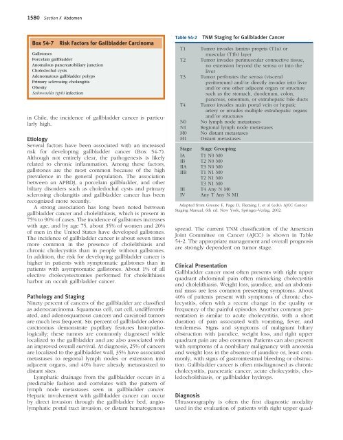
Box <strong>54</strong>-7 Risk Factors for Gallbladder Carcinoma<br />
Gallstones<br />
Porcelain gallbladder<br />
Anomalous pancreatobiliary junction<br />
<strong>Ch</strong>oledochal cysts<br />
Adenomatous gallbladder polyps<br />
Primary sclerosing cholangitis<br />
Obesity<br />
Salmonella typhi infection<br />
in <strong>Ch</strong>ile, the incidence of gallbladder cancer is particularly<br />
high.<br />
Etiology<br />
Several factors have been associated with an increased<br />
risk for developing gallbladder cancer (Box <strong>54</strong>-7).<br />
Although not entirely clear, the pathogenesis is likely<br />
related to chronic infl ammation. Among these factors,<br />
gallstones are the most common because of the high<br />
prevalence in the general population. The association<br />
between an APBDJ, a porcelain gallbladder, and other<br />
biliary disorders such as choledochal cysts and primary<br />
sclerosing cholangitis and gallbladder cancer has been<br />
recognized more recently.<br />
A strong association has long been noted between<br />
gallbladder cancer and cholelithiasis, which is present in<br />
75% to 90% of cases. The incidence of gallstones increases<br />
with age, and by age 75, about 35% of women and 20%<br />
of men in the United States have developed gallstones.<br />
The incidence of gallbladder cancer is about seven times<br />
more common in the presence of cholelithiasis and<br />
chronic cholecystitis than in people without gallstones.<br />
In addition, the risk for developing gallbladder cancer is<br />
higher in patients with symptomatic gallstones than in<br />
patients with asymptomatic gallstones. About 1% of all<br />
elective cholecystectomies performed for cholelithiasis<br />
harbor an occult gallbladder cancer.<br />
Pathology and Staging<br />
Ninety percent of cancers of the gallbladder are classifi ed<br />
as adenocarcinoma. Squamous cell, oat cell, undifferentiated,<br />
and adenosquamous cancers and carcinoid tumors<br />
are much less frequent. Six percent of gallbladder adenocarcinomas<br />
demonstrate papillary features histopathologically;<br />
these tumors are commonly diagnosed while<br />
localized to the gallbladder and are also associated with<br />
an improved overall survival. At diagnosis, 25% of cancers<br />
are localized to the gallbladder wall, 35% have associated<br />
metastases to regional lymph nodes or extension into<br />
adjacent organs, and 40% have already metastasized to<br />
distant sites.<br />
Lymphatic drainage from the gallbladder occurs in a<br />
predictable fashion and correlates with the pattern of<br />
lymph node metastases seen in gallbladder cancer.<br />
Hepatic involvement with gallbladder cancer can occur<br />
by direct invasion through the gallbladder bed, angiolymphatic<br />
portal tract invasion, or distant hematogenous<br />
Table <strong>54</strong>-2 TNM Staging for Gallbladder Cancer<br />
T1 Tumor invades lamina propria (T1a) or<br />
muscular (T1b) layer<br />
T2 Tumor invades perimuscular connective tissue,<br />
no extension beyond the serosa or into the<br />
liver<br />
T3 Tumor perforates the serosa (visceral<br />
peritoneum) and/or directly invades into liver<br />
and/or one other adjacent organ or structure<br />
such as the stomach, duodenum, colon,<br />
pancreas, omentum, or extrahepatic bile ducts<br />
T4 Tumor invades main portal vein or hepatic<br />
artery or invades multiple extrahepatic organs<br />
and/or structures<br />
N0 No lymph node metastases<br />
N1 Regional lymph node metastases<br />
M0 No distant metastases<br />
M1 Distant metastases<br />
Stage Stage Grouping<br />
IA T1 N0 M0<br />
IB T2 N0 M0<br />
IIA T3 N0 M0<br />
IIB T1 N1 M0<br />
T2 N1 M0<br />
T3 N1 M0<br />
III T4 Any N M0<br />
IV Any T Any N M1<br />
Adapted from Greene F, Page D, Fleming I, et al (eds): AJCC Cancer<br />
Staging Manual, 6th ed. New York, Springer-Verlag, 2002.<br />
spread. The current TNM classifi cation of the American<br />
Joint Committee on Cancer (AJCC) is shown in Table<br />
<strong>54</strong>-2. The appropriate management and overall prognosis<br />
are strongly dependent on tumor stage.<br />
Clinical Presentation<br />
Gallbladder cancer most often presents with right upper<br />
quadrant abdominal pain often mimicking cholecystitis<br />
and cholelithiasis. Weight loss, jaundice, and an abdominal<br />
mass are less common presenting symptoms. About<br />
40% of patients present with symptoms of chronic cholecystitis,<br />
often with a recent change in the quality or<br />
frequency of the painful episodes. Another common presentation<br />
is similar to acute cholecystitis, with a short<br />
duration of pain associated with vomiting, fever, and<br />
tenderness. Signs and symptoms of malignant biliary<br />
obstruction with jaundice, weight loss, and right upper<br />
quadrant pain are also common. Patients can also present<br />
with symptoms of a nonbiliary malignancy with anorexia<br />
and weight loss in the absence of jaundice or, least commonly,<br />
with signs of gastrointestinal bleeding or obstruction.<br />
Gallbladder cancer is often misdiagnosed as chronic<br />
cholecystitis, pancreatic cancer, acute cholecystitis, choledocholithiasis,<br />
or gallbladder hydrops.<br />
Diagnosis<br />
Ultrasonography is often the fi rst diagnostic modality<br />
used in the evaluation of patients with right upper quad-