View/Open - Università degli Studi di Milano-Bicocca
View/Open - Università degli Studi di Milano-Bicocca
View/Open - Università degli Studi di Milano-Bicocca
You also want an ePaper? Increase the reach of your titles
YUMPU automatically turns print PDFs into web optimized ePapers that Google loves.
Bonetti et. al., PLoS Genet. 2010 May; 6(5): e1000966.<br />
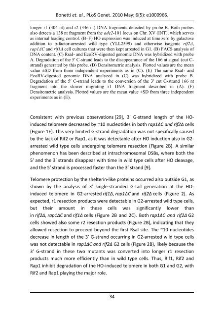
longer r1 (304 nt) and r2 (346 nt) DNA fragments detected by probe B. Both probes<br />
also detects a 138 nt fragment from the ade2-101 locus on Chr. XV (INT), which serves<br />
as internal loa<strong>di</strong>ng control. (B–F) HO expression was induced at time zero by galactose<br />
ad<strong>di</strong>tion to α-factor-arrested wild type (YLL2599) and otherwise isogenic rif2Δ,<br />
rap1ΔC and rif1Δ cell cultures that were then kept arrested in G1. (B) FACS analysis of<br />
DNA content. (C) RsaI- and EcoRV-<strong>di</strong>gested genomic DNA was hybri<strong>di</strong>zed with probe<br />
A. Degradation of the 5′ C-strand leads to the <strong>di</strong>sappearance of the 166 nt signal (cut Cstrand)<br />
generated by this probe. (D) Densitometric analysis. Plotted values are the mean<br />
value ±SD from three independent experiments as in (C). (E) The same RsaI- and<br />
EcoRV-<strong>di</strong>gested genomic DNA analyzed in (C) was hybri<strong>di</strong>zed with probe B.<br />
Degradation of the 5′ C-strand leads to the conversion of the 3′ cut G-strand 166 nt<br />
fragment into the slower migrating r1 DNA fragment described in (A). (F)<br />
Densitometric analysis. Plotted values are the mean value ±SD from three independent<br />
experiments as in (E).<br />
Consistent with previous observations [29], 3′ G-strand length of the HOinduced<br />
telomere decreased by ~10 nucleotides in both rap1ΔC and rif2Δ cells<br />
(Figure 1E). This very limited G-strand degradation was not specifically caused<br />
by the lack of Rif2 or Rap1, as it was detectable after HO induction also in G2arrested<br />
wild type cells undergoing telomere resection (Figure 2B). A similar<br />
phenomenon has been described at intrachromosomal DSBs, where both the<br />
5′ and the 3′ strands <strong>di</strong>sappear with time in wild type cells after HO cleavage,<br />
and the 5′ strand is processed faster than the 3′ strand [9].<br />
Telomere protection by the shelterin-like proteins occurred also outside G1, as<br />
shown by the analysis of 3′ single-stranded G-tail generation at the HOinduced<br />
telomere in G2-arrested rif1Δ, rap1ΔC and rif2Δ cells (Figure 2). As<br />
expected, r1 resection products were detectable in G2-arrested wild type cells,<br />
but their amount in these cells was significantly lower than<br />
in rif2Δ, rap1ΔC and rif1Δ cells (Figure 2B and 2C). Both rap1ΔC and rif2Δ G2<br />
cells showed also some r2 resection products (Figure 2B), in<strong>di</strong>cating that they<br />
allowed resection to proceed beyond the first RsaI site. The ~10 nucleotides<br />
decrease in length of the 3′ G-strand occurring in G2-arrested wild type cells<br />
was not detectable in rap1ΔC and rif2Δ G2 cells (Figure 2B), likely because the<br />
3′ G-strand in these two mutants was converted into longer r1 resection<br />
products much more efficiently than in wild type cells. Thus, Rif1, Rif2 and<br />
Rap1 inhibit degradation of the HO-induced telomere in both G1 and G2, with<br />
Rif2 and Rap1 playing the major role.<br />
34


