View/Open - Università degli Studi di Milano-Bicocca
View/Open - Università degli Studi di Milano-Bicocca
View/Open - Università degli Studi di Milano-Bicocca

Create successful ePaper yourself
Turn your PDF publications into a flip-book with our unique Google optimized e-Paper software.
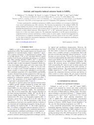
Bonetti et. al., PLoS Genet. 2010 May; 6(5): e1000966.<br />
(E) RsaI-<strong>di</strong>gested genomic DNA was hybri<strong>di</strong>zed with the single-stranded riboprobe A<br />
described in Figure 1A, which anneals to the 5′ C-strand and reveals an uncut 460 nt<br />
DNA fragment (uncut). After HO cleavage, this fragment is converted into a 304 nt<br />
fragment (cut) detected by the same probe (cut C-strand). (F) Densitometric analysis.<br />
Plotted values are the mean value ±SD from three independent experiments as in (E).<br />
(G) The system used to generate an HO-induced DSB. Hybri<strong>di</strong>zation of EcoRV<strong>di</strong>gested<br />
genomic DNA with a probe that anneals to the 5′ strand to a site located 215 nt<br />
from the HO cutting site reveals a 430 nt HO-cut 5′-strand fragment. Loss of the 5′<br />
strand beyond the hybri<strong>di</strong>zation region leads to <strong>di</strong>sappearance of the signal generated<br />
by the probe. (H–L) HO expression was induced at time zero by galactose ad<strong>di</strong>tion to<br />
α-factor-arrested wild type (YLL2600) and otherwise isogenic yku70Δ and dnl4Δ cells,<br />
all carrying the system in (G). Cells were then kept arrested in G1. (H) FACS analysis<br />
of DNA content. (I) EcoRV-<strong>di</strong>gested genomic DNA was hybri<strong>di</strong>zed with the probe<br />
in<strong>di</strong>cated in (G). The INT band, correspon<strong>di</strong>ng to a chromosome IV sequence, serves as<br />
internal loa<strong>di</strong>ng control. (L) Densitometric analysis. Plotted values are the mean value<br />
±SD from three independent experiments as in (I).<br />
Rif1, Rif2, and Rap1 limit resection at a de novo telomere in<br />
yku70Δ G1 cells<br />
Interestingly, G1-arrested yku70Δ cells converted the 5′ C-strand fragment of<br />
the HO-induced telomere into <strong>di</strong>screte smaller DNA fragments (Figure 3E),<br />
suggesting that C-strand degradation under these con<strong>di</strong>tions is limited to the<br />
terminal part. In order to confirm this observation, we monitored the 3′ Gstrand<br />
of the HO-induced telomere in yku70Δ cells. As shown in Figure 4A and<br />
4B, the 3′ cut G-strand was not converted into the longer resection products r1<br />
and r2 in G1-arrested yku70Δ cells. Therefore, exonucleolytic degradation <strong>di</strong>d<br />
not proceed beyond the EcoRV site located 166 bp from the HO site.<br />
Thus, other proteins might limit resection of the HO-induced telomere in G1<br />
even in the absence of Yku70, and the shelterin-like proteins appear to exert<br />
this effect. In fact, 3′-ended r1 resection products were clearly detectable in<br />
G1-arrested yku70Δ rif2Δ, yku70Δ rap1ΔC and, although to a lesser extent,<br />
yku70Δ rif1Δ cells (Figure 4A and 4B). Furthermore, the smaller C-strand<br />
fragments that accumulated in G1-arrested yku70Δ cells were only slightly<br />
detectable in similarly treated yku70Δ rif2Δ(Figure 4C) and yku70Δ rap1ΔC<br />
cells (data not shown), in<strong>di</strong>cating that 5′ C-strand degradation in these cells<br />
had proceeded beyond 166 bp from the HO site. Thus, Rap1, Rif2 and, to a<br />
lesser extent, Rif1 limit telomeric ssDNA generation in G1 cells lacking Yku.<br />
38