nr. 477 - 2011 - Institut for Natur, Systemer og Modeller (NSM)
nr. 477 - 2011 - Institut for Natur, Systemer og Modeller (NSM)
nr. 477 - 2011 - Institut for Natur, Systemer og Modeller (NSM)

Create successful ePaper yourself
Turn your PDF publications into a flip-book with our unique Google optimized e-Paper software.
36 The DuCa Model<br />
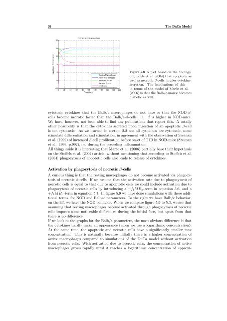
Figure 5.8 A plot based on the findings<br />
of Stoffels et al. (2004) that apoptotic as<br />
well as necrotic β-cells implies cytokine<br />
secretion. The implications of this<br />
in terms of the model of Marée et al.<br />
(2006) is that the Balb/c-mouse becomes<br />
diabetic as well.<br />
cytotoxic cytokines that the Balb/c macrophages do not have or that the NOD-βcells<br />
become necrotic faster than the Balb/c-β-cells; i.e. d is higher in NOD-mice.<br />
We have, however, not been able to find any publications that report this. A totally<br />
other possibility is that the cytokines secreted upon ingestion of an apoptotic β-cell<br />
is not cytotoxic. As we learned in section 2.2 not all cytokines are cytotoxic, some<br />
stimulate differentiation and stimulation, in agreement with the observation of Sreenan<br />
et al. (1999) of increased β-cell proliferation be<strong>for</strong>e onset of T1D in NOD-mice (Sreenan<br />
et al., 1999, p.992), i.e. during the preceding inflammation.<br />
All things aside it is interesting that Marée et al. (2006) partially base their hypothesis<br />
on the Stoffels et al. (2004) article, without mentioning that according to Stoffels et al.<br />
(2004) phagocytosis of apoptotic cells also leads to release of cytokines.<br />
Activation by phagocytosis of necrotic β-cells<br />
A curious thing is that the resting macrophages do not become activated via phagocytosis<br />
of necrotic β-cells. If we assume that the activation rate due to phagocytosis of<br />
necrotic cells is equal to that due to apoptotic cells we could include activation due to<br />
phagocytosis of necrotic cells by introducing a −f1MBn-term in equation 5.6, and a<br />
+f1MBn-term in equation 5.7. In figure 5.9 we have done simulations with these additional<br />
terms, <strong>for</strong> NOD and Balb/c parameters. To the right we have Balb/c behavior,<br />
on the left we have the NOD behavior. When we compare figure 5.9 to 5.3, we see that<br />
assuming that resting macrophages become activated through phagocytosis of necrotic<br />
cells imposes some noticeable differences during the initial face, but apart from that<br />
there is no difference.<br />
If we look at the graphs <strong>for</strong> the Balb/c parameters, the most obvious difference is that<br />
the cytokines hardly make an appearance (when we use a l<strong>og</strong>arithmic concentration).<br />
At the same time, the apoptotic and necrotic cells have a significantly smaller max<br />
concentration. This is naturally because initially there is a higher concentration of<br />
active macrophages compared to simulations of the DuCa model without activation<br />
from necrotic cells. With activation due to necrotic cells, the concentration of active<br />
macrophages grows rapidly until it reaches a l<strong>og</strong>arithmic concentration of approxi-