

MASTER THESIS Biomimetic potential of sponge ... - IAP/TU Wien
MASTER THESIS Biomimetic potential of sponge ... - IAP/TU Wien
MASTER THESIS Biomimetic potential of sponge ... - IAP/TU Wien

Create successful ePaper yourself
Turn your PDF publications into a flip-book with our unique Google optimized e-Paper software.
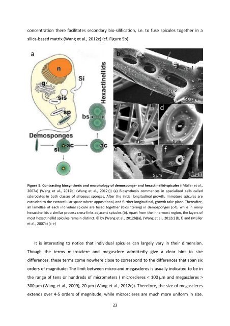
concentration there facilitates secondary bio-silification, i.e. to fuse spicules together in a<br />
silica-based matrix (Wang et al., 2012c) (cf. Figure 5b).<br />
Figure 5: Contrasting biosynthesis and morphology <strong>of</strong> demo<strong>sponge</strong>- and hexactinellid-spicules ((Müller et al.,<br />
2007a) (Wang et al., 2012b) (Wang et al., 2012c)) (a) Biosynthesis commences in specialized cells called<br />
sclerocytes in both classes <strong>of</strong> siliceous <strong>sponge</strong>s. After the initial longitudinal growth, immature spicules are<br />
extruded to the extracellular space where appositional, and further longitudinal, growth take place. Thereafter,<br />
all lamellae <strong>of</strong> each individual spicule are fused together (biosintering) in demo<strong>sponge</strong>s (c-f), while in many<br />
hexactinellids a similar process cross-links adjacent spicules (b). Apart from the innermost region, the layers <strong>of</strong><br />
most hexactinellid spicules remain distinct. © by (Wang et al., 2012b)(a), (Wang et al., 2012c) (b, f) and (Müller<br />
et al., 2007a) (c-e)<br />
It is interesting to notice that individual spicules can largely vary in their dimension.<br />
Though the terms microsclere and megasclere admittedly give a clear hint to size<br />
differences, these terms come nowhere close to correspond to the differences that span six<br />
orders <strong>of</strong> magnitude: The limit between micro-and megascleres is usually indicated to be in<br />
the range <strong>of</strong> tens or hundreds <strong>of</strong> micrometers ( microscleres < 100 µm and megascleres ><br />
300 µm (Wang et al., 2009), 20 µm (Wang et al., 2012c)). Therefore, the size <strong>of</strong> megascleres<br />
extends over 4-5 orders <strong>of</strong> magnitude, while microscleres are much more uniform in size.<br />
23


