

MASTER THESIS Biomimetic potential of sponge ... - IAP/TU Wien
MASTER THESIS Biomimetic potential of sponge ... - IAP/TU Wien
MASTER THESIS Biomimetic potential of sponge ... - IAP/TU Wien

You also want an ePaper? Increase the reach of your titles
YUMPU automatically turns print PDFs into web optimized ePapers that Google loves.
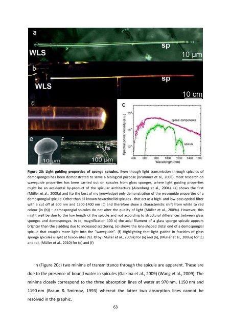
Figure 20: Light guiding properties <strong>of</strong> <strong>sponge</strong> spicules. Even though light transmission through spicules <strong>of</strong><br />
demo<strong>sponge</strong>s has been demonstrated to serve a biological purpose (Brümmer et al., 2008), most research on<br />
waveguide properties has been carried out on spicules from glass <strong>sponge</strong>s, where light guiding properties<br />
might be an accidental by-product <strong>of</strong> the spicular architecture (Aizenberg et al., 2004). (a) shows the first<br />
(Müller et al., 2009a) and (to the best <strong>of</strong> my knowledge) only demonstration <strong>of</strong> the waveguide properties <strong>of</strong> a<br />
demospongial spicule. Other than all known hexactinellid spicules - that act as a high- and low-pass optical filter<br />
with a cut <strong>of</strong>f at 600 nm and 1300-1400 nm (c) and therefore show a characteristic shift from white to red<br />
colour (in (b)) – demospongial spicules do not alter the quality <strong>of</strong> light (Müller et al., 2009a). However, this<br />
might well be due to the low length <strong>of</strong> the spicule and not according to structural differences between glass<br />
<strong>sponge</strong>s and demo<strong>sponge</strong>s. In (d, magnification 100 x) the axial filament <strong>of</strong> a glass <strong>sponge</strong> spicule appears<br />
brighter than the cladding due to increased scattering. (e) shows the lens-shaped distal end <strong>of</strong> a demospongial<br />
spicule that couples more light into the “waveguide”. (f) Highlighting that light guided in fascicles <strong>of</strong> glass<br />
<strong>sponge</strong> spicules is split at fusion sites (fs). © by (Müller et al., 2009a) for (a) and (b), (Müller et al., 2006a) for (c)<br />
and (d), (Müller et al., 2010) for (e) and (f)<br />
In (Figure 20c) two minima <strong>of</strong> transmittance through the spicule are apparent. These are<br />
due to the presence <strong>of</strong> bound water in spicules (Galkina et al., 2009) (Wang et al., 2009). The<br />
minima closely correspond to the three absorption lines <strong>of</strong> water at 970 nm, 1150 nm and<br />
1190 nm (Braun & Smirnov, 1993) whereat the latter two absorption lines cannot be<br />
resolved in the graphic.<br />
63


