

MASTER THESIS Biomimetic potential of sponge ... - IAP/TU Wien
MASTER THESIS Biomimetic potential of sponge ... - IAP/TU Wien
MASTER THESIS Biomimetic potential of sponge ... - IAP/TU Wien

You also want an ePaper? Increase the reach of your titles
YUMPU automatically turns print PDFs into web optimized ePapers that Google loves.
their environment (Inagaki et al., 2003). Inside the cells (sclerocytes) silica is accumulated in<br />
membrane-bound bound vesicles (silicasomes) where the initial phase <strong>of</strong> spicule synthesis occurs.<br />
In parallel, the proteins yielding the axial filament are synthesized, processed and<br />
imported into silicasomes. Within these vesicles, silicateins, silintaphins and silicases<br />
assemble into the axial filaments. For the self-assembly <strong>of</strong> the silicateins into oligomers<br />
(Figure 13b-d) a fractal mechanism has been proposed (Murr & Morse, 2005). The<br />
silicasomes are associated with filaments that likely ly direct them towards the cellular<br />
membrane and are crucial for the extrusion <strong>of</strong> the immature spicule into the extracellular<br />
matrix later on (Wang et al., 2011c).<br />
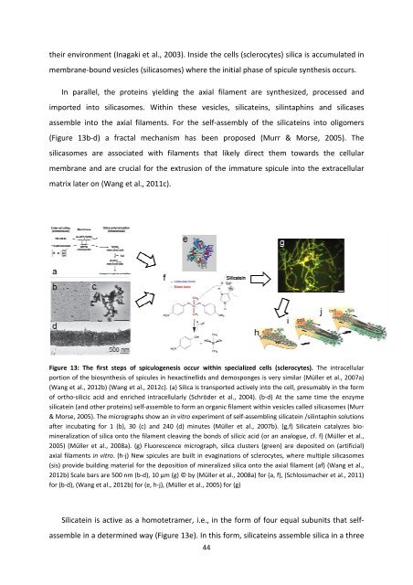
Figure 13: The first steps <strong>of</strong> spiculogenesis occur within specialized cells (sclerocytes). The intracellular<br />
portion <strong>of</strong> the biosynthesis <strong>of</strong> spicules in hexactinellids and demo<strong>sponge</strong>s is very similar (Müller et al., 2007a)<br />
(Wang et al., 2012b) (Wang et al., 2012c). (a) Silica is transported actively into the cell, presumably in the form<br />
<strong>of</strong> ortho-silicic acid and enriched intracellularly (Schröder et al., 2004). (b-d) At the same time the enzyme<br />
silicatein (and other proteins) self-assemble e to form an organic filament within vesicles called silicasomes (Murr<br />
& Morse, 2005). The micrographs show an in vitro experiment <strong>of</strong> self-assembling silicatein /silintaphin solutions<br />
after incubating for 1 (b), 30 (c) and 240 (d) minutes (Müller et al., 2007b). (g,f) Silicatein catalyzes bio-<br />
mineralization <strong>of</strong> silica onto the filament cleaving the bonds <strong>of</strong> silicic acid (or an analogue, cf. f) (Müller et al.,<br />
2005) (Müller et al., 2008a). (g) Fluorescence micrograph, silica clusters (green) are deposited on (artificial)<br />
axial filaments in vitro. (h-j) New spicules are built in evaginations <strong>of</strong> sclerocytes, where multiple silicasomes<br />
(sis) provide building material for the deposition <strong>of</strong> mineralized silica onto the axial filament (af) (Wang et al.,<br />
2012b) Scale bars are 500 nm (b-d), 10 µm (g) © by (Müller et al., 2008a) for (a, f), (Schlossmacher et al., 2011)<br />
for (b-d), (Wang et al., 2012b) for (e, h-j), (Müller et al., 2005) for (g)<br />
Silicatein is active as a homotetramer, i.e., in the form <strong>of</strong> four equal subunits that self-<br />
assemble in a determined way (Figure 13e). In this form, silicateins assemble silica in a three<br />
44


