

Complete issue - IMA Fungus
Complete issue - IMA Fungus
Complete issue - IMA Fungus

Create successful ePaper yourself
Turn your PDF publications into a flip-book with our unique Google optimized e-Paper software.
Gelatinomyces siamensis gen. sp. nov. on bamboo in Thailand<br />
ARTICLE<br />
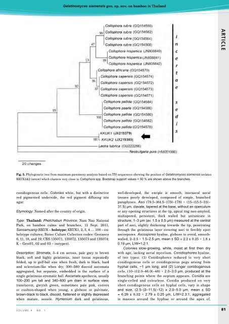
Fig. 5. Phylogenetic tree from maximum parsimony analysis based on ITS sequences showing the position of Gelatinomyces siamensis isolates<br />
KKUK1&2 (arrow) which clusters very close to Collophora spp. Bootstrap support values > 50 % are shown above the branches.<br />
conidiogenous cells. Colonies white, but with a distinctive<br />
red pigmented underside, the red pigment diffusing into<br />
agar.<br />
Etymology: Named after the country of origin.<br />
Type: Thailand: Phetchabun Province: Nam Nao National<br />
Park, on bamboo culms and branches, 11 Sept. 2011,<br />
Sanoamuang (KKUK – holotype; KKUK1, 2, 3, 4…. 100 – exholotype<br />
cultures; Biotec Culture Collection codes: Gesiasco<br />
6, 11, 18, and 19; CBS 135071, 135072, 135073 and 135074;<br />
K – Gesi01, 02 and 03 – isotypes).<br />
Description: Stromata, 3–4 cm across, pale grey to brown<br />
black, soft and highly gelatinous, inner t<strong>issue</strong> repeatedly<br />
folded, up to golf-ball size when fresh, dark to black, hard<br />
and sclerotium-like when dry; 300–560 discoid ascomata<br />
aggregated, but separate, embedded in the surface of a<br />
single gelatinous stromatic ball. Ascomata apothecia, usually<br />
100–200 µm tall and 340–600 µm diam in surface view,<br />
translucent, greyish green, sometimes pale pink, convex<br />
or cushion-shaped when young, ± globose or pulvinate,<br />
brown-black to black, discoid, flattened or slightly depressed<br />
when mature, sessile. Hymenium dark and gelatinous,<br />
well-developed, the exciple is smooth, interascal ascal<br />
t<strong>issue</strong>s poorly developed, composed of simple, branched<br />
paraphyses. Asci (79.5–)84.5–175(–178) × (15–)15.5–31(–<br />
31.5) µm, clavate, tapered at the base, without an operculum<br />
or any opening structures at the tip, apical ring non-amyloid,<br />
multispored, persistent, thick walled but unitunicate in<br />
structure, 1–3 µm (av. 1.5 ± 0.5 µm) measured at the central<br />
part of asci, slightly thickening towards the tip, penetrating<br />
through the gelatinous layer covering asci to forcibly eject<br />
ascospores. Ascospores hyaline, globose to ovoid, smoothwalled,<br />
2–2.5 × 1.5–2.5 µm, mean ± SD = 2.2 ± 0.25 × 1.8 ±<br />
0.19 µm, L/W=1.2:1.<br />
Colonies slow-growing, white, moist at first then dry<br />
with age, lacking aerial mycelium. Conidiophores hyaline,<br />
of two types: (1) Conidiophores reduced to very short<br />
conidiogenous cells or conidiogenous pegs arising from<br />
hyphal cells, ~1 µm long; and (2) Longer conidiogenous<br />
cells, (10–)12.0–46.0(–48) × 2.0–3.0 µm, produced at the<br />
branching points where the septum appears. Conidia are<br />
single-celled and colourless. Conidia produced on very<br />
short conidiogenous cells on hyphal cells, vary in shape<br />
and size, (2.5–)3–11.5(–12) x 2.0–5.0 µm, mean ± SD<br />
= 6.29 ± 0.32 × 2.79 ± 0.25 µm, L/W=2.3:1, aggregated<br />
in masses around the hyphae or around the apex of,<br />
volume 4 · no. 1<br />
81


