

Complete issue - IMA Fungus
Complete issue - IMA Fungus
Complete issue - IMA Fungus

You also want an ePaper? Increase the reach of your titles
YUMPU automatically turns print PDFs into web optimized ePapers that Google loves.
Re-evaluation of Arthrinium (syn. Apiospora)<br />
ARTICLE<br />
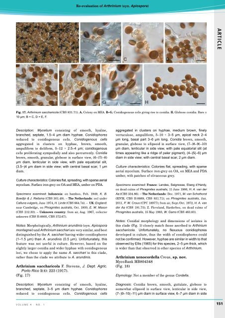
Fig. 17. Arthrinium saccharicola (CBS 831.71). A. Colony on MEA. B–G. Conidiogenous cells giving rise to conidia. H. Globose conidia. Bars =<br />
10 µm; B = C, D = E, F.<br />
Description: Mycelium consisting of smooth, hyaline,<br />
branched, septate, 1.5–4 µm diam hyphae. Conidiophores<br />
reduced to conidiogenous cells. Conidiogenous cells<br />
aggregated in clusters on hyphae, brown, smooth,<br />
ampulliform to doliiform, 5–12 × 2.5–4 µm; conidiogenous<br />
cells proliferating sympodially and also percurrently. Conidia<br />
brown, smooth, granular, globose in surface view, (6–)7(–8)<br />
µm diam, lenticular in side view, with pale equatorial slit,<br />
(3.5–)4 µm diam in side view; with central basal scar, 1 µm<br />
diam.<br />
Culture characteristics: Colonies flat, spreading, with sparse aerial<br />
mycelium. Surface iron-grey on OA and MEA, umber on PDA.<br />
Specimens examined: Indonesia: on bamboo, Feb. 1949, K. B.<br />
Boedijn & J. Reitsma (CBS 301.49). – The Netherlands: soil under<br />
Calluna vulgaris, June 1974, H. Linde (CBS 664.74). – UK: England:<br />
near Cambridge, on Phragmites australis, Oct. 1930, E. W. Mason<br />
(CBS 212.30). – Unknown country: from air, Aug. 1967, collector<br />
unknown (CBS H-8805, CBS 372.67).<br />
Notes: Morphologically, Arthinium arundinis (syn. Apiospora<br />
montagnei) and Arthrinium sacchari are very similar, and best<br />
distinguished by the A. sacchari having wider conidiophores<br />
(1–1.5 µm) than A. arundinis (0.5 µm). Unfortunately, this<br />
feature was not useful in culture. However, based on the<br />
slightly larger conidia and wider hyphae with conidiogenous<br />
loci, we chose to apply the name A. sacchari to this clade,<br />
rather than the clade we attribute to A. arundinis.<br />
Arthrinium saccharicola F. Stevens, J. Dept. Agric.<br />
Porto Rico 1(4): 223 (1917).<br />
(Fig. 17)<br />
Description: Mycelium consisting of smooth, hyaline,<br />
branched, septate, 3–5 µm diam hyphae. Conidiophores<br />
reduced to conidiogenous cells. Conidiogenous cells<br />
aggregated in clusters on hyphae, medium brown, finely<br />
verruculose, ampulliform, 5–10 × 3–5 µm, apical neck 2–4<br />
µm long, basal part 3–6 µm long. Conidia brown, smooth,<br />
granular, globose to ellipsoid in surface view, (7–)8–9(–10)<br />
µm diam, lenticular in side view, with pale equatorial slit (at<br />
times appearing like a ridge of paler pigment), (4–)5(–6) µm<br />
diam in side view; with central basal scar, 2 µm diam.<br />
Culture characteristics: Colonies flat, spreading, with sparse<br />
aerial mycelium. Surface iron-grey on OA, on MEA and PDA<br />
umber, with patches of olivaceous grey.<br />
Specimens examined: France: Landes, Seignosse, Etang d’Hardy,<br />
on dead culms of Phragmites australis, 11 June 1986, H. A. van der<br />
Aa (CBS 334.86). – The Netherlands: Dec. 1971, M. van Schothorst<br />
(RIVM, CBS H-8889, CBS 831.71); on Phragmites australis, Jan.<br />
2011, P. W. Crous (CPC 18977); from air, Sept./Oct. 1972, H. A. van<br />
der Aa (CBS 191.73); Z. Flevoland, Harderbos, on dead culms of<br />
Phragmites australis, 15 May 1983, W. Gams (CBS 463.83).<br />
Notes: Conidial morphology and dimensions of isolates in<br />
this clade (Fig. 1) closely match those ascribed to Arthinium<br />
saccharicola. Unfortunately, no flexuous conidiophores<br />
developed in culture, thus the width of conidiophores could<br />
not be confirmed. However, hyphae are similar in width to that<br />
observed by Ellis (1965) for this species, 2–5 µm thick, which<br />
is wider than that observed in other species of Arthrinium.<br />
Arthrinium xenocordella Crous, sp. nov.<br />
MycoBank MB804348<br />
(Fig. 18)<br />
Etymology: Not a member of the genus Cordella.<br />
Diagnosis: Conidia brown, smooth, guttulate, globose to<br />
somewhat ellipsoid in surface view, lenticular in side view,<br />
(7–)9–10(–11) µm diam in surface view, 6–7 µm diam in side<br />
volume 4 · no. 1<br />
151


