

Complete issue - IMA Fungus
Complete issue - IMA Fungus
Complete issue - IMA Fungus

Create successful ePaper yourself
Turn your PDF publications into a flip-book with our unique Google optimized e-Paper software.
Roux et al.<br />
ARTICLE<br />
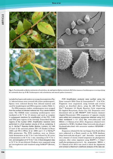
Fig. 1. Puccinia psidii on Myrtus communis in South Africa. A. Leaf spot on Myrtus communis. B. Yellow masses of urediniospores covering a dying<br />
M. communis shoot tip. C–D. Urediniospores with echinilations and smooth pathes (tonsures).<br />
included leaf spots and cankers on young shoots/petioles (Fig.<br />
1). Infected t<strong>issue</strong>s were covered with yellow urediniospores.<br />
Spores were collected directly from infected material and<br />
used for morphological and DNA sequence studies (Table 1).<br />
For DNA sequence studies, urediniospores were scraped<br />
from the surface of infected material into 18 µL sterile SABAX<br />
water. The SABAX water containing urediniospores were<br />
incubated at 94 ºC for 10 minutes and used as template<br />
in subsequent reactions for amplification of the ITS1, 5.8S<br />
and ITS2 gene regions of the Internally Transcribed Spacer<br />
regions of the nuclear rDNA. Amplification reactions were<br />
performed in a final reaction volume of 25 µL containing: 5<br />
µL 5× MyTaq Reaction Buffer (Bioline, London), 0.2 mM<br />
of each of the universal primers ITS 1-F (Gardes & Bruns<br />
1993) and ITS 4 (White et al. 1990) and 1 U of MyTaq<br />
DNA polymerase. The PCR conditions were as follows:<br />
Initial denaturation at 94 ºC for 3 min followed by 30 cycles<br />
of denaturation at 94 ºC for 1 min, annealing at 53 ºC for 1<br />
min, and elongation at 72 ºC for 1 min. A final elongation step<br />
at 72 ºC for 10 min followed. Products were separated using<br />
gel electrophoresis and visualised using GelRed (Biotium,<br />
CA).<br />
PCR Amplification products were purified using the<br />
Zymo research DNA Clean & Concentration - 5 kit (CA).<br />
Fragments were sequenced, using forward and reverse<br />
primers as described above, using the ABI Prism ® Big<br />
Dye TM Terminator 3.0 Ready Reaction Cycle sequencing<br />
Kit (Applied Biosystems, Foster City, CA). Sequences were<br />
determined with an ABI PRISM 3100 Genetic Analyzer<br />
(Applied Biosystems). DNA sequences of opposite strands<br />
were edited and consensus sequences obtained using CLC<br />
Main workbench v. 6.1 (CLC Bio, www.clcbio.com) and<br />
MEGA v. 5 (Tamura et al. 2011). Sequences obtained were<br />
submitted to NCBI’s GenBank (http://www.ncbi.nlm.nih.gov/<br />
Genbank/index.html) with accession numbers KF220289 –<br />
KF220293.<br />
Sequences obtained for the rust fungus from South Africa<br />
were subjected to a Blastn search on the NCBI database<br />
(http://www.ncbi.nlm.nih.gov) and thereafter incorporated<br />
into a dataset of closely related sequences for phylogenetic<br />
analyses. After online alignment using MAFFT v. 7 (http://<br />
mafft.cbrc.jp/alignment/server/), the programme MEGA v.<br />
5.1 (Tamura et al. 2011) was used to check the alignments<br />
and conduct a Maximum Likelihood analysis of the data set.<br />
156 ima fUNGUS


