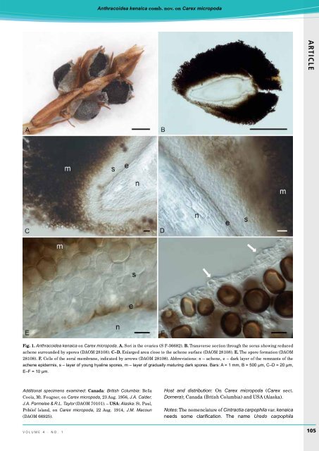
Piątek ARTICLE Table 1. Spore size range, and mean spore sizes with standard deviation of Anthracoidea kenaica specimens examined in this study. Spore size range (µm) Average spore size with standard deviation (µm) Specimen (14.5–)15.0–20.5(–21.5) × 12.0–17.5(–18.5) 17.0–20.5(–22.0) × 12.0– 18.0(–20.5) (14.0–)15.0–20.5(–21.0) × (11.5–)12.0–17.5(–18.5) 18.1 ± 1.6 × 15.2 ± 1.7 USA, Alaska, Kenai Peninsula, Head of Palmer Creek Valley, 26 July 1951, J.A. Calder 6229 (DAOM 28108 – holotype) 19.2 ± 1.3 × 16.1 ± 1.8 Same locality, date and collector (S F-36682 – isotype) 18.2 ± 1.7 × 15.2 ± 1.8 USA, Alaska, St. Paul, Pribilof Island, 22 Aug. 1914, J.M. Macoun (DAOM 66925) (14.5–)17.0–20.5(–22.0) × 13.5–18.5(–19.0) 18.5 ± 1.2 × 15.9 ± 1.4 Canada, British Columbia, Bella Coola, Mt. Fougner, 23 Aug. 1956, J.A. Calder, J.A. Parmelee & R.L. Taylor (DAOM 70101) to boiling point and cooled, then examined under a Nikon Eclipse 80i light microscope. LM micrographs were taken with a Nikon DS-Fi1 camera. Spores were measured using NIS-Elements BR v. 3.0 imaging software. Spore size range, mean spore size, and standard deviation of 50 measured spores of each investigated specimen were calculated (Table 1). The species description includes combined values from all measured specimens. The spores were measured in plane view and measurements were adjusted to the nearest 0.5 µm. Spore size ranges were assigned to one of the three groups distinguished by Savile (1952): (1) small-sized spores, 13–21(–23) × 9–17(–20) µm; (2) medium-sized spores, 15– 25(–27) × 10–21 µm; and (3) large-sized spores, 18–33 × 13–28 µm. For scanning electron microscopy (SEM), spores taken directly from dried specimens were dusted onto carbon tabs and fixed to an aluminium stub with double-sided transparent tape. The stubs were sputter-coated with carbon using a Cressington sputter-coater and viewed under a Hitachi S-4700 scanning electron microscope, with a working distance of ca. 11 mm. SEM micrographs were taken in the Laboratory of Field Emission Scanning Electron Microscopy and Microanalysis at the Institute of Geological Sciences of Jagiellonian University (Kraków). RESULTS Detailed morphological characteristics of the holotype, isotype, and two non-type specimens of Cintractia carpophila var. kenaica are embraced in the species description and illustrated (Figs 1–2). The internal soral structure in the holotype was typical of species of Anthracoidea in that the spores were produced directly on the outer surface of the achene, and not within U-shaped cavities embedded in sterile stroma, a characteristic of Cintractia (Kukkonen 1963, Kukkonen & Vaissalo 1964, Piepenbring 2000). This indicated this smut fungus was better placed in Anthracoidea, as was suggested in other studies (Kukkonen 1963, Zambettakis 1978, Piepenbring 2000). The spores were uniform in shape and size ranges between collections (Table 1). My examination of specimens of Cintractia carpophila var. kenaica matched well the short description given by Savile (1952), although the spore surface was not smooth as stated in the protologue, but smooth or very finely punctate in LM, and very finely verruculose in SEM. The very fine ornamentation of spores was probably outside the limits of resolution of Savile’s light microscope. In general, the present examination confirms the decision of Savile (1952) to consider this smut as distinct. However, a specific status seems to be appropriate for this taxon. This is in line with the conclusion of Kukkonen (1963), who, however, did not formally make the transfer. Accordingly, a new combination is necessary. TAXONOMY Anthracoidea kenaica (Savile) M. Piątek, comb. nov. MycoBank: MB804512 (Figs 1–2) Basionym: Cintractia carpophila var. kenaica Savile, Can. J. Bot. 30: 419 (1952). Synonym: Anthracoidea heterospora var. kenaica (Savile) Zambett., Bull. trimest. Soc. mycol. Fr. 94: 177 (1978), nom. inval. (Art. 41.5). Type: USA: Alaska: Kenai Peninsula, Head of Palmer Creek Valley, 60°49’N, 149°33’W, on Carex micropoda (syn. Carex pyrenaica subsp. micropoda), 26 July 1951, J.A. Calder 6229 (DAOM 28108 – holotype, S F-36682 – isotype). Description: Sori in all or single ovaries of the inflorescence, black, globose or ovoid, about 1–1.5 mm diam, at first covered by a silvery membrane and perigynium that later ruptures revealing agglutinated spores, powdery on the surface, the sori are partly hidden by the perigynium and scales. Sori develop around reduced achenes that are consecutively surrounded by a thin dark layer of the remnants of achene epidermis, a hyaline layer of sporogeneous hyphae with young spores, a layer of gradually maturing dark spores, and a thin membrane of host origin. Spores usually more or less flattened, chestnut brown, reddish brown to dark brown, quite regular in shape and size, globose, subglobose or broadly ellipsoidal, small, (14.0–)15.0–20.5(–22.0) × (11.5–)12.0–18.5(–20.5) µm [av. ± SD, 18.5 ± 1.5 × 15.6 ± 1.7 µm, n = 200/4], rarely enclosed by a thin, hyaline, mucilaginous sheath; wall even, 1.0–1.5 µm thick, somewhat darker than the rest of the spore, without protuberances and light-refractive spots, but with 2–5 distinct internal swellings; surface smooth or very finely punctate in LM, spore profile smooth, surface very finely verruculose in SEM. 104 ima fUNGUS
Anthracoidea kenaica comb. nov. on Carex micropoda ARTICLE Fig. 1. Anthracoidea kenaica on Carex micropoda. A. Sori in the ovaries (S F-36682). B. Transverse section through the sorus showing reduced achene surrounded by spores (DAOM 28108). C–D. Enlarged area close to the achene surface (DAOM 28108). E. The spore formation (DAOM 28108). F. Cells of the soral membrane, indicated by arrows (DAOM 28108). Abbreviations: n – achene, e – dark layer of the remnants of the achene epidermis, s – layer of young hyaline spores, m – layer of gradually maturing dark spores. Bars: A = 1 mm, B = 500 µm, C–D = 20 µm, E–F = 10 µm. Additional specimens examined: Canada: British Columbia: Bella Coola, Mt. Fougner, on Carex micropoda, 23 Aug. 1956, J.A. Calder, J.A. Parmelee & R.L. Taylor (DAOM 70101). – USA: Alaska: St. Paul, Pribilof Island, on Carex micropoda, 22 Aug. 1914, J.M. Macoun (DAOM 66925). Host and distribution: On Carex micropoda (Carex sect. Dornera); Canada (British Columbia) and USA (Alaska). Notes: The nomenclature of Cintractia carpophila var. kenaica needs some clarification. The name Uredo carpophila volume 4 · no. 1 105