

Complete issue - IMA Fungus
Complete issue - IMA Fungus
Complete issue - IMA Fungus

You also want an ePaper? Increase the reach of your titles
YUMPU automatically turns print PDFs into web optimized ePapers that Google loves.
Piątek<br />
ARTICLE<br />
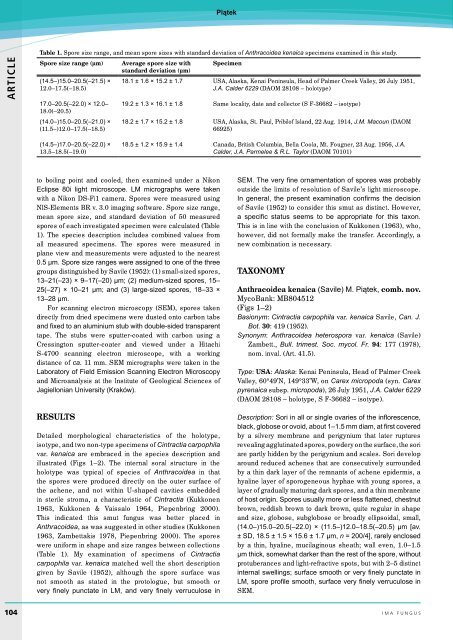
Table 1. Spore size range, and mean spore sizes with standard deviation of Anthracoidea kenaica specimens examined in this study.<br />
Spore size range (µm) Average spore size with<br />
standard deviation (µm)<br />
Specimen<br />
(14.5–)15.0–20.5(–21.5) ×<br />
12.0–17.5(–18.5)<br />
17.0–20.5(–22.0) × 12.0–<br />
18.0(–20.5)<br />
(14.0–)15.0–20.5(–21.0) ×<br />
(11.5–)12.0–17.5(–18.5)<br />
18.1 ± 1.6 × 15.2 ± 1.7 USA, Alaska, Kenai Peninsula, Head of Palmer Creek Valley, 26 July 1951,<br />
J.A. Calder 6229 (DAOM 28108 – holotype)<br />
19.2 ± 1.3 × 16.1 ± 1.8 Same locality, date and collector (S F-36682 – isotype)<br />
18.2 ± 1.7 × 15.2 ± 1.8 USA, Alaska, St. Paul, Pribilof Island, 22 Aug. 1914, J.M. Macoun (DAOM<br />
66925)<br />
(14.5–)17.0–20.5(–22.0) ×<br />
13.5–18.5(–19.0)<br />
18.5 ± 1.2 × 15.9 ± 1.4 Canada, British Columbia, Bella Coola, Mt. Fougner, 23 Aug. 1956, J.A.<br />
Calder, J.A. Parmelee & R.L. Taylor (DAOM 70101)<br />
to boiling point and cooled, then examined under a Nikon<br />
Eclipse 80i light microscope. LM micrographs were taken<br />
with a Nikon DS-Fi1 camera. Spores were measured using<br />
NIS-Elements BR v. 3.0 imaging software. Spore size range,<br />
mean spore size, and standard deviation of 50 measured<br />
spores of each investigated specimen were calculated (Table<br />
1). The species description includes combined values from<br />
all measured specimens. The spores were measured in<br />
plane view and measurements were adjusted to the nearest<br />
0.5 µm. Spore size ranges were assigned to one of the three<br />
groups distinguished by Savile (1952): (1) small-sized spores,<br />
13–21(–23) × 9–17(–20) µm; (2) medium-sized spores, 15–<br />
25(–27) × 10–21 µm; and (3) large-sized spores, 18–33 ×<br />
13–28 µm.<br />
For scanning electron microscopy (SEM), spores taken<br />
directly from dried specimens were dusted onto carbon tabs<br />
and fixed to an aluminium stub with double-sided transparent<br />
tape. The stubs were sputter-coated with carbon using a<br />
Cressington sputter-coater and viewed under a Hitachi<br />
S-4700 scanning electron microscope, with a working<br />
distance of ca. 11 mm. SEM micrographs were taken in the<br />
Laboratory of Field Emission Scanning Electron Microscopy<br />
and Microanalysis at the Institute of Geological Sciences of<br />
Jagiellonian University (Kraków).<br />
RESULTS<br />
Detailed morphological characteristics of the holotype,<br />
isotype, and two non-type specimens of Cintractia carpophila<br />
var. kenaica are embraced in the species description and<br />
illustrated (Figs 1–2). The internal soral structure in the<br />
holotype was typical of species of Anthracoidea in that<br />
the spores were produced directly on the outer surface of<br />
the achene, and not within U-shaped cavities embedded<br />
in sterile stroma, a characteristic of Cintractia (Kukkonen<br />
1963, Kukkonen & Vaissalo 1964, Piepenbring 2000).<br />
This indicated this smut fungus was better placed in<br />
Anthracoidea, as was suggested in other studies (Kukkonen<br />
1963, Zambettakis 1978, Piepenbring 2000). The spores<br />
were uniform in shape and size ranges between collections<br />
(Table 1). My examination of specimens of Cintractia<br />
carpophila var. kenaica matched well the short description<br />
given by Savile (1952), although the spore surface was<br />
not smooth as stated in the protologue, but smooth or<br />
very finely punctate in LM, and very finely verruculose in<br />
SEM. The very fine ornamentation of spores was probably<br />
outside the limits of resolution of Savile’s light microscope.<br />
In general, the present examination confirms the decision<br />
of Savile (1952) to consider this smut as distinct. However,<br />
a specific status seems to be appropriate for this taxon.<br />
This is in line with the conclusion of Kukkonen (1963), who,<br />
however, did not formally make the transfer. Accordingly, a<br />
new combination is necessary.<br />
TAXONOMY<br />
Anthracoidea kenaica (Savile) M. Piątek, comb. nov.<br />
MycoBank: MB804512<br />
(Figs 1–2)<br />
Basionym: Cintractia carpophila var. kenaica Savile, Can. J.<br />
Bot. 30: 419 (1952).<br />
Synonym: Anthracoidea heterospora var. kenaica (Savile)<br />
Zambett., Bull. trimest. Soc. mycol. Fr. 94: 177 (1978),<br />
nom. inval. (Art. 41.5).<br />
Type: USA: Alaska: Kenai Peninsula, Head of Palmer Creek<br />
Valley, 60°49’N, 149°33’W, on Carex micropoda (syn. Carex<br />
pyrenaica subsp. micropoda), 26 July 1951, J.A. Calder 6229<br />
(DAOM 28108 – holotype, S F-36682 – isotype).<br />
Description: Sori in all or single ovaries of the inflorescence,<br />
black, globose or ovoid, about 1–1.5 mm diam, at first covered<br />
by a silvery membrane and perigynium that later ruptures<br />
revealing agglutinated spores, powdery on the surface, the sori<br />
are partly hidden by the perigynium and scales. Sori develop<br />
around reduced achenes that are consecutively surrounded<br />
by a thin dark layer of the remnants of achene epidermis, a<br />
hyaline layer of sporogeneous hyphae with young spores, a<br />
layer of gradually maturing dark spores, and a thin membrane<br />
of host origin. Spores usually more or less flattened, chestnut<br />
brown, reddish brown to dark brown, quite regular in shape<br />
and size, globose, subglobose or broadly ellipsoidal, small,<br />
(14.0–)15.0–20.5(–22.0) × (11.5–)12.0–18.5(–20.5) µm [av.<br />
± SD, 18.5 ± 1.5 × 15.6 ± 1.7 µm, n = 200/4], rarely enclosed<br />
by a thin, hyaline, mucilaginous sheath; wall even, 1.0–1.5<br />
µm thick, somewhat darker than the rest of the spore, without<br />
protuberances and light-refractive spots, but with 2–5 distinct<br />
internal swellings; surface smooth or very finely punctate in<br />
LM, spore profile smooth, surface very finely verruculose in<br />
SEM.<br />
104 ima fUNGUS


