

Complete issue - IMA Fungus
Complete issue - IMA Fungus
Complete issue - IMA Fungus

Create successful ePaper yourself
Turn your PDF publications into a flip-book with our unique Google optimized e-Paper software.
Re-evaluation of Arthrinium (syn. Apiospora)<br />
ARTICLE<br />
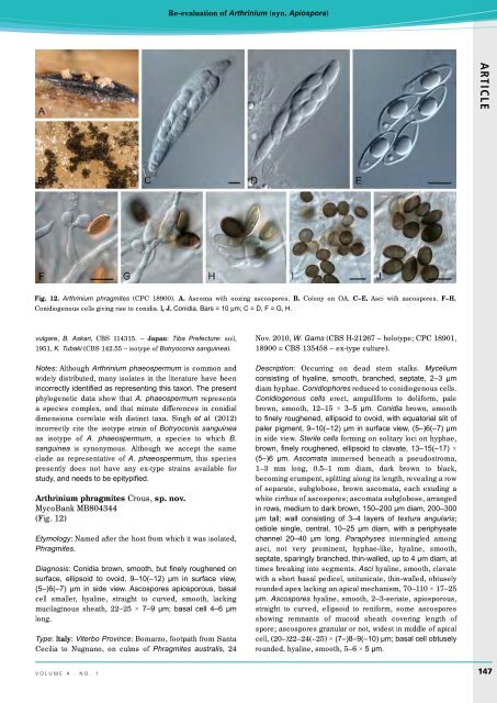
Fig. 12. Arthrinium phragmites (CPC 18900). A. Ascoma with oozing ascospores. B. Colony on OA. C–E. Asci with ascospores. F–H.<br />
Conidiogenous cells giving rise to conidia. I, J. Conidia. Bars = 10 µm; C = D, F = G, H.<br />
vulgare, B. Askari, CBS 114315. – Japan: Tiba Prefecture: soil,<br />
1951, K. Tubaki (CBS 142.55 – isotype of Botryoconis sanguinea).<br />
Notes: Although Arthrinium phaeospermum is common and<br />
widely distributed, many isolates in the literature have been<br />
incorrectly identified as representing this taxon. The present<br />
phylogenetic data show that A. phaeospermum represents<br />
a species complex, and that minute differences in conidial<br />
dimensions correlate with distinct taxa. Singh et al. (2012)<br />
incorrectly cite the isotype strain of Botryoconis sanguinea<br />
as isotype of A. phaeospermum, a species to which B.<br />
sanguinea is synonymous. Although we accept the same<br />
clade as representative of A. phaeospermum, this species<br />
presently does not have any ex-type strains available for<br />
study, and needs to be epitypified.<br />
Arthrinium phragmites Crous, sp. nov.<br />
MycoBank MB804344<br />
(Fig. 12)<br />
Etymology: Named after the host from which it was isolated,<br />
Phragmites.<br />
Diagnosis: Conidia brown, smooth, but finely roughened on<br />
surface, ellipsoid to ovoid, 9–10(–12) µm in surface view,<br />
(5–)6(–7) µm in side view. Ascospores apiosporous, basal<br />
cell smaller, hyaline, straight to curved, smooth, lacking<br />
mucilaginous sheath, 22–25 × 7–9 µm; basal cell 4–6 µm<br />
long.<br />
Type: Italy: Viterbo Province: Bomarzo, footpath from Santa<br />
Cecilia to Nugnano, on culms of Phragmites australis, 24<br />
Nov. 2010, W. Gams (CBS H-21267 – holotype; CPC 18901,<br />
18900 = CBS 135458 – ex-type culture).<br />
Description: Occurring on dead stem stalks. Mycelium<br />
consisting of hyaline, smooth, branched, septate, 2–3 µm<br />
diam hyphae. Conidiophores reduced to conidiogenous cells.<br />
Conidiogenous cells erect, ampulliform to doliiform, pale<br />
brown, smooth, 12–15 × 3–5 µm. Conidia brown, smooth<br />
to finely roughened, ellipsoid to ovoid, with equatorial slit of<br />
paler pigment, 9–10(–12) µm in surface view, (5–)6(–7) µm<br />
in side view. Sterile cells forming on solitary loci on hyphae,<br />
brown, finely roughened, ellipsoid to clavate, 13–15(–17) ×<br />
(5–)6 µm. Ascomata immersed beneath a pseudostroma,<br />
1–3 mm long, 0.5–1 mm diam, dark brown to black,<br />
becoming erumpent, splitting along its length, revealing a row<br />
of separate, subglobose, brown ascomata, each exuding a<br />
white cirrhus of ascospores; ascomata subglobose, arranged<br />
in rows, medium to dark brown, 150–200 µm diam, 200–300<br />
µm tall; wall consisting of 3–4 layers of textura angularis;<br />
ostiole single, central, 10–25 µm diam, with a periphysate<br />
channel 20–40 µm long. Paraphyses intermingled among<br />
asci, not very prominent, hyphae-like, hyaline, smooth,<br />
septate, sparingly branched, thin-walled, up to 4 µm diam, at<br />
times breaking into segments. Asci hyaline, smooth, clavate<br />
with a short basal pedicel, unitunicate, thin-walled, obtusely<br />
rounded apex lacking an apical mechanism, 70–110 × 17–25<br />
µm. Ascospores hyaline, smooth, 2–3-seriate, apiosporous,<br />
straight to curved, ellipsoid to reniform, some ascospores<br />
showing remnants of mucoid sheath covering length of<br />
spore; ascospores granular or not, widest in middle of apical<br />
cell, (20–)22–24(–25) × (7–)8–9(–10) µm; basal cell obtusely<br />
rounded, hyaline, smooth, 5–6 × 5 µm.<br />
volume 4 · no. 1<br />
147


