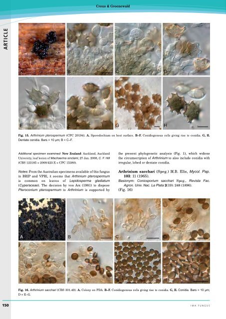
Crous & Groenewald ARTICLE Fig. 15. Arthrinium pterospermum (CPC 20194). A. Sporodochium on host surface. B–F. Conidiogenous cells giving rise to conidia. G, H. Dentate conidia. Bars = 10 µm; B = C–F. Additional specimen examined: New Zealand: Auckland, Auckland University, leaf lesion of Machaerina sinclairii, 27 Jan. 2008, C. F. Hill (CBS 123185 = 2008/423-X = CPC 15380). Notes: From the Australian specimens available of this fungus in BRIP and VPRI, it seems that Arthinium pterospermum is common on leaves of Lepidosperma gladiatum (Cyperaceae). The decision by von Arx (1981) to dispose Pteroconium pterospermum to Arthrinium is supported by the present phylogenetic analysis (Fig. 1), which widens the circumscription of Arthrinium to also include conidia with irregular, lobed or dentate conidia. Arthrinium sacchari (Speg.) M.B. Ellis, Mycol. Pap. 103: 11 (1965). Basionym: Coniosporium sacchari Speg., Revista Fac. Agron. Univ. Nac. La Plata 2(19): 248 (1896). (Fig. 16) Fig. 16. Arthrinium sacchari (CBS 301.49). A. Colony on PDA. B–F. Conidiogenous cells giving rise to conidia. G, H. Conidia. Bars = 10 µm; D = E–G. 150 ima fUNGUS
Re-evaluation of Arthrinium (syn. Apiospora) ARTICLE Fig. 17. Arthrinium saccharicola (CBS 831.71). A. Colony on MEA. B–G. Conidiogenous cells giving rise to conidia. H. Globose conidia. Bars = 10 µm; B = C, D = E, F. Description: Mycelium consisting of smooth, hyaline, branched, septate, 1.5–4 µm diam hyphae. Conidiophores reduced to conidiogenous cells. Conidiogenous cells aggregated in clusters on hyphae, brown, smooth, ampulliform to doliiform, 5–12 × 2.5–4 µm; conidiogenous cells proliferating sympodially and also percurrently. Conidia brown, smooth, granular, globose in surface view, (6–)7(–8) µm diam, lenticular in side view, with pale equatorial slit, (3.5–)4 µm diam in side view; with central basal scar, 1 µm diam. Culture characteristics: Colonies flat, spreading, with sparse aerial mycelium. Surface iron-grey on OA and MEA, umber on PDA. Specimens examined: Indonesia: on bamboo, Feb. 1949, K. B. Boedijn & J. Reitsma (CBS 301.49). – The Netherlands: soil under Calluna vulgaris, June 1974, H. Linde (CBS 664.74). – UK: England: near Cambridge, on Phragmites australis, Oct. 1930, E. W. Mason (CBS 212.30). – Unknown country: from air, Aug. 1967, collector unknown (CBS H-8805, CBS 372.67). Notes: Morphologically, Arthinium arundinis (syn. Apiospora montagnei) and Arthrinium sacchari are very similar, and best distinguished by the A. sacchari having wider conidiophores (1–1.5 µm) than A. arundinis (0.5 µm). Unfortunately, this feature was not useful in culture. However, based on the slightly larger conidia and wider hyphae with conidiogenous loci, we chose to apply the name A. sacchari to this clade, rather than the clade we attribute to A. arundinis. Arthrinium saccharicola F. Stevens, J. Dept. Agric. Porto Rico 1(4): 223 (1917). (Fig. 17) Description: Mycelium consisting of smooth, hyaline, branched, septate, 3–5 µm diam hyphae. Conidiophores reduced to conidiogenous cells. Conidiogenous cells aggregated in clusters on hyphae, medium brown, finely verruculose, ampulliform, 5–10 × 3–5 µm, apical neck 2–4 µm long, basal part 3–6 µm long. Conidia brown, smooth, granular, globose to ellipsoid in surface view, (7–)8–9(–10) µm diam, lenticular in side view, with pale equatorial slit (at times appearing like a ridge of paler pigment), (4–)5(–6) µm diam in side view; with central basal scar, 2 µm diam. Culture characteristics: Colonies flat, spreading, with sparse aerial mycelium. Surface iron-grey on OA, on MEA and PDA umber, with patches of olivaceous grey. Specimens examined: France: Landes, Seignosse, Etang d’Hardy, on dead culms of Phragmites australis, 11 June 1986, H. A. van der Aa (CBS 334.86). – The Netherlands: Dec. 1971, M. van Schothorst (RIVM, CBS H-8889, CBS 831.71); on Phragmites australis, Jan. 2011, P. W. Crous (CPC 18977); from air, Sept./Oct. 1972, H. A. van der Aa (CBS 191.73); Z. Flevoland, Harderbos, on dead culms of Phragmites australis, 15 May 1983, W. Gams (CBS 463.83). Notes: Conidial morphology and dimensions of isolates in this clade (Fig. 1) closely match those ascribed to Arthinium saccharicola. Unfortunately, no flexuous conidiophores developed in culture, thus the width of conidiophores could not be confirmed. However, hyphae are similar in width to that observed by Ellis (1965) for this species, 2–5 µm thick, which is wider than that observed in other species of Arthrinium. Arthrinium xenocordella Crous, sp. nov. MycoBank MB804348 (Fig. 18) Etymology: Not a member of the genus Cordella. Diagnosis: Conidia brown, smooth, guttulate, globose to somewhat ellipsoid in surface view, lenticular in side view, (7–)9–10(–11) µm diam in surface view, 6–7 µm diam in side volume 4 · no. 1 151