

Complete issue - IMA Fungus
Complete issue - IMA Fungus
Complete issue - IMA Fungus

Create successful ePaper yourself
Turn your PDF publications into a flip-book with our unique Google optimized e-Paper software.
Crous & Groenewald<br />
ARTICLE<br />
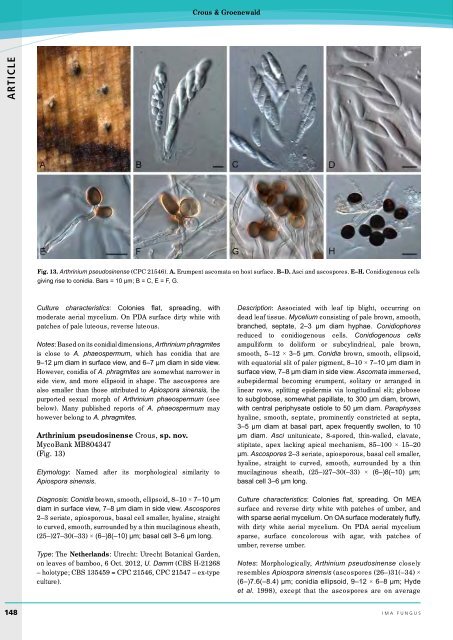
Fig. 13. Arthrinium pseudosinense (CPC 21546). A. Erumpent ascomata on host surface. B–D. Asci and ascospores. E–H. Conidiogenous cells<br />
giving rise to conidia. Bars = 10 µm; B = C, E = F, G.<br />
Culture characteristics: Colonies flat, spreading, with<br />
moderate aerial mycelium. On PDA surface dirty white with<br />
patches of pale luteous, reverse luteous.<br />
Notes: Based on its conidial dimensions, Arthrinium phragmites<br />
is close to A. phaeospermum, which has conidia that are<br />
9–12 µm diam in surface view, and 6–7 µm diam in side view.<br />
However, conidia of A. phragmites are somewhat narrower in<br />
side view, and more ellipsoid in shape. The ascospores are<br />
also smaller than those attributed to Apiospora sinensis, the<br />
purported sexual morph of Arthrinium phaeospermum (see<br />
below). Many published reports of A. phaeospermum may<br />
however belong to A. phragmites.<br />
Arthrinium pseudosinense Crous, sp. nov.<br />
MycoBank MB804347<br />
(Fig. 13)<br />
Etymology: Named after its morphological similarity to<br />
Apiospora sinensis.<br />
Diagnosis: Conidia brown, smooth, ellipsoid, 8–10 × 7–10 µm<br />
diam in surface view, 7–8 µm diam in side view. Ascospores<br />
2–3 seriate, apiosporous, basal cell smaller, hyaline, straight<br />
to curved, smooth, surrounded by a thin mucilaginous sheath,<br />
(25–)27–30(–33) × (6–)8(–10) µm; basal cell 3–6 µm long.<br />
Type: The Netherlands: Utrecht: Utrecht Botanical Garden,<br />
on leaves of bamboo, 6 Oct. 2012, U. Damm (CBS H-21268<br />
– holotype; CBS 135459 = CPC 21546, CPC 21547 – ex-type<br />
culture).<br />
Description: Associated with leaf tip blight, occurring on<br />
dead leaf t<strong>issue</strong>. Mycelium consisting of pale brown, smooth,<br />
branched, septate, 2–3 µm diam hyphae. Conidiophores<br />
reduced to conidiogenous cells. Conidiogenous cells<br />
ampulliform to doliiform or subcylindrical, pale brown,<br />
smooth, 5–12 × 3–5 µm. Conidia brown, smooth, ellipsoid,<br />
with equatorial slit of paler pigment, 8–10 × 7–10 µm diam in<br />
surface view, 7–8 µm diam in side view. Ascomata immersed,<br />
subepidermal becoming erumpent, solitary or arranged in<br />
linear rows, splitting epidermis via longitudinal slit; globose<br />
to subglobose, somewhat papillate, to 300 µm diam, brown,<br />
with central periphysate ostiole to 50 µm diam. Paraphyses<br />
hyaline, smooth, septate, prominently constricted at septa,<br />
3–5 µm diam at basal part, apex frequently swollen, to 10<br />
µm diam. Asci unitunicate, 8-spored, thin-walled, clavate,<br />
stipitate, apex lacking apical mechanism, 85–100 × 15–20<br />
µm. Ascospores 2–3 seriate, apiosporous, basal cell smaller,<br />
hyaline, straight to curved, smooth, surrounded by a thin<br />
mucilaginous sheath, (25–)27–30(–33) × (6–)8(–10) µm;<br />
basal cell 3–6 µm long.<br />
Culture characteristics: Colonies flat, spreading. On MEA<br />
surface and reverse dirty white with patches of umber, and<br />
with sparse aerial mycelium. On OA surface moderately fluffy,<br />
with dirty white aerial mycelium. On PDA aerial mycelium<br />
sparse, surface concolorous with agar, with patches of<br />
umber, reverse umber.<br />
Notes: Morphologically, Arthinium pseudosinense closely<br />
resembles Apiospora sinensis (ascospores (26–)31(–34) ×<br />
(6–)7.6(–8.4) µm; conidia ellipsoid, 9–12 × 6–8 µm; Hyde<br />
et al. 1998), except that the ascospores are on average<br />
148 ima fUNGUS


