

Complete issue - IMA Fungus
Complete issue - IMA Fungus
Complete issue - IMA Fungus

Create successful ePaper yourself
Turn your PDF publications into a flip-book with our unique Google optimized e-Paper software.
Re-evaluation of Arthrinium (syn. Apiospora)<br />
ARTICLE<br />
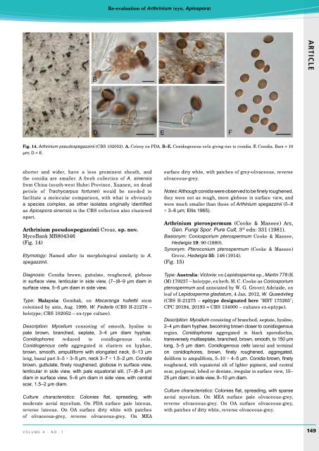
Fig. 14. Arthrinium pseudospegazzinii (CBS 102052). A. Colony on PDA. B–E. Conidiogenous cells giving rise to conidia. F. Conidia. Bars = 10<br />
µm; D = E.<br />
shorter and wider, have a less prominent sheath, and<br />
the conidia are smaller. A fresh collection of A. sinensis<br />
from China (south-west Huhei Province, Xuanen, on dead<br />
petiole of Trachycarpus fortunei) would be needed to<br />
facilitate a molecular comparison, with what is obviously<br />
a species complex, as other isolates originally identified<br />
as Apiospora sinensis in the CBS collection also clustered<br />
apart.<br />
Arthrinium pseudospegazzinii Crous, sp. nov.<br />
MycoBank MB804346<br />
(Fig. 14)<br />
Etymology: Named after its morphological similarity to A.<br />
spegazzinii.<br />
Diagnosis: Conidia brown, guttulate, roughened, globose<br />
in surface view, lenticular in side view, (7–)8–9 µm diam in<br />
surface view, 5–6 µm diam in side view.<br />
Type: Malaysia: Gombak, on Macaranga hullettii stem<br />
colonised by ants, Aug. 1999, W. Federle (CBS H-21276 –<br />
holotype; CBS 102052 – ex-type culture).<br />
Description: Mycelium consisting of smooth, hyaline to<br />
pale brown, branched, septate, 3–4 µm diam hyphae.<br />
Conidiophores reduced to conidiogenous cells.<br />
Conidiogenous cells aggregated in clusters on hyphae,<br />
brown, smooth, ampulliform with elongated neck, 8–13 µm<br />
long, basal part 3–5 × 3–5 µm, neck 3–7 × 1.5–2 µm. Conidia<br />
brown, guttulate, finely roughened, globose in surface view,<br />
lenticular in side view, with pale equatorial slit, (7–)8–9 µm<br />
diam in surface view, 5–6 µm diam in side view, with central<br />
scar, 1.5–2 µm diam.<br />
Culture characteristics: Colonies flat, spreading, with<br />
moderate aerial mycelium. On PDA surface pale luteous,<br />
reverse luteous. On OA surface dirty white with patches<br />
of olivaceous-grey, reverse olivaceous-grey. On MEA<br />
surface dirty white, with patches of grey-olivaceous, reverse<br />
olivaceous-grey.<br />
Notes: Although conidia were observed to be finely roughened,<br />
they were not as rough, more globose in surface view, and<br />
were much smaller than those of Arthinium spegazzinii (5–8<br />
× 3–6 µm; Ellis 1965).<br />
Arthrinium pterospermum (Cooke & Massee) Arx,<br />
Gen. Fungi Spor. Pure Cult, 3 rd edn: 331 (1981).<br />
Basionym: Coniosporium pterospermum Cooke & Massee,<br />
Hedwigia 19: 90 (1880).<br />
Synonym: Pteroconium pterospermum (Cooke & Massee)<br />
Grove, Hedwigia 55: 146 (1914).<br />
(Fig. 15)<br />
Type: Australia: Victoria: on Lepidosperma sp., Martin 778 (K<br />
(M) 179237 – holotype, ex herb. M. C. Cooke as Coniosporium<br />
pterospermum and annotated by W. G. Grove); Adelaide, on<br />
leaf of Lepidosperma gladiatum, 4 Jan. 2012, W. Quaedvlieg<br />
(CBS H-21275 – epitype designated here "MBT 175265";<br />
CPC 20194, 20193 = CBS 134000 – cultures ex-epitype).<br />
Description: Mycelium consisting of branched, septate, hyaline,<br />
2–4 µm diam hyphae, becoming brown closer to conidiogenous<br />
region. Conidiophores aggregated in black sporodochia,<br />
transversely multiseptate, branched, brown, smooth, to 150 µm<br />
long, 3–5 µm diam. Conidiogenous cells lateral and terminal<br />
on conidiophores, brown, finely roughened, aggregated,<br />
doliiform to ampulliform, 5–10 × 4–5 µm. Conidia brown, finely<br />
roughened, with equatorial slit of lighter pigment, and central<br />
scar, polygonal, lobed or dentate, irregular in surface view, 15–<br />
25 µm diam; in side view, 8–10 µm diam.<br />
Culture characteristics: Colonies flat, spreading, with sparse<br />
aerial mycelium. On MEA surface pale olivaceous-grey,<br />
reverse olivaceous-grey. On OA surface olivaceous-grey,<br />
with patches of dirty white, reverse olivaceous-grey.<br />
volume 4 · no. 1<br />
149


