Crous & Groenewald ARTICLE Fig. 8. Arthrinium malaysianum (CBS 102053). A. Colony on OA. B–E. Conidiogenous cells giving rise to conidia. F. Globose conidia in surface view. Bars = 10 µm. S. Lee (CBS H-21271 – holotype; CBS 113333 – ex-type culture). Description: Mycelium consisting of smooth, hyaline, branched, septate, 3–5 µm diam hyphae. Conidiophores reduced to conidiogenous cells. Conidiogenous cells aggregated in clusters on hyphae, pale brown, smooth, doliiform to subcylindrical, 5–12 × 4–5 µm. Conidia brown, smooth, finely guttulate, globose to ellipsoid in surface view, lenticular in side view, with pale equatorial slit, (8–)9–10 × 7–8(–9) µm in surface view, 4–5 µm diam in side view, with central scar, 1.5–2 µm diam. Culture characteristics: Colonies flat, spreading, with moderate aerial mycelium. On PDA, MEA and OA surface dirty white, reverse pale luteous to sienna. Additional specimens examined: Croatia: Adriatic Coast, unknown alga, E. Eguereva (CBS 117206). – South Africa: Western Cape Province: Jonkershoek Nature Reserve, dead culms of Cannomois virgata, 15 July 2001, S. Lee (CBS 113332; Helderberg Nature Reserve, dead culms of Restio quadratus, 13 Apr. 2002, S. Lee (CBS 113335). – Sweden: Uppland: Börstil par., on Juncus gerardi, 2 Aug. 1990, K. & L. Holm (CBS 114734 = UPSC 3251). Notes: Arthrinium kogelbergense is morphologically close to A. phaeospermum, which has conidia that are slightly longer, (9–)10(–12) µm diam in surface view, and wider, 6–7 µm diam in side view. Arthrinium malaysianum Crous, sp. nov. MycoBank MB804342 (Fig. 8) Etymology: Named after the country where one of the strains was collected, Malaysia. Diagnosis: Conidia brown, smooth, globose in surface view, lenticular in side view, 5–6 diam in surface view, 3–4 µm diam in side view. Type: Malaysia: Gombak, on Macaranga hullettii stem colonised by ants, Aug. 1999, W. Federle (CBS H-21269 – holotype; CBS 102053 – ex-type culture). Description: Mycelium consisting of smooth, hyaline, branched, septate, 2–3 µm diam hyphae. Conidiophores reduced to conidiogenous cells. Conidiogenous cells aggregated in clusters on hyphae, hyaline to pale brown, smooth, doliiform to clavate to ampulliform, 4–7 × 3–5 µm. Conidia brown, smooth, globose in surface view, lenticular in side view, with pale equatorial slit, 5–6 µm diam in surface view, 3–4 µm diam in side view. Culture characteristics: Colonies flat, spreading, with fluffy aerial mycelium. On PDA surface dirty white, with patches of iron-grey due to sporulation, reverse luteous to sienna. Additional specimen examined: Unknown country: stem base of Cinnamomum camphora, CBS 251.29. Notes: Conidial dimensions are close to, but slightly longer than those of Arthrinium euphorbiae, (4–)4.7(–5.5) µm in surface view, (3–)3.2(–4) µm in side view (from Euphorbia, collected in Zambia; Ellis 1965). Arthrinium malaysianum is the second species collected from the same source, namely Macaranga hullettii stems colonised by ants in Malaysia (see CBS 102052). Arthrinium marii Larrondo & Calvo, Mycologia 82: 397 (1990). (Fig. 9) Type: Spain: Barcelona, from beach sand, Nov. 1990, J.V. Larrondo & A. Calvo (IMI 326872 – holotype; CBS 497.90 = MUCL 31300 – ex-type culture). Description: Mycelium consisting of smooth, hyaline, branched, septate, 1.5–4 µm diam hyphae. Conidiophores reduced to conidiogenous cells. Conidiogenous cells aggregated in 144 ima fUNGUS
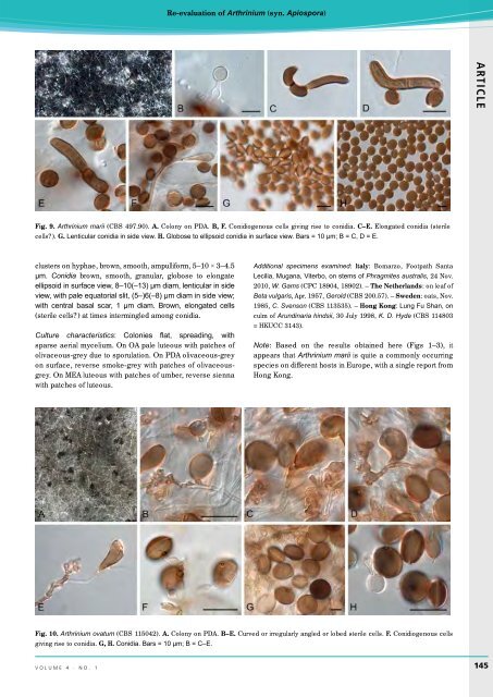
Re-evaluation of Arthrinium (syn. Apiospora) ARTICLE Fig. 9. Arthrinium marii (CBS 497.90). A. Colony on PDA. B, F. Conidiogenous cells giving rise to conidia. C–E. Elongated conidia (sterile cells?). G. Lenticular conidia in side view. H. Globose to ellipsoid conidia in surface view. Bars = 10 µm; B = C, D = E. clusters on hyphae, brown, smooth, ampulliform, 5–10 × 3–4.5 µm. Conidia brown, smooth, granular, globose to elongate ellipsoid in surface view, 8–10(–13) µm diam, lenticular in side view, with pale equatorial slit, (5–)6(–8) µm diam in side view; with central basal scar, 1 µm diam. Brown, elongated cells (sterile cells?) at times intermingled among conidia. Culture characteristics: Colonies flat, spreading, with sparse aerial mycelium. On OA pale luteous with patches of olivaceous-grey due to sporulation. On PDA olivaceous-grey on surface, reverse smoke-grey with patches of olivaceousgrey. On MEA luteous with patches of umber, reverse sienna with patches of luteous. Additional specimens examined: Italy: Bomarzo, Footpath Santa Lecilia, Mugana, Viterbo, on stems of Phragmites australis, 24 Nov. 2010, W. Gams (CPC 18904, 18902). – The Netherlands: on leaf of Beta vulgaris, Apr. 1957, Gerold (CBS 200.57). – Sweden: oats, Nov. 1985, C. Svenson (CBS 113535). – Hong Kong: Lung Fu Shan, on culm of Arundinaria hindsii, 30 July 1998, K. D. Hyde (CBS 114803 = HKUCC 3143). Note: Based on the results obtained here (Figs 1–3), it appears that Arthrinium marii is quite a commonly occurring species on different hosts in Europe, with a single report from Hong Kong. Fig. 10. Arthrinium ovatum (CBS 115042). A. Colony on PDA. B–E. Curved or irregularly angled or lobed sterile cells. F. Conidiogenous cells giving rise to conidia. G, H. Conidia. Bars = 10 µm; B = C–E. volume 4 · no. 1 145