

Complete issue - IMA Fungus
Complete issue - IMA Fungus
Complete issue - IMA Fungus

Create successful ePaper yourself
Turn your PDF publications into a flip-book with our unique Google optimized e-Paper software.
Crous & Groenewald<br />
ARTICLE<br />
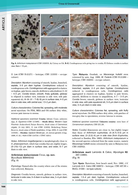
Fig. 8. Arthrinium malaysianum (CBS 102053). A. Colony on OA. B–E. Conidiogenous cells giving rise to conidia. F. Globose conidia in surface<br />
view. Bars = 10 µm.<br />
S. Lee (CBS H-21271 – holotype; CBS 113333 – ex-type<br />
culture).<br />
Description: Mycelium consisting of smooth, hyaline, branched,<br />
septate, 3–5 µm diam hyphae. Conidiophores reduced to<br />
conidiogenous cells. Conidiogenous cells aggregated in clusters<br />
on hyphae, pale brown, smooth, doliiform to subcylindrical, 5–12<br />
× 4–5 µm. Conidia brown, smooth, finely guttulate, globose<br />
to ellipsoid in surface view, lenticular in side view, with pale<br />
equatorial slit, (8–)9–10 × 7–8(–9) µm in surface view, 4–5 µm<br />
diam in side view, with central scar, 1.5–2 µm diam.<br />
Culture characteristics: Colonies flat, spreading, with moderate<br />
aerial mycelium. On PDA, MEA and OA surface dirty white,<br />
reverse pale luteous to sienna.<br />
Additional specimens examined: Croatia: Adriatic Coast, unknown<br />
alga, E. Eguereva (CBS 117206). – South Africa: Western Cape<br />
Province: Jonkershoek Nature Reserve, dead culms of Cannomois<br />
virgata, 15 July 2001, S. Lee (CBS 113332; Helderberg Nature<br />
Reserve, dead culms of Restio quadratus, 13 Apr. 2002, S. Lee (CBS<br />
113335). – Sweden: Uppland: Börstil par., on Juncus gerardi, 2 Aug.<br />
1990, K. & L. Holm (CBS 114734 = UPSC 3251).<br />
Notes: Arthrinium kogelbergense is morphologically close to<br />
A. phaeospermum, which has conidia that are slightly longer,<br />
(9–)10(–12) µm diam in surface view, and wider, 6–7 µm<br />
diam in side view.<br />
Arthrinium malaysianum Crous, sp. nov.<br />
MycoBank MB804342<br />
(Fig. 8)<br />
Etymology: Named after the country where one of the strains<br />
was collected, Malaysia.<br />
Diagnosis: Conidia brown, smooth, globose in surface view,<br />
lenticular in side view, 5–6 diam in surface view, 3–4 µm diam<br />
in side view.<br />
Type: Malaysia: Gombak, on Macaranga hullettii stem<br />
colonised by ants, Aug. 1999, W. Federle (CBS H-21269 –<br />
holotype; CBS 102053 – ex-type culture).<br />
Description: Mycelium consisting of smooth, hyaline,<br />
branched, septate, 2–3 µm diam hyphae. Conidiophores<br />
reduced to conidiogenous cells. Conidiogenous cells<br />
aggregated in clusters on hyphae, hyaline to pale brown,<br />
smooth, doliiform to clavate to ampulliform, 4–7 × 3–5 µm.<br />
Conidia brown, smooth, globose in surface view, lenticular<br />
in side view, with pale equatorial slit, 5–6 µm diam in surface<br />
view, 3–4 µm diam in side view.<br />
Culture characteristics: Colonies flat, spreading, with fluffy<br />
aerial mycelium. On PDA surface dirty white, with patches of<br />
iron-grey due to sporulation, reverse luteous to sienna.<br />
Additional specimen examined: Unknown country: stem base of<br />
Cinnamomum camphora, CBS 251.29.<br />
Notes: Conidial dimensions are close to, but slightly longer<br />
than those of Arthrinium euphorbiae, (4–)4.7(–5.5) µm in<br />
surface view, (3–)3.2(–4) µm in side view (from Euphorbia,<br />
collected in Zambia; Ellis 1965). Arthrinium malaysianum is<br />
the second species collected from the same source, namely<br />
Macaranga hullettii stems colonised by ants in Malaysia (see<br />
CBS 102052).<br />
Arthrinium marii Larrondo & Calvo, Mycologia 82:<br />
397 (1990).<br />
(Fig. 9)<br />
Type: Spain: Barcelona, from beach sand, Nov. 1990, J.V.<br />
Larrondo & A. Calvo (IMI 326872 – holotype; CBS 497.90 =<br />
MUCL 31300 – ex-type culture).<br />
Description: Mycelium consisting of smooth, hyaline, branched,<br />
septate, 1.5–4 µm diam hyphae. Conidiophores reduced<br />
to conidiogenous cells. Conidiogenous cells aggregated in<br />
144 ima fUNGUS


