E-<strong>International</strong> <strong>Scientific</strong> <strong>Research</strong> Journal ISSN: 2094-1749 Volume: 3 Issue: 2, 2011 Kong, X. L., Huang, L. C. L., Hsu, C. M., Chen, W. H., Han, C. and Chang, H. C. (2005). Highaffinity capture of proteins by diamond nanop<strong>article</strong>s for mass spectrometric analysis,” Anal. Chem. 77, 259-265. Lewis, E.B. (1960). A new standard food medium. D. I. S. 34: 117--118. Lim, T.S., Fu, C.-C., Lee, K.-C., Lee, H.-Y., Chen, K., Cheng, W.-F., Pai, W. W., Chang, H.-C. and Fann, W. (2009). Fluorescence enhancement and lifetime modification of single nanodiamonds near a nanocrystalline silver surface. Phys. Chem. Chem. Phys. 14;11(10):1508- 14. Liu, H., Ye, T. and Mao, C. (2007). Fluorescent Carbon Nanop<strong>article</strong>s Derived from Candle Soot. Angew. Chem. 46 (34), 6473-6475 Liu, H.Y. ,and Vu, T.Q.( 2007). Quantum Dot Hybrid Gel Blotting: A Technique for Identifying Quantum Dot-Protein/Protein-Protein Interactions .Nano Lett. 7:1044–1049. Mochalin, V. and Gogotsi, Y.( 2009). Wet Chemistry Route to Hydrophobic Blue Fluorescent Nanodiamond. J. Am. Chem. Soc.131, 4594-95. Medintz, I. L., Uyeda, H. T., Goldman, E. R. and Mattoussi, H. (2005). Quantum dot bioconjugates for imaging, labelling and sensing. Nat. Mater. 4(6):435-46. Ray, S. C., Saha, A., Jana, N. R. and Sarkar R.(2009). Fluorescent Carbon Nanop<strong>article</strong>s: Syn<strong>the</strong>sis, Characterization, and Bioimaging Application, J. Phys. Chem. C.113, 18546–18551 Selvi, B. R., Jagadeesan, D., Suma, B. S., Nagashankar, G., Arif, M., Balasubramanyam, K., swaramoorthy, M. and Kundu, T. K. (2008). Intrinsically Fluorescent Carbon Nanosp<strong>here</strong>s as a Nuclear Targeting Vector: Delivery of Membrane-Impermeable Molecule to Modulate Gene Expression In Vivo. Nano lett., 8(10):3182-85. Sun, Y. P., Zhou, B., Lin, Y., Wang, W., Fernando, K. A. S., Pathak, P., Meziani, M. J., Harruff, B. A., Wang, X., Wang, H., Luo, P. G., Yang, H., Kose, M. E., Chen, B., Veca, L. M. ans Xie, S. Y. (2006). Quantum-sized carbon dots for bright and colorful photoluminescence. J. Am. Chem. Soc. 128(24):7756. Zhou, J., Booker, C., Li, R., Zhou, X., Sham, T. K., Sun, X. and Ding, Z. (2007). An electrochemical avenue to blue luminescent nanocrystals from multiwalled carbon nanotubes (MWCNTs). J. Am. Chem. Soc. 129(4):744-5. Zhao, Q.-L., Zhang, Z.-L., Huang, B.-H., Peng, J., Zhang, M. and Pang, D.-W. (2008). Facile preparation of low cytotoxicity fluorescent carbon nanocrystals by electrooxidation of graphite. Chem. Commun. 41, 5116-5118. 105
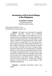
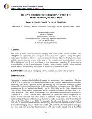
E-<strong>International</strong> <strong>Scientific</strong> <strong>Research</strong> Journal ISSN: 2094-1749 Volume: 3 Issue: 2, 2011 Figure Legends Figure 1. Schematic diagram of WSCQDs treated drosophila (fluorescing) and untreated drosophila (not fluorescing). Figure 2. AFM topography images of water soluble C-Dots(left) and HRTEM image C.Dots (right). Figure 3. Fluorescence images of various developmental stages of drosophila treated with WSCQDs. From left to right egg, larva, pupa, female imago and male imago respectively. Figure 4. Various internal organs of D. melanogaster larva treated with water soluble quantum dots. In vivo image, merge of three lights (488, 561 and 633nm). W<strong>here</strong> at-atrium, bn-brain, asanterior spiracle, tc-trachea, pxpharynx, sd- salivary duct, sg-salivary gland, Ep-esophagus, pvcproventiculus, gc-gastric ceaca, mg-midgut, mi-mid intestine, gd-gonad, utr-ureter, mtmalpighian tubule, hg-hind gut, as- anus. Scale bar 0.5mm. Figure-1 106