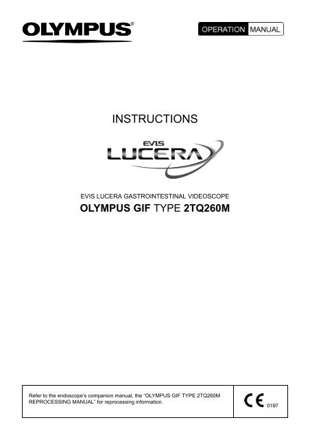
INSTRUCTIONS - Olympus
INSTRUCTIONS - Olympus
INSTRUCTIONS - Olympus

You also want an ePaper? Increase the reach of your titles
YUMPU automatically turns print PDFs into web optimized ePapers that Google loves.
<strong>INSTRUCTIONS</strong><br />
EVIS LUCERA GASTROINTESTINAL VIDEOSCOPE<br />
OLYMPUS GIF TYPE 2TQ260M<br />
Refer to the endoscope’s companion manual, the “OLYMPUS GIF TYPE 2TQ260M<br />
REPROCESSING MANUAL” for reprocessing information.
Contents<br />
Contents<br />
Symbols......................................................................................... 1<br />
Important Information – Please Read Before Use ..................... 2<br />
Intended use ............................................................................................ 2<br />
Applicability of endoscopy and endoscopic treatment ............................. 2<br />
Instruction manual..................................................................................... 3<br />
User qualifications .................................................................................... 3<br />
Instrument compatibility ........................................................................... 3<br />
Reprocessing before the first use/reprocessing and storage after use..... 4<br />
Spare equipment ...................................................................................... 4<br />
Maintenance management ....................................................................... 4<br />
Prohibition of improper repair and modification ........................................ 4<br />
Signal words.............................................................................................. 5<br />
Warnings and cautions ............................................................................. 5<br />
Examples of inappropriate handling ......................................................... 8<br />
Chapter 1 Checking the Package Contents............................ 9<br />
Chapter 2 Instrument Nomenclature and Specifications ...... 12<br />
2.1 Nomenclature.................................................................................. 12<br />
2.2 Endoscope functions....................................................................... 14<br />
2.3 Specifications.................................................................................. 17<br />
2.4 Attaching the chain for water-resistant cap (MAJ-1119) ................. 19<br />
Chapter 3 Preparation and Inspection .................................... 22<br />
3.1 Preparation of the equipment.......................................................... 23<br />
3.2 Inspection of the endoscope ........................................................... 24<br />
3.3 Preparation and inspection of accessories ..................................... 29<br />
3.4 Attaching accessories to the endoscope ........................................ 33<br />
3.5 Inspection and connection of ancillary equipment .......................... 36<br />
3.6 Inspection of the endoscopic system .............................................. 39<br />
Chapter 4 Operation ................................................................. 45<br />
4.1 Insertion .......................................................................................... 47<br />
4.2 Using endo-therapy accessories..................................................... 53<br />
4.3 Withdrawal of the endoscope.......................................................... 59<br />
4.4 Transportation of the endoscope .................................................... 59<br />
EVIS LUCERA GIF TYPE 2TQ260M OPERATION MANUAL<br />
i
Contents<br />
Chapter 5 Troubleshooting ...................................................... 61<br />
5.1 Troubleshooting guide .................................................................... 61<br />
5.2 Withdrawal of the endoscope with any abnormality........................ 66<br />
5.3 Returning the endoscope for repair................................................. 68<br />
Appendix........................................................................................ 69<br />
System chart ............................................................................................ 69<br />
EMC information........................................................................................ 77<br />
ii<br />
EVIS LUCERA GIF TYPE 2TQ260M OPERATION MANUAL
Symbols<br />
Symbols<br />
The meaning(s) of the symbol(s) shown on the package with the components,<br />
the back cover of this instruction manual and/or this instrument are as follows:<br />
Refer to instructions.<br />
Endoscope<br />
TYPE BF applied part<br />
Manufacturer<br />
Authorized representative in the European Community<br />
EVIS LUCERA GIF TYPE 2TQ260M OPERATION MANUAL<br />
1
Important Information – Please Read Before Use<br />
Important Information – Please Read<br />
Before Use<br />
Intended use<br />
This instrument has been designed to be used with an <strong>Olympus</strong> video system<br />
center, light source, documentation equipment, video monitor, endo-therapy<br />
accessories such as a biopsy forceps and other ancillary equipment.<br />
Use the EVIS LUCERA GASTROINTESTINAL VIDEOSCOPE GIF-2TQ260M<br />
for endoscopy and endoscopic surgery within the upper digestive tract (including<br />
the esophagus, stomach and duodenum).<br />
Do not use this instrument for any purpose other than its intended use. Select<br />
the endoscope to be used according to the purpose of the intended endoscopy<br />
and endoscopic treatment based on full understanding of the endoscope<br />
specifications in this instruction manual.<br />
Applicability of endoscopy and endoscopic treatment<br />
If there is an official standard on the applicability of endoscopy and endoscopic<br />
treatment that is defined by the hospital’s administration or other official<br />
institutions such as academic societies on endoscopy, follow that standard.<br />
Before starting endoscopy and endoscopic treatment, thoroughly evaluate its<br />
properties, purposes, effects, and possible risk (their natures, extent and<br />
probability). Perform endoscopy and endoscopic treatment only when its<br />
potential benefits are greater than its risks.<br />
Fully explain to the patient the potential benefits and risks of the endoscopy and<br />
endoscopic treatment as well as any examination/treatment methods that can be<br />
performed in its place, and perform the endoscopy and endoscopic treatment<br />
only after obtaining the consent of the patient.<br />
Even after starting the endoscopy and endoscopic treatment, continue to<br />
evaluate the potential benefits and risks, and immediately stop the<br />
endoscopy/treatment and take proper measures if the risks to the patient<br />
become greater than the potential benefits.<br />
2<br />
EVIS LUCERA GIF TYPE 2TQ260M OPERATION MANUAL
Important Information – Please Read Before Use<br />
Instruction manual<br />
This instruction manual contains essential information on using this instrument<br />
safely and effectively. Before use, thoroughly review this manual and the<br />
manuals of all equipment which will be used during the procedure and use the<br />
equipment as instructed.<br />
Note that the complete instruction manual set for this endoscope consists of this<br />
manual and the “OLYMPUS GIF TYPE 2TQ260M REPROCESSING MANUAL”<br />
accompanied the endoscope at shipment.<br />
Keep this and all related instruction manuals in a safe, accessible location.<br />
If you have any questions or comments about any information in this manual,<br />
please contact <strong>Olympus</strong>.<br />
User qualifications<br />
If there is an official standard on the qualification of endoscopy and endoscopic<br />
treatment that is defined by the medical administration or other official<br />
institutions such as the academic society on endoscopy, follow the standard. If<br />
there is no official qualification standard, the operator of this instrument must be<br />
a physician approved by the medical safety manager of the hospital or person in<br />
charge of the department (department of internal medicine, etc.). The medical<br />
safety manager of the hospital or person in charge of the department should<br />
select a physician who is capable of safely performing the planned endoscopy<br />
and endoscopic treatment by following the official guidelines set by the academic<br />
society on endoscopy, etc., and considering the difficulty of each type of<br />
endoscopy and endoscopic treatment.<br />
Instrument compatibility<br />
Refer to the “System chart” in the Appendix to confirm that this instrument is<br />
compatible with the ancillary equipment being used. Using incompatible<br />
equipment can result in patient or operator injury and/or equipment damage.<br />
EVIS LUCERA GIF TYPE 2TQ260M OPERATION MANUAL<br />
3
Important Information – Please Read Before Use<br />
Reprocessing before the first use/reprocessing and<br />
storage after use<br />
This instrument was not cleaned, disinfected or sterilized before shipment.<br />
Before using this instrument for the first time, reprocess it according to the<br />
instructions given in the endoscope’s companion manual, the “OLYMPUS GIF<br />
TYPE 2TQ260M REPROCESSING MANUAL”. After using this instrument,<br />
reprocess and store it according to the instructions given in the endoscope’s<br />
companion reprocessing manual. Improper and/or incomplete reprocessing or<br />
storage can present an infection control risk, cause equipment damage or<br />
reduce performance.<br />
Spare equipment<br />
Be sure to prepare another endoscope to avoid that the examination will be<br />
interrupted due to equipment failure or malfunction.<br />
Maintenance management<br />
The probability of failure of endoscope and ancillary equipment increase as the<br />
total operation cause and/or total operating hours increase. In addition to the<br />
inspection before each procedure, the person in charge of medical equipment<br />
maintenance in each hospital should inspect the items specified in this manual<br />
periodically. An endoscope with which an irregularity is suspected should not be<br />
used, but should be inspected by following Section 5.1, “Troubleshooting guide”.<br />
If the irregularity is still suspected after inspection, contact <strong>Olympus</strong> before use.<br />
Prohibition of improper repair and modification<br />
Never repair by persons other than <strong>Olympus</strong>-qualified technicians or modify the<br />
instrument, as this may result in injury of the patient or operator as well as<br />
damage to the equipment.<br />
4<br />
EVIS LUCERA GIF TYPE 2TQ260M OPERATION MANUAL
Important Information – Please Read Before Use<br />
Signal words<br />
The following signal words are used throughout this manual:<br />
Indicates a potentially hazardous situation which, if not<br />
avoided, could result in death or serious injury.<br />
Indicates a potentially hazardous situation which, if not<br />
avoided, may result in minor or moderate injury. It may also<br />
be used to alert against unsafe practices or potential<br />
equipment damage.<br />
Indicates additional helpful information.<br />
Warnings and cautions<br />
Follow the warnings and cautions given below when handling this instrument.<br />
This information is to be supplemented by the warnings and cautions given in<br />
each chapter.<br />
• After using this instrument, reprocess and store it according<br />
to the instructions given in the endoscope’s companion<br />
manual, the “OLYMPUS GIF TYPE 2TQ260M<br />
REPROCESSING MANUAL”. Using improperly or<br />
incompletely reprocessed or stored instruments may cause<br />
patient cross-contamination and/or infection.<br />
• Before endoscopy, remove any metallic objects (watch,<br />
glasses, necklace, etc.) from the patient. If high-frequency<br />
cauterization becomes necessary, performing the treatment<br />
while the patient wears a metallic object may cause burns<br />
around the metallic object.<br />
• Do not strike, bend, hit, pull, twist, or drop the endoscope’s<br />
distal end, insertion tube, bending section, control section,<br />
universal cord, or endoscope connector of the endoscope<br />
with excessive force. The endoscope may be damaged and<br />
could cause patient injury, burns, bleeding and/or<br />
perforations. It could also cause parts of the endoscope to fall<br />
off inside the patient.<br />
EVIS LUCERA GIF TYPE 2TQ260M OPERATION MANUAL<br />
5
Important Information – Please Read Before Use<br />
• Never perform angulation control forcibly or abruptly. Never<br />
forcefully pull, twist or rotate the angulated bending section.<br />
Patient injury, bleeding and/or perforation can result. It may<br />
also become impossible to straighten the bending section<br />
during an examination.<br />
• Never insert or withdraw the endoscope’s insertion tube while<br />
the bending section is locked in position. Patient injury,<br />
bleeding and/or perforation can result.<br />
• Never operate the bending section, feed air or perform<br />
suction, insert or withdraw the endoscope’s insertion tube,<br />
use endo-therapy accessories without viewing the<br />
endoscopic image. Never use endo-therapy accessories<br />
without viewing the endoscopic image. Patient injury,<br />
bleeding and/or perforation can result.<br />
• Never operate the bending section, feed air or perform<br />
suction, insert or withdraw the endoscope’s insertion tube<br />
while the image is frozen. Never use endo-therapy<br />
accessories while the image is frozen. Patient injury, bleeding<br />
and/or perforation can result.<br />
• Never insert or withdraw the insertion tube with forcefully or<br />
suddenly. Patient injury, bleeding and/or perforation can<br />
result.<br />
• Do not touch the light guide of the endoscope connector<br />
immediately after removing it from the light source because it<br />
is extremely hot. Operator or patient burns can result.<br />
• When the endoscopic image does not appear on the monitor,<br />
the CCD may have been damaged. Turn the video system<br />
center OFF immediately. Continued power supply in such a<br />
case will cause the distal end to become hot and could cause<br />
operator and/or patient burns.<br />
• When it is difficult to insert the endoscope, do not force<br />
inserting the endoscope but stop the endoscopy. Forcible<br />
insertion can result in patient injury, bleeding and/or<br />
perforation.<br />
• Do not pull the universal cord during an examination. The<br />
endoscope connector will be pulled out from the output<br />
socket of the light source and the endoscopic image will not<br />
be visible.<br />
• Do not coil the insertion tube or universal cord into a diameter<br />
of less than 12 cm. Equipment damage can result.<br />
6<br />
EVIS LUCERA GIF TYPE 2TQ260M OPERATION MANUAL
Important Information – Please Read Before Use<br />
• Do not touch the electrical contacts inside the electrical<br />
connector. CCD damage may result.<br />
• Do not apply shock to the distal end of the insertion tube,<br />
particularly the objective lens surface at the distal end. Visual<br />
abnormalities may result.<br />
• Do not twist or bend the bending section with your hands.<br />
Equipment damage may result.<br />
• Do not squeeze the bending section forcefully. The covering<br />
of the bending section may stretch or break and cause water<br />
leaks.<br />
• Turn the video system center OFF before connecting or<br />
disconnecting the videoscope cable from the electrical<br />
connector on the endoscope. Turn the video system center<br />
ON or OFF only when the videoscope cable is connected to<br />
both the video system center and electrical connector on the<br />
endoscope. Failure to do so can result in equipment damage,<br />
including destruction of the CCD.<br />
• The endoscope’s remote switches cannot be removed from<br />
the control section. Pressing, pulling or twisting them with<br />
excessive force can break the switches and/or may cause<br />
water leaks.<br />
• If remote switch 1 does not return to the OFF position after<br />
being pressed strongly from the side, gently pull the switch<br />
upwards to return it to the OFF position.<br />
• Do not hit or bend the electrical contacts on the endoscope<br />
connector. The connection to the light source may be<br />
impaired and faulty contact can result.<br />
• Do not attempt to bend the endoscope’s insertion tube with<br />
excessive force. Otherwise, the insertion tube may be<br />
damaged.<br />
• Electromagnetic interference may occur on this instrument<br />
near equipment marked with the following symbol or other<br />
portable and mobile RF (Radio Frequency) communications<br />
equipment such as cellular phones. If electromagnetic<br />
interference occurs, mitigation measures may be necessary,<br />
such as reorienting or relocating this instrument, or shielding<br />
the location.<br />
EVIS LUCERA GIF TYPE 2TQ260M OPERATION MANUAL<br />
7
Important Information – Please Read Before Use<br />
The endoscope contains a memory chip that stores<br />
information about the endoscope and communicates this<br />
information to the CV-260.<br />
Examples of inappropriate handling<br />
Details on clinical endoscopic technique are the responsibility of trained<br />
specialists. Patient safety in endoscopic examinations and endoscopic treatment<br />
can be ensured through appropriate handling by the physician and the medical<br />
facility. Examples of inappropriate handling are given below.<br />
• Over-insufflating the lumen may cause patient pain, injury, bleeding<br />
and/or perforation.<br />
• Applying suction with the distal end in prolonged contact with the<br />
mucosal surface, with higher suction pressure than required or with<br />
prolonged suction time may cause bleeding and/or lesions.<br />
• The endoscope has not been designed for use in retroflexed<br />
observation in other part than the stomach. Performing retroflexed<br />
observation in a narrow tube may make it impossible to straighten the<br />
endoscope again and to withdraw the endoscope. Retroflexed<br />
observation in other part than the stomach should be performed only<br />
when the usefulness is larger than the danger.<br />
• Inserting, withdrawing and using endo-therapy accessories without a<br />
clear endoscopic image may cause patient injury, burns, bleeding<br />
and/or perforation.<br />
• Inserting or withdrawing the endoscope, feeding air or applying suction<br />
or operating the bending section without a clear endoscopic image may<br />
cause patient injury, bleeding and/or perforation.<br />
• Never perform forcible angulation control of the first bending section and<br />
the second bending section simultaneously in the patient’s body cavity.<br />
Otherwise, the mucosal surface may be damaged or the endoscope<br />
may not be withdrawn.<br />
8<br />
EVIS LUCERA GIF TYPE 2TQ260M OPERATION MANUAL
Chapter 1 Checking the Package Contents<br />
Chapter 1<br />
Checking the Package<br />
Contents<br />
Match all items in the package with the components shown below. Inspect each<br />
item for damage. If the instrument is damaged, a component is missing or you<br />
have any questions, do not use the instrument; immediately contact <strong>Olympus</strong>.<br />
This instrument was not disinfected or sterilized before shipment. Before using<br />
this instrument for the first time, reprocess it according to the instructions given<br />
in the endoscope’s companion manual, the “OLYMPUS GIF TYPE 2TQ260M<br />
REPROCESSING MANUAL”.<br />
Endoscope<br />
Channel cleaning brush<br />
(BW-20T)<br />
Water-resistant cap<br />
(MH-553)<br />
Injection tube (MH-946)<br />
EVIS LUCERA GIF TYPE 2TQ260M OPERATION MANUAL<br />
9
Chapter 1 Checking the Package Contents<br />
Channel-opening cleaning<br />
brush (MH-507)<br />
AW channel cleaning adapter<br />
(MH-948)<br />
Suction valve<br />
(MH-443, 2 pcs)<br />
Air/water valve<br />
(MH-438, 2 pcs)<br />
Channel plug (MH-944)<br />
Biopsy valve (MAJ-419)<br />
Caps for biopsy valve<br />
(10 pcs)<br />
Chain for water-resistant cap<br />
(MAJ-1119)<br />
Channel connection tube<br />
(MAJ-420)<br />
Mouthpiece<br />
(MB-142, 2 pcs)<br />
Auxiliary water tube<br />
(MAJ-855)<br />
Operation manual<br />
Reprocessing<br />
manual<br />
Instructions manual<br />
(Caution for the angulation)<br />
10<br />
EVIS LUCERA GIF TYPE 2TQ260M OPERATION MANUAL
Chapter 1 Checking the Package Contents<br />
EVIS LUCERA GIF TYPE 2TQ260M OPERATION MANUAL<br />
11
Chapter 2 Instrument Nomenclature and Specifications<br />
Chapter 2<br />
Instrument Nomenclature<br />
and Specifications<br />
2.1 Nomenclature<br />
Universal cord<br />
20. Auxiliary water inlet<br />
1. Suction connector<br />
5. Electrical connector<br />
Auxiliary water inlet cap<br />
(MAJ-215)<br />
Air pipe<br />
2. S-cord connector mount<br />
3. Air supply connector<br />
Light guide<br />
3. Water supply connector<br />
Product name and serial number<br />
Electrical contacts<br />
4. Endoscope connector<br />
12<br />
EVIS LUCERA GIF TYPE 2TQ260M OPERATION MANUAL
Chapter 2 Instrument Nomenclature and Specifications<br />
22. Neutral index for the second<br />
bending section<br />
18. UP/DOWN angulation control knob<br />
for the second bending section<br />
19. UP/DOWN angulation lock for the<br />
second bending section<br />
7. UP/DOWN angulation lock<br />
for the first bending section<br />
6. UP/DOWN angulation control knob<br />
for the first bending section<br />
8. Suction valve<br />
(MH-443)<br />
9. Air/water valve<br />
(MH-438)<br />
Suction cylinder<br />
Air/water cylinder<br />
17. RIGHT/LEFT angulation control<br />
knob for the first bending section<br />
16. RIGHT/LEFT angulation lock<br />
for the first bending section<br />
Grip section<br />
Instrument channel ports<br />
Control<br />
section<br />
15. Color code<br />
Yellow: A<br />
Yellow: B<br />
Boot<br />
21. Suction<br />
switchover lever<br />
Biopsy valve (MAJ-419)<br />
10. Instrument channels<br />
11. Insertion tube<br />
limit mark<br />
Working length<br />
14. Remote switches<br />
Distal end<br />
Insertion tube<br />
13. Second bending section<br />
12. First bending<br />
section<br />
EVIS LUCERA GIF TYPE 2TQ260M OPERATION MANUAL<br />
13
Chapter 2 Instrument Nomenclature and Specifications<br />
2.2 Endoscope functions<br />
1. Suction connector<br />
This connector connects the endoscope to the suction tube of the suction<br />
pump.<br />
2. S-cord connector mount<br />
This mount connects the endoscope with the <strong>Olympus</strong> electrosurgical unit<br />
via the S-cord. The S-cord conducts leakage current from the endoscope to<br />
the electrosurgical unit. To connect the S-cord, refer to the instruction<br />
manual for the electrosurgical unit. Connect the fitting part of the chain for<br />
water-resistant cap as required.<br />
3. Water supply connector and air supply connector<br />
These connectors connect the endoscope to the water container via the<br />
water container tube, to supply water to the distal end of the endoscope.<br />
4. Endoscope connector<br />
This connector connects the endoscope to the output socket of the light<br />
source and transmits light from the light source to the endoscope.<br />
5. Electrical connector<br />
This connector connects the endoscope to the video system center via the<br />
videoscope cable. This endoscope contains a memory chip that stores<br />
information about the endoscope and communicates this information to the<br />
video system center CV-260. For more details, refer to the instruction<br />
manual of the CV-260.<br />
6. UP/DOWN angulation control knob for the first bending section<br />
When this knob is turned in the “ U” direction, the first bending section<br />
moves UP; when the knob is turned in the “D ” direction, the first bending<br />
section moves DOWN.<br />
7. UP/DOWN angulation lock for the first bending section<br />
Moving this lock in the “F ” direction frees angulation. Moving the lock in<br />
the opposite direction locks the first bending section at any desired position.<br />
8. Suction valve (MH-443)<br />
This valve is depressed to activate suction. The valve is used to remove any<br />
fluid, debris, flatus or air from the patient.<br />
9. Air/water valve (MH-438)<br />
The hole in this valve is covered to insufflate air and the valve is depressed<br />
to feed water for lens washing. It also can be used to feed air to remove any<br />
fluid or debris adhering to the objective lens.<br />
10. Instrument channels<br />
The instrument channels function as:<br />
−<br />
−<br />
−<br />
channel for the insertion of endo-therapy accessories<br />
suction channel<br />
fluid feed channel (from a syringe via the biopsy valve)<br />
14<br />
EVIS LUCERA GIF TYPE 2TQ260M OPERATION MANUAL
Chapter 2 Instrument Nomenclature and Specifications<br />
11. Insertion tube limit mark<br />
This mark shows the maximum point to which the endoscope may be<br />
inserted into the patient’s body.<br />
12. First bending section<br />
This section moves the distal end of the endoscope when the UP/DOWN<br />
and RIGHT/LEFT angulation control knobs for the first bending section are<br />
operated.<br />
13. Second bending section<br />
This section moves the distal end of the endoscope when the UP/DOWN<br />
angulation control knob for the second bending section is operated. The<br />
angulation in the RIGHT/LEFT directions of this section cannot be controlled<br />
by a knob, but it can be bended by applying an external force.<br />
14. Remote switches 1 to 4<br />
The functions of the remote switches 1 to 4 can be selected on the video<br />
system center. When selecting the functions, refer to the instruction manual<br />
for the video system center.<br />
15. Color code<br />
This code is used to quickly determine the compatibility of endo-therapy<br />
accessories. The endoscope can be used with endo-therapy accessories<br />
that have the same color code.<br />
Yellow (A):<br />
Yellow (B):<br />
Denotes the ø 3.2 mm instrument channel (A channel)<br />
Denotes the ø 3.2 mm instrument channel (B channel)<br />
16. RIGHT/LEFT angulation lock for the first bending section<br />
Turning this lock in the “F ” direction frees RIGHT/LEFT angulation of the<br />
first bending section. Turning the lock in the opposite direction locks the first<br />
bending section at any desired position.<br />
17. RIGHT/LEFT angulation control knob for the first bending section<br />
When this knob is turned in the “R ” direction, the first bending section<br />
moves RIGHT; when the knob is turned in the “ L” direction, the first<br />
bending section moves LEFT.<br />
18. UP/DOWN angulation control knob for the second bending section<br />
When this knob is turned in the “ U2” direction, the second bending<br />
section moves UP; when the knob is turned in the “D2 ” direction, the<br />
second bending section moves DOWN.<br />
19. UP/DOWN angulation lock for the second bending section<br />
Turning this lock knob in the direction of “F ” frees the UP/DOWN<br />
angulation of the second bending section. Turning it in the opposite direction<br />
locks the UP/DOWN angulation of the second bending section<br />
approximately at the desired position.<br />
EVIS LUCERA GIF TYPE 2TQ260M OPERATION MANUAL<br />
15
Chapter 2 Instrument Nomenclature and Specifications<br />
20. Auxiliary water inlet<br />
This inlet is connected to the auxiliary water tube. Feed water from this inlet<br />
through the auxiliary water channel when necessary (e.g. when blood<br />
adheres to mucosa in the patient’s body cavity). When the auxiliary water<br />
inlet is not being used, make sure that it is covered by the auxiliary water<br />
inlet cap.<br />
21. Suction switchover lever<br />
It is possible to aspirate from only the A: ø 3.2 instrument channel by turning<br />
to the direction of “A” and pushing the suction valve. And also, it is possible<br />
to aspirate from two channels of the A: ø 3.2 instrument channel and the B:<br />
ø 3.2 instrument channel at the same time by turning to the direction of “AB”<br />
and pushing the suction valve. The suction switchover lever certainly<br />
adjusts the position to the stopper of the biopsy valve. When the suction<br />
switchover lever is not attached properly, only the A: ø 3.2 instrument<br />
channel can be used for suction.<br />
22. Neutral index for the second bending section<br />
Aligning the index on the UP/DOWN angulation control knob for the second<br />
bending section with that on the casing sets the second section almost<br />
straight.<br />
16<br />
EVIS LUCERA GIF TYPE 2TQ260M OPERATION MANUAL
Chapter 2 Instrument Nomenclature and Specifications<br />
2.3 Specifications<br />
Environment<br />
Operating<br />
environment<br />
Ambient temperature<br />
Relative humidity 30 – 85%<br />
10 – 40°C (50 – 104°F)<br />
Atmospheric pressure<br />
700 – 1060 hPa<br />
(0.7 – 1.1 kgf/cm 2 )<br />
(10.2 – 15.4 psia)<br />
Transportation and<br />
storage<br />
environment<br />
Ambient temperature –47 to 70°C (–52.6 to 158°F)<br />
Relative humidity 10 – 95%<br />
Atmospheric pressure 700 – 1060 hPa<br />
(0.7 – 1.1 kgf/cm 2 )<br />
(10.2 – 15.4 psia)<br />
EVIS LUCERA GIF TYPE 2TQ260M OPERATION MANUAL<br />
17
Chapter 2 Instrument Nomenclature and Specifications<br />
Specifications<br />
Endoscope functions<br />
Model<br />
GIF-2TQ260M<br />
Optical system Field of view 140°<br />
Insertion tube<br />
Direction of view<br />
Depth of field<br />
Distal end outer<br />
diameter<br />
Distal end enlarged<br />
Forward viewing<br />
3 – 100 mm<br />
ø11.7mm<br />
1. Air/water nozzle<br />
2. Light guide lens<br />
3. Objective lens<br />
4. Instrument channel outlet<br />
5. Auxiliary water channel<br />
3.<br />
UP<br />
5.<br />
1.<br />
RIGHT<br />
4. (B)<br />
4. (A)<br />
DOWN<br />
LEFT<br />
2.<br />
Instrument<br />
channel<br />
Insertion tube outer<br />
diameter<br />
Working length<br />
Channel inner<br />
diameter<br />
Minimum visible<br />
distance<br />
Direction from which<br />
endo-therapy<br />
accessories enter<br />
and exit the<br />
endoscopic image<br />
ø11.7mm<br />
1030 mm<br />
A: ø 3.2 mm<br />
B: ø 3.2 mm<br />
5 mm from the distal end<br />
Air flow rate 25 cm 3 /s<br />
Note: Standard when CLV-260 (high air<br />
pressure) is used.<br />
FIrst bending<br />
section<br />
Second<br />
bending<br />
section<br />
Total length<br />
Angulation range UP 210°, DOWN 180°<br />
RIGHT 100°, LEFT 100°<br />
Angulation range UP 70°, DOWN 70°<br />
1395 mm<br />
18<br />
EVIS LUCERA GIF TYPE 2TQ260M OPERATION MANUAL
Chapter 2 Instrument Nomenclature and Specifications<br />
Medical Device<br />
Directive<br />
This device complies with the requirements<br />
of Directive 93/42/EEC concerning medical<br />
devices. Classification: Class II a<br />
EMC<br />
Applied standard;<br />
IEC 60601-1-2: 2001<br />
This instrument complies with the<br />
standards listed in the left column.<br />
CISPR 11 of emission:<br />
Group 1, Class B<br />
Year of<br />
manufacture<br />
Degree of<br />
protection<br />
against<br />
electric shock<br />
2512345<br />
This instrument complies with the EMC<br />
standard for medical electrical equipment;<br />
edition 2 (IEC 60601-1-2: 2001). However,<br />
when connecting to an instrument that<br />
complies with the EMC standard for<br />
medical electrical equipment; edition 1<br />
(IEC 60601-1-2: 1993), the whole system<br />
complies with edition 1.<br />
The last digit of the year of manufacture is<br />
the second digit of the serial number.<br />
TYPE BF applied part<br />
2.4 Attaching the chain for water-resistant cap<br />
(MAJ-1119)<br />
• Do not lift the endoscope by the chain for water-resistant cap.<br />
Otherwise, operator and/or patient injury can result, or the<br />
endoscope and/or water-resistant cap may be damaged<br />
when fitting part comes off from the S-cord connector mount.<br />
• Do not connect the fitting part to any mount but the S-cord<br />
connector mount. Connecting the fitting part to suction<br />
connector may cause unreliable connection of the suction<br />
tube to the suction connector. It may also cause departing of<br />
the suction tube from the endoscope and the patient debris to<br />
spray.<br />
EVIS LUCERA GIF TYPE 2TQ260M OPERATION MANUAL<br />
19
Chapter 2 Instrument Nomenclature and Specifications<br />
• The chain for water-resistant cap and water-resistant cap<br />
cannot be ultrasonic cleaning and may be damaged if they<br />
are subjected to ultrasonic cleaning. Only in the case of<br />
connecting the water-resistant cap with the chain to<br />
endoscopes, they are allowed to be cleaned by ultrasonic<br />
cleaning with endoscope reprocessor (OER, OER-A).<br />
• When attaching the water-resistant cap to the electrical<br />
connector, do not pinch the chain for water-resistant cap by<br />
the electrical connector of endoscope and the water-resistant<br />
cap. Otherwise, equipment damage may result.<br />
• The chain for water-resistant cap and water-resistant cap<br />
cannot be ETO gas sterilized and may be damaged if they<br />
are subjected to ETO gas sterilization. If the water-resistant<br />
cap is connected to the endoscope using the chain, be sure<br />
to remove the chain for water-resistant cap and the<br />
water-resistant cap from the endoscope before proceeding to<br />
ETO gas sterilization.<br />
• The chain for water-resistant cap and water-resistant cap<br />
cannot be steam sterilization (autoclaving) and may be<br />
damaged severely if they are subjected to steam sterilization<br />
(autoclaving).<br />
Notch<br />
Chain part<br />
Fitting part<br />
Connecting plate<br />
Hole<br />
Figure 2.1<br />
To ensure that you do not forget to attach the water-resistant<br />
cap, it is recommended to connect it to the endoscope’s<br />
S-cord connector mount using the chain for water-resistant<br />
cap.<br />
1. Confirm that the chain for water-resistant cap is free from cracks, flaws,<br />
wear, deformation or other damage.<br />
20<br />
EVIS LUCERA GIF TYPE 2TQ260M OPERATION MANUAL
Chapter 2 Instrument Nomenclature and Specifications<br />
2. Align the notch on the connecting plate with the pin on the venting connector<br />
of the water-resistant cap (MH-553, see Figure 2.2).<br />
3. Insert the connecting plate to the venting connector (see Figure 2.2).<br />
4. Confirm that the connecting plate is reliably inserted to the foot of the<br />
venting connector and can be smoothly rotated.<br />
5. Insert the hole on the fitting part to the S-cord connector mount of the<br />
endoscope connector (see Figure 2.3).<br />
6. Confirm that the fitting part is reliably inserted to the foot of the S-cord<br />
connector mount and can be smoothly rotated.<br />
Connecting plate<br />
Notch<br />
Pin<br />
Venting connector<br />
Water-resistant cap<br />
Figure 2.2<br />
Fitting part<br />
Hole<br />
S-cord connector<br />
mount<br />
Figure 2.3<br />
After this page, detaching condition of the chain for<br />
water-resistant cap (MAJ-1119) is explained.<br />
EVIS LUCERA GIF TYPE 2TQ260M OPERATION MANUAL<br />
21
Chapter 3 Preparation and Inspection<br />
Chapter 3<br />
Preparation and Inspection<br />
Before each case, prepare and inspect this instrument as instructed below.<br />
Inspect other equipment to be used with this instrument as instructed in their<br />
respective instruction manuals. If this instrument malfunctions, do not use it.<br />
Return it to <strong>Olympus</strong> for repair as described in Section 5.3, “Returning the<br />
endoscope for repair”.<br />
If the irregularities are suspected after inspection, follow the instructions given in<br />
Chapter 5, “Troubleshooting”.<br />
• Using an endoscope that is not functioning properly may<br />
compromise patient or operator safety and may result in<br />
more severe equipment damage.<br />
• This instrument was not cleaned, disinfected or sterilized<br />
before shipment. Before using this instrument for the first<br />
time, reprocess it according to the instructions given in the<br />
endoscope’s companion manual, the “OLYMPUS GIF TYPE<br />
2TQ260M REPROCESSING MANUAL”.<br />
22<br />
EVIS LUCERA GIF TYPE 2TQ260M OPERATION MANUAL
Chapter 3 Preparation and Inspection<br />
3.1 Preparation of the equipment<br />
Prepare the equipment shown in Figure 3.1 (for compatibility, see the “System<br />
chart” in the Appendix) and personal protective equipment, such as eye wear,<br />
face mask, moisture-resistant clothing and chemical-resistant gloves, before<br />
each use. Refer to the respective instruction manuals for each piece of<br />
equipment.<br />
Video monitor<br />
Video system center<br />
Light source<br />
Water container<br />
Suction pump<br />
Endoscope<br />
Endo-therapy accessories Auxiliary water tube Mouthpiece<br />
• Paper towels • Trays • Lint-free cloths • Personal protective equipment<br />
Figure 3.1<br />
EVIS LUCERA GIF TYPE 2TQ260M OPERATION MANUAL<br />
23
Chapter 3 Preparation and Inspection<br />
3.2 Inspection of the endoscope<br />
Clean and disinfect or sterilize the endoscope as described in the “OLYMPUS<br />
GIF TYPE 2TQ260M REPROCESSING MANUAL”. Then remove the<br />
water-resistant cap from the endoscope connector.<br />
Inspection of the endoscope<br />
1. Inspect the control section and the endoscope connector for excessive<br />
scratching, deformation, loose parts or other irregularities.<br />
2. Inspect the boot and the insertion tube near the boot for bends, twists or<br />
other irregularities.<br />
3. Inspect the external surface of the entire insertion tube including the<br />
bending section and the distal end for dents, bulges, swelling, scratching,<br />
holes, sagging, transformation, bends, adhesion of foreign bodies, dropout<br />
of parts, any protruding objects or other irregularities.<br />
4. Holding the insertion tube gently with a hand, carefully run the fingertips<br />
over the entire length of the insertion tube in both directions (See Figure<br />
3.2). Confirm that there is no object stopping the hand or protrusion of<br />
metallic wire from the inside on and around the insertion tube and that the<br />
insertion tube is not abnormally rigid.<br />
Figure 3.2<br />
24<br />
EVIS LUCERA GIF TYPE 2TQ260M OPERATION MANUAL
Chapter 3 Preparation and Inspection<br />
5. Using both hands, bend the insertion tube of the endoscope into a<br />
semicircle. Then, moving your hands as shown by the arrows, confirm that<br />
the entire insertion tube can be smoothly bent to form a semicircle and that<br />
the insertion tube is enough pliable (see Figure 3.3). Follow the above<br />
checks on both the softest and stuffiest conditions when the endoscope has<br />
the flexibility adjustment mechanism.<br />
Figure 3.3<br />
6. Gently hold the midpoint of the second bending section and a point 20 cm<br />
from the distal end. Push and pull gently to confirm that the border between<br />
the second bending section and the insertion tube is not loose. And also,<br />
gently hold the midpoint of the first bending section and the midpoint of the<br />
second bending section. Push and pull gently to confirm that the border<br />
between the first bending section and the second bending section is not<br />
loose.<br />
7. Inspect the objective lens and light guide lens at the distal end of the<br />
endoscope’s insertion tube for scratching, cracks, stains, gaps around the<br />
lens or other irregularities.<br />
8. Inspect the air/water nozzle at the distal end of the endoscope’s insertion<br />
tube for abnormal swelling, bulges, dents or other irregularities.<br />
EVIS LUCERA GIF TYPE 2TQ260M OPERATION MANUAL<br />
25
Chapter 3 Preparation and Inspection<br />
Inspection of the bending mechanisms<br />
Perform the following inspections while the bending section is straight.<br />
• If the movement of the UP/DOWN angulation lock,<br />
RIGHT/LEFT angulation lock and their angulation control<br />
knobs are loose and/or not smooth, or the bending section<br />
does not angulate smoothly, the bending mechanism may be<br />
abnormal. In this case, do not use the endoscope because it<br />
may be impossible to straighten the bending section during<br />
an examination.<br />
• If any irregularity is suspected with either the first or second<br />
bending section, do not use the endoscope. Because even if<br />
one bending section functions normally, the overall<br />
angulation mechanism may not work properly, causing injury,<br />
bleeding or perforation in the patient’s body cavity.<br />
Inspection for smooth operation<br />
1. Confirm that both the UP/DOWN angulation lock for the first bending<br />
section, RIGHT/LEFT angulation lock for the first bending section and<br />
UP/DOWN angulation lock for the second bending section move all the way<br />
in the “F ” position.<br />
2. Turn the angulation control knobs of the first bending section and the<br />
second bending section slowly in each direction until they stop, and return to<br />
respective neutral positions. Confirm that the bending section angulates<br />
smoothly and correctly, and confirm that maximum angulation can be<br />
achieved and return to respective neutral positions.<br />
26<br />
EVIS LUCERA GIF TYPE 2TQ260M OPERATION MANUAL
Chapter 3 Preparation and Inspection<br />
3. When the angulation control knobs of the first bending section and the<br />
second bending section to their respective neutral positions (as shown in<br />
Figure 3.4), confirm that the bending section with an approximately straight<br />
condition.<br />
Neutral index on the<br />
UP/DOWN angulation<br />
control knobs for the<br />
second bending section.<br />
Neutral index on<br />
the casing<br />
Neutral position<br />
of both the<br />
UP/DOWN,<br />
RIGHT/LEFT<br />
angulation<br />
control knobs of<br />
the first bending<br />
section.<br />
Figure 3.4<br />
Aligning the index on the UP/DOWN angulation control knob<br />
for the second bending section with that on the casing sets<br />
the second bending section almost straight.<br />
Inspection of the UP/DOWN angulation mechanism for the<br />
first bending section<br />
1. Move the UP/DOWN angulation lock for the first bending section all the way<br />
in the opposite direction of the “F ” mark. Then turn the UP/DOWN<br />
angulation control knob for the first bending section in the “ U” or the “D ”<br />
direction until it stops.<br />
2. Confirm that the angle of the first bending section is roughly stabilized when<br />
the UP/DOWN angulation control knob for the first bending section is<br />
released.<br />
3. Confirm that the first bending section straightens out automatically when the<br />
UP/DOWN angulation lock for the first bending section is moved all the way<br />
in the “F ” direction and the UP/DOWN angulation control knob for the first<br />
bending section is released.<br />
EVIS LUCERA GIF TYPE 2TQ260M OPERATION MANUAL<br />
27
Chapter 3 Preparation and Inspection<br />
Inspection of the RIGHT/LEFT angulation mechanism for<br />
the first bending section<br />
1. Turn the RIGHT/LEFT angulation lock for the first bending section all the<br />
way in the opposite direction of the “F ” mark. Then turn the RIGHT/LEFT<br />
angulation control knob for the first bending section in the “R ” or the “ L”<br />
direction until it stops.<br />
2. Confirm that the angle of the first bending section is roughly stabilized when<br />
the RIGHT/LEFT angulation control knob for the first bending section is<br />
released.<br />
3. Confirm that the bending section straightens out automatically when the<br />
RIGHT/LEFT angulation lock for the first bending section is turned in the<br />
“F ” direction and the RIGHT/LEFT angulation control knob for the<br />
bending section is released.<br />
Inspection of the UP/DOWN angulation mechanism for the<br />
second bending section<br />
1. Move the UP/DOWN angulation lock for the second bending section all the<br />
way in the opposite direction of the “F ” mark. Then turn the UP/DOWN<br />
angulation control knob for the second bending section in the “ U2” or the<br />
“D2 ” direction until it stops.<br />
2. Confirm that the angle of the second bending section is roughly stabilized<br />
when the UP/DOWN angulation control knob for the second bending section<br />
is released.<br />
3. Confirm that the second bending section straightens out automatically when<br />
the UP/DOWN angulation lock for the second bending section is moved all<br />
the way in the “F ” direction and the UP/DOWN angulation control knob for<br />
the second bending section is released.<br />
28<br />
EVIS LUCERA GIF TYPE 2TQ260M OPERATION MANUAL
Chapter 3 Preparation and Inspection<br />
3.3 Preparation and inspection of accessories<br />
Clean and disinfect or sterilize the air/water valve, suction valve, biopsy valve<br />
and auxiliary water tube as described in the “OLYMPUS GIF TYPE 2TQ260M<br />
REPROCESSING MANUAL”.<br />
Inspection of the air/water and suction valves<br />
Confirm that the top hole of the air/water valve is not blocked.<br />
If the hole is blocked, air is fed continuously and patient pain,<br />
bleeding and/or perforation can result.<br />
1. Confirm that the holes of the valves are not blocked (see Figures 3.5 and<br />
3.6).<br />
2. Confirm that the valves are not deformed or cracked (see Figures 3.5 and<br />
3.6).<br />
3. Check for excessive scratching or tears in the air/water valve’s seals (see<br />
Figure 3.5).<br />
Spring<br />
Hole<br />
Seals<br />
Skirt<br />
Hole<br />
Air/water valve (MH-438)<br />
Figure 3.5<br />
Spring<br />
Skirt<br />
Hole<br />
Suction valve (MH-443)<br />
Figure 3.6<br />
EVIS LUCERA GIF TYPE 2TQ260M OPERATION MANUAL<br />
29
Chapter 3 Preparation and Inspection<br />
The air/water and suction valves are consumables. If the<br />
inspection of the air/water or suction valve reveals any<br />
irregularities, use new valves.<br />
Inspection of the biopsy valve<br />
• Failure to attach the caps to the main body can reduce the<br />
efficacy of the endoscope’s suction system and may cause<br />
patient debris to leak or spray from the endoscope.<br />
• The biopsy valve is a consumable item that should be<br />
inspected before each use. Replace it with a new one if<br />
irregularities are observed by following inspection. An<br />
irregular, abnormal or damaged valve can reduce the efficacy<br />
of the endoscope’s suction system, and may leak or spray<br />
patient debris or fluids, posing an infection control risk.<br />
• When the cap is damaged, exchange it with a new one.<br />
• Do not apply lubricant to the suction switchover lever.<br />
Applying lubricant (e.g. silicon oil) could impair the function of<br />
the biopsy valve.<br />
• Do not apply excessive force to the suction switchover lever.<br />
Damage of the lever may result.<br />
The biopsy valve is composed of three parts: main body,<br />
caps and suction switchover lever. Two caps are necessary<br />
to cover both instrument channel ports.<br />
30<br />
EVIS LUCERA GIF TYPE 2TQ260M OPERATION MANUAL
Chapter 3 Preparation and Inspection<br />
1. Confirm that the slits and holes on the biopsy valves have no splits, cracks,<br />
deformation, discoloration or other damage (see Figure 3.7).<br />
Normal<br />
Outside of Cap<br />
Abnormal<br />
Splits, Cracks<br />
Slit<br />
Inside of Cap<br />
Discoloration<br />
Main Body<br />
Hole<br />
Splits, Cracks<br />
Figure 3.7<br />
2. Firmly install two cap parts on the main body as shown in Figure 3.8.<br />
Be sure to push the suction switchover lever all the way into<br />
the biopsy valve. Otherwise, when turning the suction<br />
switchover, the suction instrument channels cannot be<br />
changed correctly.<br />
EVIS LUCERA GIF TYPE 2TQ260M OPERATION MANUAL<br />
31
Chapter 3 Preparation and Inspection<br />
3. Insert the suction switchover lever into the main body until it is stopped.<br />
Rotate it until it is positioned in the stopper of the valve (see Figure 3.8).<br />
4. Attach the caps to the main body (see Figure 3.8).<br />
Cap<br />
Suction<br />
switchover lever<br />
Stopper<br />
Main body<br />
Cap part<br />
Slit<br />
Figure 3.8<br />
5. Confirm that the suction switchover lever cannot come off easily and the<br />
lever can rotate smoothly.<br />
Inspection of the auxiliary water inlet cap<br />
Inspect the auxiliary water inlet cap for dents, cracks or other irregularities.<br />
Inspection of the auxiliary water tube<br />
Inspect the auxiliary water tube is free from cracks, scratches, flaws and<br />
other damages.<br />
Inspection of the mouthpiece<br />
Do not use a mouthpiece that is damaged, deformed or<br />
reveals other irregularities. Doing so may cause patient injury<br />
and/or equipment damage.<br />
Placing the mouthpiece in the patient’s mouth before the<br />
procedure prevents the patient from biting and/or damaging<br />
the endoscope’s insertion tube.<br />
32<br />
EVIS LUCERA GIF TYPE 2TQ260M OPERATION MANUAL
Chapter 3 Preparation and Inspection<br />
1. Confirm that the mouthpiece is free from cracks, deformation or<br />
discoloration (see Figure 3.9).<br />
Opening<br />
Main body<br />
Outer flange<br />
Figure 3.9<br />
2. Using your fingers, check for excessive scratching or other irregularities on<br />
all surface of the mouthpiece (see Figure 3.9).<br />
3.4 Attaching accessories to the endoscope<br />
The air/water valve and the suction valve do not require<br />
lubrication. Lubricants can cause swelling of the valves’<br />
seals, which will impair valve function.<br />
Attaching the suction valve<br />
1. Align the two metal ridges on the underside of the suction valve with the two<br />
holes in the suction cylinder.<br />
2. Attach the suction valve to the suction cylinder of the endoscope (see<br />
Figures 3.10 and 3.11). Confirm that the valve fits properly without any<br />
bulging of the skirt and that the valve does not rotate.<br />
EVIS LUCERA GIF TYPE 2TQ260M OPERATION MANUAL<br />
33
Chapter 3 Preparation and Inspection<br />
Skirt<br />
Suction cylinder<br />
Side view<br />
Two metal ridges<br />
Suction cylinder<br />
Bottom view<br />
Top view<br />
Two holes<br />
Figure 3.10<br />
The suction valve will make a whistling noise when it is dry;<br />
this does not indicate a malfunction.<br />
Attaching the air/water valve<br />
1. Attach the air/water valve to the air/water cylinder of the endoscope (see<br />
Figure 3.11).<br />
2. Confirm that the valve fits properly without any bulging of the skirt.<br />
Air/water valve<br />
Suction valve<br />
Skirt<br />
Suction cylinder<br />
Air/water cylinder<br />
Figure 3.11<br />
The air/water valve may stick at first, but it should operate<br />
smoothly after is depressed a few times.<br />
34<br />
EVIS LUCERA GIF TYPE 2TQ260M OPERATION MANUAL
Chapter 3 Preparation and Inspection<br />
Attaching the biopsy valve<br />
If a biopsy valve is not properly connected to instrument<br />
channel port, it can reduce the efficacy of the endoscope’s<br />
suction system and may cause patient debris to leak or spray<br />
from the endoscope.<br />
Attach the biopsy valve to the instrument channel ports of the endoscope as<br />
shown in Figure 3.12. Confirm that the biopsy valve fits properly.<br />
Biopsy valve<br />
Instrument channel ports<br />
Figure 3.12<br />
• Biopsy valve MAJ-419 should be used with this endoscope.<br />
• Biopsy valve MB-358 can be also used, but the aspiration is<br />
performed through only one (“A” channel) of both instrument<br />
channels.<br />
EVIS LUCERA GIF TYPE 2TQ260M OPERATION MANUAL<br />
35
Chapter 3 Preparation and Inspection<br />
3.5 Inspection and connection of ancillary<br />
equipment<br />
Inspection of ancillary equipment<br />
• Attach the water container to the specified receptacle on the<br />
trolley or the light source. If the water container is attached<br />
anywhere else, water may drip from the water container’s<br />
water supply tube, and equipment malfunction can result.<br />
• Take care not to spill water from the water container’s<br />
connection adapter when detaching the connection adapter<br />
from the endoscope. Spilled water could splash on the<br />
equipment, and it may cause equipment malfunction.<br />
Prepare and inspect the light source, video system center, video monitor,<br />
water container, suction pump and endo-therapy accessories as described<br />
in their respective instruction manuals.<br />
Connection of the endoscope and ancillary equipment<br />
Firmly connect the suction tube from the suction pump to the<br />
suction connector on the endoscope connector. If the suction<br />
tube is not attached properly, debris may drip from the tube<br />
and can present an infection control risk, damage and/or<br />
reduce performance.<br />
1. If the ancillary equipment is ON, turn it OFF.<br />
2. Insert the endoscope connector completely into the scope socket (output<br />
socket when using the CLV-U20/U40) of the light source.<br />
3. Connect the water container’s connection adapter to the air supply<br />
connector and water supply connector (see Figure 3.13).<br />
4. Confirm that the water container’s connection adapter fits properly and that<br />
water container’s connection adapter does not rotate.<br />
36<br />
EVIS LUCERA GIF TYPE 2TQ260M OPERATION MANUAL
Chapter 3 Preparation and Inspection<br />
(1) (2) (3) (4)<br />
Air supply connector<br />
Water container’s connection adapter<br />
Water supply connector<br />
Endoscope connector<br />
Figure 3.13<br />
5. Align the mark on the videoscope cable LUCERA/200 with mark 1 on the<br />
endoscope connector and push it in until it stops (see Figure 3.14).<br />
Mark 2 (Gray)<br />
Mark (Gray)<br />
Mark 1 (Gray)<br />
Figure 3.14<br />
6. Turn the connector of the videoscope cable clockwise until it stops (see<br />
Figure 3.14).<br />
7. Confirm that the mark on the videoscope cable is aligned with mark 2 on the<br />
endoscope connector.<br />
EVIS LUCERA GIF TYPE 2TQ260M OPERATION MANUAL<br />
37
Chapter 3 Preparation and Inspection<br />
8. Connect the suction tube from the suction pump to the suction connector on<br />
the endoscope connector (see Figure 3.15).<br />
Suction pump<br />
Suction tube<br />
Figure 3.15<br />
9. Open the auxiliary water inlet cap (see Figure 3.16).<br />
10. Connect the auxiliary water tube to the auxiliary water inlet on the<br />
endoscope connector and turn it clockwise until it stops (see Figure 3.16).<br />
Auxiliary water tube<br />
Auxiliary water inlet<br />
Auxiliary water inlet cap<br />
Endoscope connector<br />
Figure 3.16<br />
38<br />
EVIS LUCERA GIF TYPE 2TQ260M OPERATION MANUAL
Chapter 3 Preparation and Inspection<br />
3.6 Inspection of the endoscopic system<br />
Inspection of the endoscopic image<br />
Do not stare directly at the distal end of the endoscope while<br />
examination light is output from it. Otherwise, eye injury may<br />
result.<br />
1. Turn ON the video system center, light source and video monitor and<br />
inspect the endoscopic image as described in their respective instruction<br />
manuals.<br />
2. While observing the palm of your hand, confirm that the examination light is<br />
output and that the endoscopic image is free from noise, blur, fog or other<br />
irregularities.<br />
3. Angulate the endoscope and confirm that the endoscopic image free from<br />
momentary disappearing or other irregularities.<br />
If the object cannot be seen clearly, wipe the objective lens<br />
using a clean, lint-free cloth moistened with 70% ethyl or<br />
isopropyl alcohol.<br />
Inspection of remote switch<br />
All remote control switches should be checked to work<br />
normally even when they are not expected for use. The<br />
endoscopic image may freeze or other irregularities may<br />
occur during examination and may cause patient injury,<br />
bleeding and/or perforation.<br />
Depress every remote control switch and confirm that the specified functions<br />
work normally.<br />
Inspection of the air feeding function<br />
1. Set the airflow regulator of the light source to “High”, as described in the<br />
light source’s instruction manual.<br />
2. Immerse the distal end of the insertion tube in sterile water to a depth of<br />
10 cm and confirm that no air bubbles are emitted when the air/water valve<br />
is not operated.<br />
EVIS LUCERA GIF TYPE 2TQ260M OPERATION MANUAL<br />
39
Chapter 3 Preparation and Inspection<br />
3. Cover the hole in the air/water valve with your finger and confirm that air<br />
bubbles are continuously emitted from the air/water nozzle.<br />
4. Uncover the hole in the air/water valve and confirm that no air bubbles are<br />
emitted from the air/water nozzle.<br />
If a stream of air bubbles is emitted from the air/water nozzle<br />
even though the air/water valve is not being operated and the<br />
distal end of the insertion tube is 10 cm or more below the<br />
surface of the sterile water, the irregularity in the air feeding<br />
function may be suspected. If the endoscope is used while<br />
air is continuously being fed, over-insufflation and patient<br />
injury may result.<br />
If air bubbles are emitted from the air/water nozzle, remove<br />
and reattach the air/water valve correctly, or replace it with a<br />
new one. If this fails to stop air bubbles from being emitted,<br />
do not use the endoscope, as there may be a malfunction.<br />
Contact <strong>Olympus</strong>.<br />
When the distal end of the insertion tube is immersed less<br />
than 10 cm below the surface of the sterile water, a small<br />
amount of air bubbles may be emitted from the air/water<br />
nozzle even when the air/water valve is not being operated.<br />
This does not indicate a malfunction.<br />
Inspection of the objective lens cleaning function<br />
Use sterile water only. Using non-sterile water may cause<br />
patient cross-contamination and infection.<br />
• When the air/water valve is depressed for the first time, it<br />
may take a few seconds before water is emitted.<br />
• If the air/water valve returns to its original position slowly after<br />
water feeding, remove the air/water valve and moisten the<br />
seals with sterile water.<br />
• During the inspection, place the distal end of the endoscope<br />
in a beaker or other container so that the floor does not get<br />
wet.<br />
1. Keep the air/water valve’s hole covered with your finger and depress the<br />
valve. Observe the endoscopic image and confirm that water flows on the<br />
entire objective lens.<br />
40<br />
EVIS LUCERA GIF TYPE 2TQ260M OPERATION MANUAL
Chapter 3 Preparation and Inspection<br />
2. Release the air/water valve. Observe the endoscopic image and confirm<br />
that the emission of water stops and that the valve returns smoothly to its<br />
original position.<br />
3. While observing the endoscopic image, feed air after feeding water by<br />
covering the hole in the air/water valve with your finger. Confirm that the<br />
emitted air removes the remaining water on the objective lens and clears the<br />
endoscopic image.<br />
Inspection of the suction function<br />
• If the suction valve does not operate smoothly, detach it and<br />
reattach it, or replace it with a new one. If the endoscope is<br />
used while the suction valve is not working properly, it may<br />
be impossible to stop suction, which could cause patient<br />
injury.<br />
If the reattached or replaced suction valve fails to operate<br />
smoothly, the endoscope may be malfunctioning; stop using<br />
it and contact <strong>Olympus</strong>.<br />
• If the biopsy valve leaks, replace it with a new one. A leaking<br />
biopsy valve can reduce the efficacy of the endoscope’s<br />
suction system, and may leak or spray patient debris or<br />
fluids, posing an infection-control risk.<br />
1. Turn the suction switchover lever on the biopsy valve so that “A” is facing<br />
upward (see Figure 3.17).<br />
2. Place the container of sterile water and the endoscope on the same height.<br />
For the inspection, adjust the suction pressure to the same level as it will be<br />
during the procedure.<br />
Figure 3.17<br />
EVIS LUCERA GIF TYPE 2TQ260M OPERATION MANUAL<br />
41
Chapter 3 Preparation and Inspection<br />
3. Immerse the distal end of the insertion tube in sterile water with the<br />
endoscope’s instrument channel port at the same height as the water level<br />
in the water container. Press the suction valve and confirm that water is<br />
continuously aspirated into the suction bottle of the suction pump.<br />
4. Release the suction valve. Confirm that suction stops and the valve returns<br />
to its original position.<br />
5. Depress the suction valve and aspirate water for one second. Then, release<br />
the suction valve for one second. Repeat this several times and confirm that<br />
no water leaks from the biopsy valve.<br />
6. Remove the distal end of the endoscope from the water. Depress the<br />
suction valve and aspirate air for a few seconds to remove any water from<br />
the instrument channel.<br />
7. Turn the suction switchover lever on the biopsy valve so that “AB” is facing<br />
upward. Repeat Steps 2. to 4. to confirm that the suction amount is larger<br />
than the time when the suction switchover lever is set to “A” upward (see<br />
Figure 3.18).<br />
Figure 3.18<br />
Inspection of the instrument channel<br />
Keep your eyes away from the distal end when inserting<br />
endo-therapy accessories. Extending the endo-therapy<br />
accessory from the distal end could cause eye injury.<br />
1. Insert the endo-therapy accessory through the biopsy valve. Confirm that<br />
the endo-therapy accessory extends smoothly from the distal end and that a<br />
foreign objects does not come out of the distal end.<br />
2. Confirm that the endo-therapy accessory is withdrawn smoothly from the<br />
biopsy valve.<br />
42<br />
EVIS LUCERA GIF TYPE 2TQ260M OPERATION MANUAL
Chapter 3 Preparation and Inspection<br />
Inspection of the auxiliary water feeding function<br />
Use sterile water only. Using non-sterile water may cause<br />
patient cross-contamination and infection.<br />
1. Attach a syringe containing sterile water or the water tube from a water<br />
pump to the luer port of the auxiliary water tube (see Figure 3.19). Feed<br />
water and confirm that water is emitted from the auxiliary water channel at<br />
the distal end of the insertion tube.<br />
2. Make sure that no water leaks at the connection between the connecting<br />
end of the auxiliary water tube and the auxiliary water inlet.<br />
3. Make sure that no water leaks at the connection between the luer port of the<br />
auxiliary water tube and the syringe or the water tube.<br />
4. Disconnect the water tube from the water pump or the syringe from the luer<br />
port of the auxiliary water tube. Make sure that no water leaks at the luer<br />
port of the auxiliary water tube and/or the distal end of the insertion tube.<br />
• If the auxiliary water channel is used for feeding water, never<br />
disconnect the auxiliary water tube during examination, leave<br />
it attached until the endoscope is precleaned. If the auxiliary<br />
water tube is detached before precleaning, water remaining<br />
in the auxiliary water channel may be spilled on the<br />
surrounding equipment. This could cause damage and/or<br />
malfunction of the equipment.<br />
• Note that the luer port on the MAJ-855 includes a one-way<br />
valve to prevent backflow – do not use the MAJ-855 without<br />
this connector in place, otherwise backflow of contaminated<br />
material may occur and equipment damage or patient injury<br />
may result.<br />
EVIS LUCERA GIF TYPE 2TQ260M OPERATION MANUAL<br />
43
Chapter 3 Preparation and Inspection<br />
Syringe<br />
or<br />
water tube from a pump<br />
Auxiliary water tube<br />
Luer port<br />
Connecting end<br />
Figure 3.19<br />
44<br />
EVIS LUCERA GIF TYPE 2TQ260M OPERATION MANUAL
Chapter 4 Operation<br />
Chapter 4<br />
Operation<br />
The operator of this instrument must be a physician or medical personnel under<br />
the supervision of a physician and must have received sufficient training in<br />
clinical endoscopic technique. This manual, therefore, does not explain or<br />
discuss clinical endoscopic procedures. It only describes basic operation and<br />
precautions related to the operation of this instrument.<br />
• Wear personal protective equipment to guard against<br />
dangerous chemicals and potentially infectious material<br />
during operation. During operation, wear appropriate<br />
personal protective equipment, such as eye wear, face mask,<br />
moisture-resistant clothing, chemical-resistant gloves that fit<br />
properly and are long enough so that your skin is not<br />
exposed.<br />
• The temperature of the distal end of the endoscope may<br />
exceed 41°C (106°F) and reach 50°C (122°F) due to intense<br />
endoscopic illumination. Surface temperatures over 41°C<br />
(106°F) may cause mucosal burns. Always use the minimum<br />
level of illumination, minimum time and suitable distance<br />
necessary for adequate viewing. Whenever possible, avoid<br />
close stationary viewing and do not leave the distal end of the<br />
endoscope close to the mucous membrane for a long time.<br />
• Whenever possible, do not leave the endoscope illuminated<br />
before and/or after an examination. Continued illumination<br />
will cause the distal end of the endoscope to become hot and<br />
could cause operator and/or patient burns.<br />
• Turn the video system center ON to operate the light source’s<br />
automatic brightness function. When the video system center<br />
is OFF, it cannot operate the light source’s automatic<br />
brightness function, and the light intensity is set to the<br />
maximum level. In this case, the distal end of the endoscope<br />
can become hot and could cause operator and/or patient<br />
burns (when using a light source expect CLV-260).<br />
EVIS LUCERA GIF TYPE 2TQ260M OPERATION MANUAL<br />
45
Chapter 4 Operation<br />
• Never insert or withdraw the endoscope under any of the<br />
following conditions. Otherwise, the patient injury, bleeding<br />
and/or perforation can result.<br />
−<br />
−<br />
−<br />
Insertion or withdrawal while the endo-therapy accessory<br />
extends from the distal end of the endoscope.<br />
Insertion or withdrawal while the first bending section and<br />
the second bending section are locked in position.<br />
Insertion or withdrawal with an excessive force, or forcible<br />
insertion or withdrawal.<br />
• If any of the following phenomena occurs during an<br />
examination, immediately stop the examination and withdraw<br />
the endoscope from the patient as described in Section 5.2,<br />
“Withdrawal of the endoscope with any abnormality”.<br />
−<br />
−<br />
−<br />
−<br />
If any abnormality is suspected with the functionality of<br />
the endoscope.<br />
If the endoscopic image on the video monitor disappears<br />
or freezes unexpectedly.<br />
If the first or second angulation control knobs are locked.<br />
If the first or second angulation control mechanisms are<br />
not functioning properly.<br />
• If an abnormal endoscopic image/function occurs and returns<br />
to normal condition by itself, the endoscope may have<br />
malfunctioned. Continuous use of such an endoscope may<br />
cause repetition of the abnormality, which may not return to<br />
normal. In this case, stop the examination immediately and<br />
slowly withdraw the endoscope while viewing the endoscopic<br />
image. Otherwise, patient injury, bleeding and/or perforation<br />
can result.<br />
46<br />
EVIS LUCERA GIF TYPE 2TQ260M OPERATION MANUAL
Chapter 4 Operation<br />
4.1 Insertion<br />
Holding and manipulating the endoscope<br />
The control section of the endoscope is designed to be held in the left hand. The<br />
air/water and suction valves can be operated using the left index finger. The<br />
UP/DOWN angulation control knob for the first bending section can be operated<br />
using the left thumb. The right hand is free to manipulate the insertion tube and<br />
the RIGHT/LEFT angulation control knob for the first bending section and the<br />
UP/DOWN angulation control knob for the second bending section (see Figure<br />
4.1).<br />
Figure 4.1<br />
Insertion of the endoscope<br />
• To prevent the patient from accidentally biting the insertion<br />
tube during an examination, it is strongly recommended that<br />
a mouthpiece be placed in the patient's mouth before<br />
inserting the endoscope.<br />
• Do not apply olive oil or products containing petroleum-based<br />
lubricants (e.g. vaseline). These products may cause<br />
stretching and deterioration of the bending section’s<br />
covering.<br />
EVIS LUCERA GIF TYPE 2TQ260M OPERATION MANUAL<br />
47
Chapter 4 Operation<br />
• Do not allow the insertion tube to be bent within a distance of<br />
10 cm or less from the junction of the boot. Insertion tube<br />
damage can occur (see Figure 4.2).<br />
> 10 cm<br />
Figure 4.2<br />
1. If necessary, apply a medical-grade, water-soluble lubricant to the insertion<br />
tube.<br />
2. Place the mouthpiece between the patient’s teeth or gums, with the outer<br />
flange on the outside of the patient’s mouth.<br />
3. Insert the distal end of the endoscope through the opening of the<br />
mouthpiece, then from the mouth to the pharynx, while viewing the<br />
endoscopic image. Do not insert the insertion tube into the mouth beyond<br />
the insertion tube limit mark.<br />
Angulation of the distal end<br />
When the second bending section is angulated, the shape of<br />
this endoscope is different from an endoscope without the<br />
second bending section. Before using this endoscope, be<br />
sure to acquire the technique of manipulating both bending<br />
sections and to comprehend the shape of the endoscope<br />
when either or both of the bending sections are angulated.<br />
Unnatural angulation may cause injury in the patient’s body<br />
cavity.<br />
Avoid forcible or excessive angulation, as this imposes load<br />
on the wire controlling the bending section and may cause<br />
stretching or tearing of the wire and trouble in the action of<br />
the bending section.<br />
48<br />
EVIS LUCERA GIF TYPE 2TQ260M OPERATION MANUAL
Chapter 4 Operation<br />
1. Operate the UP/DOWN angulation control knob for the first bending section,<br />
the RIGHT/LEFT angulation control knob for the first bending section and<br />
UP/DOWN angulation control knob for the second bending section as<br />
necessary to guide the distal end for insertion and observation.<br />
2. The endoscope’s UP/DOWN angulation lock for the first bending section,<br />
the RIGHT/LEFT angulation lock for the first bending section and the<br />
UP/DOWN angulation lock for the second bending section are used to hold<br />
the angulated distal end in position.<br />
• When passing an endo-therapy accessory through the<br />
channel while the UP/DOWN angulation lock for the first<br />
bending section, the RIGHT/LEFT angulation lock for the first<br />
bending section and the UP/DOWN angulation lock for the<br />
second bending section are locked, the angle of the distal<br />
end may change. When it is necessary to keep the<br />
angulation stationary, hold the angulation control knobs in<br />
place with your hand.<br />
• When operating the UP/DOWN angulation lock for the first<br />
bending section, and the RIGHT/LEFT angulation lock for the<br />
first bending section, hold the angulation control knob<br />
stationary with your finger. If this is not done, the angulation<br />
will change.<br />
• The angulation angle of the second bending section may be<br />
altered when its UP/DOWN angulation lock knob for the<br />
second bending section, readjust the UP/DOWN angulation<br />
control knob to set the desired angle.<br />
Air/water feeding and suction<br />
Air/water feeding<br />
• If the sterile water level in the water container is too low, then<br />
air, not water, will be supplied. In this case, turn OFF the<br />
airflow regulator on the light source and add sterile water till<br />
the upper bound of the specified water level in the water<br />
container.<br />
• If air/water feeding does not stop, turn OFF the airflow<br />
regulator on the light source and replace the air/water valve<br />
with a new one.<br />
EVIS LUCERA GIF TYPE 2TQ260M OPERATION MANUAL<br />
49
Chapter 4 Operation<br />
• Before using a syringe to inject liquid through the biopsy<br />
valve, detach the valve’s cap from the main body. Then insert<br />
the syringe straight into the valve and inject the liquid. If the<br />
cap is not detached and/or the syringe is not inserted<br />
straight, the biopsy valve could be damaged, which could<br />
reduce the efficacy of the endoscope’s suction system, and<br />
may leak or spray patient debris or fluids, posing an<br />
infection-control risk.<br />
• If the biopsy valve is left uncapped during the procedure,<br />
debris or fluids could leak or spray from it, posing an infection<br />
control risk. When the valve is uncapped, place a piece of<br />
sterile gauze over it to prevent leakage.<br />
1. Cover the air/water valve’s hole to feed air from the air/water nozzle at the<br />
distal end (see Figure 4.3).<br />
2. Depress the air/water valve to feed water onto the objective lens (see Figure<br />
4.3).<br />
Suction valve<br />
Air/water valve<br />
Figure 4.3<br />
Suction<br />
• Avoid aspirating solid matter or thick fluids; channel or valve<br />
clogging can occur. If the suction valve gets clogged and<br />
suction cannot be stopped, disconnect the suction tube from<br />
the suction connector on the endoscope connector. Turn the<br />
suction pump OFF, detach the suction valve and remove<br />
solid matter or thick fluids.<br />
50<br />
EVIS LUCERA GIF TYPE 2TQ260M OPERATION MANUAL
Chapter 4 Operation<br />
• When aspirating, maintain the suction pressure at the lowest<br />
level necessary to perform the procedure. Excessive suction<br />
pressure could cause aspiration of and/or injury to the<br />
mucous membrane. In addition, patient fluids could leak or<br />
spray from the biopsy valve, posing an infection-control risk.<br />
• When aspirating, attach the two caps to the main body of the<br />
biopsy valve. The uncapped biopsy valve can reduce the<br />
efficacy of the endoscope’s suction system, and may leak or<br />
spray patient debris or fluids, posing an infection control risk.<br />
During the procedure, take notice that the suction bottle does<br />
not fill completely. Aspirating fluids into a full bottle may<br />
cause the suction pump to malfunction.<br />
Depress the suction valve to aspirate excess fluid or other debris obscuring the<br />
endoscopic image (see Figure 4.3). When the suction switchover lever is turned<br />
so that “A” is facing upward (see Figure 4.4), aspiration is performed only<br />
through the instrument channel “A”. When the lever is turned so that “AB” is<br />
facing upward (see Figure 4.5), aspiration is performed through both instrument<br />
channels simultaneously.<br />
Figure 4.4<br />
EVIS LUCERA GIF TYPE 2TQ260M OPERATION MANUAL<br />
51
Chapter 4 Operation<br />
Figure 4.5<br />
• Performing both air feeding and suction at the same time<br />
sometimes makes it easier to remove water droplets from the<br />
objective lens surface.<br />
• The aspiration through both instrument channels is<br />
more-effective than aspiration through only one channel.<br />
Auxiliary water feeding<br />
Use sterile water only. Using non-sterile water may cause<br />
patient cross-contamination and infection.<br />
• Never disconnect the auxiliary water tube from the auxiliary<br />
water inlet during examination and until the endoscope is<br />
precleaned. If the auxiliary water tube is detached before<br />
precleaning, water remaining in the auxiliary water channel<br />
may be spilled on the equipment. This could cause damage<br />
and/or malfunction of the equipment.<br />
• When the auxiliary water tube is not connected to the<br />
auxiliary water inlet, be sure to have the auxiliary water inlet<br />
cap attached to the auxiliary water inlet. Otherwise, patient<br />
debris etc. that back flowed may drip out of the auxiliary<br />
water inlet.<br />
1. Attach a syringe containing sterile water or the water tube from a water<br />
pump to the luer port of the auxiliary water tube. Feed water.<br />
2. When disconnecting the syringe or the water tube from the water pump<br />
during examination, disconnect it directly from the luer port but leave the<br />
auxiliary water tube itself attached.<br />
52<br />
EVIS LUCERA GIF TYPE 2TQ260M OPERATION MANUAL
Chapter 4 Operation<br />
Observation of the endoscopic image<br />
Refer to the light source’s instruction manual for instructions on how to adjust the<br />
brightness.<br />
4.2 Using endo-therapy accessories<br />
For more information on combining the endoscope with particular endo-therapy<br />
accessories, refer to the “System chart” in the Appendix and the instruction<br />
manuals of the accessories. Refer to the instruction manuals of the accessories<br />
for instructions on how to operate the accessories.<br />
• When using endo-therapy accessories, keep the distance<br />
between the distal end of the endoscope and the mucous<br />
membrane greater than the endoscope’s minimum visible<br />
distance so that the endo-therapy accessory remains visible<br />
in the endoscopic image. If the distal end of the endoscope is<br />
placed closer than its own minimum visible distance, the<br />
position of the accessory cannot be seen in the endoscopic<br />
image, which could cause serious injury and/or equipment<br />
damage. The minimum visible distance depends on the type<br />
of endoscope being used. Refer to the Section 2.3,<br />
“Specifications”.<br />
• When inserting or withdrawing an endo-therapy accessory,<br />
confirm that its distal end is closed or completely retracted<br />
into the sheath. Slowly insert or withdraw the endo-therapy<br />
accessory straight into/from the slit of the biopsy valve.<br />
Otherwise, the biopsy valve may be damaged and pieces of it<br />
could fall off.<br />
• If the insertion or withdrawal of endo-therapy accessories is<br />
difficult, straighten the bending section as much as possible<br />
without losing the endoscopic image. Inserting or<br />
withdrawing endo-therapy accessories with excessive force<br />
may damage the instrument channel or endo-therapy<br />
accessories cause some parts to fall off and/or cause patient<br />
injury.<br />
• If the distal end of an endo-therapy accessory is not visible in<br />
the endoscopic image, do not open the distal end or extend<br />
the needle of the instrument. This could cause patient injury,<br />
bleeding, perforation and/or equipment damage.<br />
EVIS LUCERA GIF TYPE 2TQ260M OPERATION MANUAL<br />
53
Chapter 4 Operation<br />
• When using distal attachment, the distal end of the<br />
endoscope becomes long and its outer diameter grows.<br />
Handle the endoscope carefully enough so as not to cause<br />
perforation. When performing the endoscopic treatment by<br />
using these equipment, judge the means from speciality<br />
angle.<br />
• When using a biopsy forceps with a needle, confirm that the<br />
needle is not bent excessively. A bent needle could protrude<br />
from the closed cups of the biopsy forceps. Using such a<br />
biopsy forceps could damage the instrument channel and/or<br />
cause patient injury.<br />
• When using an injector, be sure not to extend or retract the<br />
needle from the catheter of the injector until the injector is<br />
extended from the distal end of the endoscope. The needle<br />
could damage the instrument channel if extended inside the<br />
channel, or if the injector is inserted or withdrawn while the<br />
needle is extended.<br />
Insertion of endo-therapy accessories into the endoscope<br />
• Do not insert endo-therapy accessories forcibly or abruptly.<br />
Otherwise, the endo-therapy accessory may extend from the<br />
distal end of the endoscope abruptly, which could cause<br />
patient injury, bleeding and/or perforation.<br />
• The force required to insert an endo-therapy accessory is<br />
reduced if the biopsy valve’s cap is detached from the main<br />
body. However, a detached cap can reduce the efficacy of<br />
the endoscope’s suction system and may cause patient<br />
debris to leak or spray from the endoscope.<br />
Whenever endo-therapy accessories are not being used,<br />
attach the cap biopsy valve’s cap to the main body.<br />
• When using the endo-therapy accessory with the cap of the<br />
biopsy valve detached, it is easier to insert the accessory.<br />
But, as a result, it can reduce efficacy of the endoscope’s<br />
suction system, and may leak or spray patient debris or<br />
fluids, posing an infection control risk. When not using the<br />
endo-therapy accessory, attach the cap to the main body of<br />
the biopsy valve.<br />
54<br />
EVIS LUCERA GIF TYPE 2TQ260M OPERATION MANUAL
Chapter 4 Operation<br />
• When the cap of the biopsy valve is detached, it may cause<br />
patient debris or fluids to leak or spray from the endoscope,<br />
posing an infection control risk. When the valve is uncapped,<br />
place a piece of sterile gauze over it to prevent leakage.<br />
• Do not let the endo-therapy accessory “hang down” from the<br />
biopsy valve. Doing so can create a space between the<br />
accessory and the valve’s slit or hole and/or damage the<br />
valve, which can reduce the efficacy of the endoscope’s<br />
suction system, and may leak or spray patient debris or<br />
fluids, posing an infection-control risk.<br />
• Hold the part which is close to the biopsy valve of the<br />
endo-therapy accessory, and insert it straight, slowly, and<br />
little by little to the biopsy valve. Otherwise, the endo-therapy<br />
accessory and/or biopsy valve could be damaged. This can<br />
reduce the efficacy of the endoscope’s suction system, and<br />
may leak or spray patient debris or fluids, posing an<br />
infection-control risk.<br />
• During operation, be careful not to touch the suction<br />
switchover lever of the biopsy valve with your body. The<br />
suction switchover lever may catch in your clothing. This<br />
could dislodge the biopsy valve and cause debris leakage.<br />
1. Select endo-therapy accessories compatible with the instrument from the<br />
“System chart” in the Appendix. And refer to the instruction manuals of the<br />
endo-therapy accessories.<br />
2. Hold or lock the angulation control knob for the first/second bending section<br />
stationary.<br />
3. Confirm that the tip of the endo-therapy accessory is closed or retracted into<br />
its sheath and insert the endo-therapy accessory slowly and straight into the<br />
slit of the biopsy valve.<br />
• Do not open the tip of the endo-therapy accessory or extend<br />
the tip of the endo-therapy accessory from its sheath in the<br />
instrument channel. The instrument channel and/or the<br />
endo-therapy accessory may become damaged.<br />
• Hold the endo-therapy accessory close to the biopsy valve<br />
and insert it straight into the biopsy valve using slow, short<br />
strokes. Otherwise, the endo-therapy accessory could bend<br />
or break.<br />
EVIS LUCERA GIF TYPE 2TQ260M OPERATION MANUAL<br />
55
Chapter 4 Operation<br />
4. Hold the endo-therapy accessory approximately 4 cm from the biopsy valve<br />
and advance it slowly and straight it into the biopsy valve using short strokes<br />
while observing the endoscopic image.<br />
When the tip of the endo-therapy accessory extends<br />
approximately 1 cm from the distal end of the endoscope, the<br />
accessory appears in the endoscopic image.<br />
Operation of endo-therapy accessories<br />
Operate the endo-therapy accessory according to the directions given in its<br />
instruction manual.<br />
Withdrawal of endo-therapy accessories<br />
• Patient debris might spray when the endo-therapy<br />
accessories are withdrawn from the biopsy valve. Apply<br />
gauze to the biopsy valve, to prevent patient debris to spray.<br />
• Do not withdraw the endo-therapy accessory if the tip is open<br />
or extended from its sheath; patient injury, bleeding,<br />
perforation and/or instrument damage may occur.<br />
• Withdraw the endo-therapy accessory slowly and straight out<br />
of the biopsy valve. Otherwise, the valve’s slit and/or hole<br />
could be damaged. This can reduce the efficacy of the<br />
endoscope’s suction system, and may leak or spray patient<br />
debris or fluids, and may cause an infection-control risk.<br />
• If the endo-therapy accessory cannot be withdrawn from the<br />
endoscope, close the endo-therapy accessory and/or retract<br />
it into its sheath, carefully withdraw both the endoscope and<br />
the endo-therapy accessory together under endoscopic<br />
observation. Take care not to cause tissue trauma.<br />
Withdraw the endo-therapy accessory slowly while the tip of the endo-therapy<br />
accessory is closed and/or retracted into its sheath.<br />
56<br />
EVIS LUCERA GIF TYPE 2TQ260M OPERATION MANUAL
Chapter 4 Operation<br />
Use of non-flammable gases<br />
Performing treatment while the intestines are filled with a<br />
flammable gas could result in an explosion, fire and/or<br />
serious patient injury. If the intestines contain a flammable<br />
gas, replace it with air or a non-flammable gas such as CO 2<br />
before performing high-frequency treatment or laser<br />
cauterization treatment.<br />
When a non-flammable gas is used, only water containers MH-970 or MAJ-902<br />
may be used with the endoscope. Carefully follow their instruction manuals.<br />
High frequency cauterization<br />
• If the intestines contain a flammable gas, replace it with air or<br />
a non-flammable gas such as CO 2 before performing high<br />
frequency cauterization treatment. Otherwise, fire or<br />
explosion could result.<br />
• Not all parts of the endoscope are electrically insulated.<br />
When applying high frequency current, there is a danger of<br />
unintentional diathermy burns. Always wear electrically<br />
insulating chemical-resistant gloves.<br />
• Never emit high frequency current before confirming the<br />
distal end of the high frequency endo-therapy accessory in<br />
the endoscope’s field of view. Also confirm that the electrode<br />
section and the mucous membrane in the vicinity of the target<br />
area are at an appropriate distance from the distal end of the<br />
endoscope. If the high frequency current is emitted while the<br />
distal end of the endo-therapy accessory is not visible or<br />
close to the distal end of the endoscope, patient injury,<br />
bleeding and/or perforation as well as equipment damage<br />
can result.<br />
Prepare, inspect and connect the electrosurgical unit and electrosurgical<br />
accessories as described in their instruction manuals.<br />
The application of high frequency current may interfere with<br />
the endoscopic image. This is normal and does not indicate a<br />
malfunction.<br />
EVIS LUCERA GIF TYPE 2TQ260M OPERATION MANUAL<br />
57
Chapter 4 Operation<br />
Laser cauterization treatment<br />
• Performing treatment while the intestines are filled with a<br />
flammable gas could result in an explosion, fire and/or<br />
serious patient injury. If the intestines contain a flammable<br />
gas, replace it with air or a non-flammable gas such as CO 2<br />
before performing laser cauterization treatment.<br />
• To avoid patient injury, burns, bleeding and/or perforation as<br />
well as damage to the endoscope, do not start laser radiation<br />
before confirming that the tip of the laser probe appears in<br />
the proper position in the endoscopic image. Keep an<br />
appropriate distance between the target and the endoscope’s<br />
distal end and always use the lowest power output possible.<br />
• Before inserting or withdrawing the laser probe, return the<br />
UP/DOWN angulation control knob for the first bending<br />
section, the RIGHT/LEFT angulation control knob for the first<br />
bending section and the UP/DOWN angulation control knob<br />
for the second bending section to their neutral positions (see<br />
Figure 3.4) so that the bending section will be straight. If it is<br />
bent, the instrument channel and/or the laser probe may be<br />
damaged.<br />
• Allow the tip of the laser probe to cool down before pulling it<br />
in the channel. If the laser probe is withdrawn while hot,<br />
channel damage may occur.<br />
• Do not use a damaged laser probe. A laser probe with a<br />
damaged sheath or distal end may cause patient injury<br />
and/or equipment damage.<br />
Prepare, inspect and connect the laser unit and laser probe as described in their<br />
instruction manuals.<br />
58<br />
EVIS LUCERA GIF TYPE 2TQ260M OPERATION MANUAL
Chapter 4 Operation<br />
4.3 Withdrawal of the endoscope<br />
If blood unexpectedly adhereds to the surface of the insertion<br />
tube of the withdraw endoscope, check the conditions of the<br />
patient carefully.<br />
1. When using the image magnification function of the video system center,<br />
release the function.<br />
2. Aspirate accumulated air, blood, mucos or other debris by depressing the<br />
suction valve.<br />
3. Turn the UP/DOWN angulation lock for the first bending section, the<br />
RIGHT/LEFT angulation lock for the first bending section and the<br />
UP/DOWN angulation lock for the second bending section to the “F ”<br />
direction to release them.<br />
4. Carefully withdraw the endoscope while observing the endoscopic image.<br />
5. Remove the mouthpiece from the patient’s mouth.<br />
4.4 Transportation of the endoscope<br />
Transporting within the hospital<br />
1. When carry the endoscope with the auxiliary water tube being connected to<br />
the auxiliary water inlet, attach the clip of the auxiliary water tube to the<br />
universal cord (see Figure 4.6).<br />
Clip<br />
Figure 4.6<br />
EVIS LUCERA GIF TYPE 2TQ260M OPERATION MANUAL<br />
59
Chapter 4 Operation<br />
2. When carrying the endoscope by hand, loop the universal cord, hold the<br />
endoscope connector together with the control section in one hand and hold<br />
the distal end of the insertion tube securely, but gently without squeezing, in<br />
the other hand (see Figure 4.7).<br />
Figure 4.7<br />
Transporting outside the hospital<br />
Transport the endoscope in the carrying case.<br />
Always clean, disinfect or sterilize the endoscope after<br />
removing it from the carrying case. If the endoscope is not<br />
cleaned, disinfected or sterilized, the patient might be<br />
infected.<br />
• The carrying case cannot be cleaned, disinfected or<br />
sterilized. Clean and disinfect or sterilize the endoscope<br />
before placing it in the carrying case.<br />
• Do not attach the water-resistant cap when transporting the<br />
endoscope, to avoid damage to the endoscope caused by<br />
changes in air pressure.<br />
60<br />
EVIS LUCERA GIF TYPE 2TQ260M OPERATION MANUAL
Chapter 5 Troubleshooting<br />
Chapter 5<br />
Troubleshooting<br />
If the endoscope is visibly damaged, does not function as expected or is found to<br />
have irregularities during the inspection described in Chapter 3, “Preparation<br />
and Inspection”, do not use the endoscope. Contact <strong>Olympus</strong>.<br />
Some problems that appear to have irregularities may be correctable by referring<br />
to Section 5.1, “Troubleshooting guide”. If the problem cannot be resolved by the<br />
described remedial action, stop using the endoscope and send it to <strong>Olympus</strong> for<br />
repair.<br />
<strong>Olympus</strong> does not repair accessory parts. If an accessory part becomes<br />
damaged, contact <strong>Olympus</strong> to purchase a new one.<br />
• Never use the endoscope on a patient if an abnormality is<br />
suspected. Damage or irregularity in the instrument may<br />
compromise patient or user safety and may result in more<br />
severe equipment damage.<br />
• If any parts of the endoscope fall off inside the patient body<br />
due to equipment damage or failure, stop using the<br />
endoscope immediately and retrieve the parts in an<br />
appropriate way.<br />
If any abnormality in the function of the endoscope and/or endoscopic image is<br />
suspected during use, stop the examination immediately and carefully withdraw<br />
the endoscope from the patient as described in Section 5.2, “Withdrawal of the<br />
endoscope with any abnormality”.<br />
5.1 Troubleshooting guide<br />
The following table shows the possible causes of and countermeasures against<br />
troubles that may occur due to equipment setting errors or deterioration of<br />
consumables.<br />
Troubles or failures due to other causes than those listed below should be<br />
serviced. As repair performed by persons who are not qualified by <strong>Olympus</strong><br />
could cause patient or user injury and/or equipment damage, be sure to contact<br />
<strong>Olympus</strong> for repair according to Section 5.3, “Returning the endoscope for<br />
repair”.<br />
EVIS LUCERA GIF TYPE 2TQ260M OPERATION MANUAL<br />
61
Chapter 5 Troubleshooting<br />
Endoscope functions<br />
Angulation<br />
Irregularity description Possible cause Solution<br />
Resistance is<br />
encountered when<br />
rotating angulation<br />
control knob(s).<br />
The UP/DOWN angulation<br />
lock for the first bending<br />
section, the RIGHT/LEFT<br />
angulation lock for the first<br />
bending section and/or the<br />
UP/DOWN angulation lock<br />
for the second bending<br />
section is (are) engaged.<br />
Rotate angulation lock(s) in<br />
the “F ” direction.<br />
Air/water feeding<br />
Irregularity description Possible cause Solution<br />
No air feeding.<br />
No water feeding.<br />
The air/water valve is<br />
sticky.<br />
The air/water valve<br />
cannot be attached.<br />
The air pump is not<br />
operating.<br />
The air/water valve is<br />
damaged.<br />
The air pump is not<br />
operating.<br />
There is no sterile water in<br />
the water container.<br />
The air/water valve is<br />
damaged.<br />
The air/water valve is dirty.<br />
The air/water valve is<br />
damaged.<br />
An incorrect air/water valve<br />
is used.<br />
The air/water valve is<br />
damaged.<br />
Press the LOW, MED or HIGH<br />
button on the light source as<br />
described in the light source’s<br />
instruction manual.<br />
Replace it with a new one.<br />
Press the LOW, MED or HIGH<br />
button on the light source as<br />
described in the light source’s<br />
instruction manual.<br />
Add sterile water till the upper<br />
bound of the specified water<br />
level in the water container.<br />
Replace it with a new one.<br />
Remove the air/water valve.<br />
Reprocess the air/water valve<br />
and then attach it again.<br />
Replace it with a new one.<br />
Use a correct air/water valve.<br />
Replace it with a new one.<br />
62<br />
EVIS LUCERA GIF TYPE 2TQ260M OPERATION MANUAL
Chapter 5 Troubleshooting<br />
Suction<br />
Irregularity description Possible cause Solution<br />
The suction is absent or<br />
insufficient.<br />
The suction valve is<br />
sticky.<br />
The suction valve cannot<br />
be attached.<br />
Liquid leaks out from the<br />
biopsy valve.<br />
The biopsy valve is not<br />
attached properly.<br />
The biopsy valve is<br />
damaged.<br />
The suction pump is not<br />
set properly.<br />
The suction valve is<br />
damaged.<br />
The suction switchover<br />
lever is not attached<br />
properly.<br />
The position of the suction<br />
switchover lever is not<br />
properly.<br />
The suction valve is dirty.<br />
The suction valve is<br />
damaged.<br />
The suction valve is<br />
damaged.<br />
An incorrect suction valve<br />
is used.<br />
The biopsy valve is<br />
damaged.<br />
The biopsy valve is not<br />
attached properly.<br />
The cap of the biopsy valve<br />
is damaged.<br />
The suction switchover<br />
lever is not attached<br />
properly.<br />
The position of the suction<br />
switchover lever is not<br />
properly.<br />
Attach it correctly.<br />
Replace it with a new one.<br />
Adjust the suction pump’s<br />
setting as described in its<br />
instruction manual.<br />
Replace it with a new one.<br />
Insert the suction switchover<br />
lever firmly into the biopsy<br />
valve until it is stopped.<br />
Rotate the suction switchover<br />
lever until it is positioned in the<br />
stopper of the biopsy valve.<br />
Remove the suction valve.<br />
Reprocess the suction valve<br />
and attach it again.<br />
Replace it with a new one.<br />
Replace it with a new one.<br />
Use a correct suction valve.<br />
Replace it with a new one.<br />
Attach it correctly.<br />
Replace it with a new one.<br />
Insert the suction switchover<br />
lever firmly into the biopsy<br />
valve until it position in the<br />
stopper of the biopsy valve.<br />
Rotate the suction switchover<br />
lever until it position in the<br />
stopper of the biopsy valve.<br />
EVIS LUCERA GIF TYPE 2TQ260M OPERATION MANUAL<br />
63
Chapter 5 Troubleshooting<br />
Image quality or brightness<br />
Irregularity description Possible cause Solution<br />
There is no video image.<br />
Not all power switches are<br />
ON.<br />
Turn ON all the power<br />
switches.<br />
An image is not clear. The objective lens is dirty. Feed water to remove mucus,<br />
etc.<br />
An image is excessively<br />
dark or bright.<br />
An image is abnormal.<br />
The light source is not set<br />
properly.<br />
An incompatible video<br />
system center is being<br />
used.<br />
An incompatible light<br />
source is being used.<br />
Adjust the light source’s<br />
setting as described in its<br />
instruction manual.<br />
Select a compatible video<br />
system center.<br />
Select a compatible light<br />
source.<br />
Auxiliary water feeding<br />
Irregularity description Possible cause Solution<br />
The auxiliary water inlet<br />
cap is leaking.<br />
The auxiliary water inlet<br />
cap is worn out.<br />
The auxiliary water inlet<br />
cap is incorrectly installed.<br />
Replace it with a new one.<br />
Install auxiliary water inlet cap<br />
correctly.<br />
Endo-therapy accessories<br />
Irregularity description Possible cause Solution<br />
Endo-therapy accessory<br />
does not pass through<br />
the instrument channel<br />
smoothly.<br />
An incompatible<br />
endo-therapy accessory is<br />
being used.<br />
Refer to the “System chart” in<br />
the Appendix and select a<br />
compatible endo-therapy<br />
accessory. Confirm that the<br />
color code on the<br />
endo-therapy accessory<br />
matches that on the<br />
endoscope.<br />
64<br />
EVIS LUCERA GIF TYPE 2TQ260M OPERATION MANUAL
Chapter 5 Troubleshooting<br />
Others<br />
Irregularity description Possible cause Solution<br />
The remote switch does<br />
not work.<br />
The wrong remote switch<br />
is operated.<br />
The remote switch function<br />
has been set improperly.<br />
Operate the correct remote<br />
switch.<br />
Set the remote switch function<br />
correctly as described in the<br />
video system center’s<br />
instruction manual.<br />
EVIS LUCERA GIF TYPE 2TQ260M OPERATION MANUAL<br />
65
Chapter 5 Troubleshooting<br />
5.2 Withdrawal of the endoscope with any<br />
abnormality<br />
If an abnormality occurs while the endoscope is in use, take a proper measure<br />
as described in either “When the endoscopic image appears on the monitor” or<br />
“When the endoscopic image does not appear on the monitor or the frozen<br />
image cannot be restored” below. After withdrawal, return the endoscope for<br />
repair as described in Section 5.3, “Returning the endoscope for repair”.<br />
If the endoscope or endo-therapy accessory cannot be<br />
withdrawn from the patient smoothly, do not attempt to<br />
forcibly withdraw it. If any irregularities are suspected,<br />
immediately contact <strong>Olympus</strong>. Forcibly withdrawing the<br />
endoscope or endo-therapy accessory may cause patient<br />
injury, bleeding and/or perforation.<br />
When the endoscopic image appears on the monitor<br />
1. Turn OFF all equipment except the video system center, light source and<br />
monitor.<br />
2. When using the image magnification function of video system center,<br />
release the function.<br />
3. When using an endo-therapy accessory, withdraw the endo-therapy<br />
accessory slowly while the tip of the endo-therapy accessory is closed<br />
and/or retracted into its sheath.<br />
4. Aspirate accumulated air, blood, mucus or other debris by depressing the<br />
suction valve.<br />
5. Turn the UP/DOWN angulation lock for the first bending section, the<br />
RIGHT/LEFT angulation lock for the first bending section and the<br />
UP/DOWN angulation lock for the second bending section to the “F ”<br />
direction to release them.<br />
6. Carefully withdraw the endoscope while observing the endoscopic image.<br />
7. Remove the mouthpiece from the patient’s mouth.<br />
66<br />
EVIS LUCERA GIF TYPE 2TQ260M OPERATION MANUAL
Chapter 5 Troubleshooting<br />
When the endoscopic image does not appear on the monitor<br />
or the frozen image cannot be restored<br />
1. Turn OFF all equipment except the video system center, the light source<br />
and the monitor.<br />
2. Turn the video system center and light source OFF and then ON again. If<br />
the endoscopic image appears or the frozen image restored, follow the<br />
procedure of step 2. and below in “When the endoscopic image appears on<br />
the monitor” on page 66.<br />
When the endoscopic image still does not appear or the frozen image<br />
cannot be restored, perform the following steps.<br />
3. Turn OFF the video system center, the light source and the monitor.<br />
4. When using an endo-therapy accessory, withdraw the endo-therapy<br />
accessory slowly while the tip of the endo-therapy accessory is closed<br />
and/or retracted into its sheath.<br />
5. Turn the UP/DOWN angulation lock for the first bending section, the<br />
RIGHT/LEFT angulation lock for the first bending section and the<br />
UP/DOWN angulation lock for the second bending section to the “F ”<br />
direction to release them.<br />
6. Turn the UP/DOWN angulation control knob for the first bending section and<br />
the RIGHT/LEFT angulation control knob for the first bending section and<br />
the UP/DOWN angulation control knob for the second bending section to<br />
their respective neutral positions (see Figure 3.4). Release the angulation<br />
control knobs and carefully withdraw the endoscope.<br />
7. Remove the mouthpiece from the patient’s mouth.<br />
EVIS LUCERA GIF TYPE 2TQ260M OPERATION MANUAL<br />
67
Chapter 5 Troubleshooting<br />
5.3 Returning the endoscope for repair<br />
Thoroughly clean and high-level disinfect or sterilize the<br />
endoscope before returning it for repair. Improperly<br />
reprocessed equipment presents an infection control risk to<br />
each person who handles the endoscope within the hospital<br />
or at <strong>Olympus</strong>.<br />
Before returning the endoscope for repair, contact <strong>Olympus</strong>. With the<br />
endoscope, include a description of the malfunction or damage and the name<br />
and telephone number of the individual at your location who is most familiar with<br />
the problem. Also include a repair purchase order.<br />
When returning the endoscope for repair, follow the instructions given in<br />
“Transporting outside the hospital” on page 60.<br />
68<br />
EVIS LUCERA GIF TYPE 2TQ260M OPERATION MANUAL
Appendix<br />
Appendix<br />
System chart<br />
The recommended combinations of equipment and accessories that can be<br />
used with this instrument are listed below. Some items may not be available in<br />
some areas. New products released after the introduction of this instrument may<br />
also be compatible for use in combination with this instrument. For further<br />
details, contact <strong>Olympus</strong>.<br />
If combination of equipment other than those shown below<br />
are used, the full responsibility is assumed by the medical<br />
treatment facility.<br />
EVIS LUCERA GIF TYPE 2TQ260M OPERATION MANUAL<br />
69
Appendix<br />
Suction pumps<br />
Auxiliary water tube<br />
(MAJ-855)<br />
OFP<br />
KV-5<br />
KV-4<br />
SSU-2<br />
Suction polyp trap<br />
(MH-14)<br />
Videoscope cable<br />
LUCERA (MAJ-1154)<br />
Videoscope cable<br />
200 (MH-977)<br />
EVIS LUCERA video<br />
system center (CV-260)<br />
EVIS video system<br />
center (CV-240)<br />
Water container<br />
(MH-884/<br />
*MH-970/<br />
MAJ-901/<br />
*MAJ-902)<br />
Mouthpiece (MB-142)<br />
Sclerotherapy tube<br />
EVIS LUCERA<br />
light source<br />
(CLV-260)<br />
EVIS universal<br />
light source<br />
(CLV-U20/U40)<br />
* Use a non flammable gas<br />
70<br />
EVIS LUCERA GIF TYPE 2TQ260M OPERATION MANUAL
Appendix<br />
Biopsy valve (MAJ-419)<br />
Electrosurgical units<br />
Endo-therapy accessories<br />
See next page.<br />
UES-20/30<br />
Electrosurgical accessories<br />
See next page.<br />
PSD-20/30/60<br />
Cleaning and disinfection equipment<br />
Channel<br />
connection tube<br />
(MAJ-420)<br />
Endoscope washer<br />
(** EW-30)<br />
Endoscope reprocessor<br />
(** OER)<br />
(***OER-A)<br />
Channel plug<br />
(MH-944)<br />
Injection tube<br />
(MH-946)<br />
Channel<br />
cleaning brush<br />
(BW-20T)<br />
AW channel cleaning<br />
adapter (MH-948)<br />
Water-resistant<br />
cap (MH-553)<br />
Maintenance unit<br />
(MU-1)<br />
Sclerotherapy<br />
balloon<br />
(MD-694)<br />
Ultrasonic cleaner<br />
(KS-2, ENDOSONIC)<br />
Leakage tester<br />
(MB-155)<br />
Channel-opening cleaning<br />
brush (MH-507)<br />
** Use cleaning tube (MAJ-918)<br />
*** Use cleaning tube (MAJ-825)<br />
*** Use retaining rack (MAJ-840)<br />
This equipment may not be available in some areas.<br />
EVIS LUCERA GIF TYPE 2TQ260M OPERATION MANUAL<br />
71
Appendix<br />
Endo-therapy accessories<br />
BIOPSY FORCEPS<br />
BIOPSY FORCEPS<br />
(Fenestrated)<br />
Single side open<br />
type<br />
Alligator jaws type<br />
Alligator jaws type<br />
GIF-2TQ260M FB-11K-1 FB-15K-1 FB-36K-1<br />
BIOPSY FORCEPS (Fenestrated)<br />
Standard type<br />
Elongated cups<br />
Elongated cups with<br />
needle<br />
with needle<br />
GIF-2TQ260M FB-25K-1 FB-21K-1 FB-24K-1 FB-23K-1<br />
BIOPSY FORCEPS (Fenestrated)<br />
Rat tooth type<br />
Swing jaws alligator<br />
type with tooth<br />
Swing jaws alligator<br />
type with tooth<br />
(Elongated cups)<br />
Swing jaws alligator<br />
type with tooth<br />
(Elongated cups<br />
with needle)<br />
GIF-2TQ260M FB-37K-1 FB-53K-1 FB-54K-1 FB-55K-1<br />
DISPOSABLE BIOPSY FORCEPS (Fenestrated)<br />
Swing jaws alligator<br />
type with tooth<br />
Swing jaws alligator<br />
type with tooth (with<br />
needle)<br />
Swing jaws<br />
Swing jaws<br />
(Elongated cups<br />
with needle)<br />
GIF-2TQ260M FB-210K FB-220K FB-230K FB-240K<br />
72<br />
EVIS LUCERA GIF TYPE 2TQ260M OPERATION MANUAL
Appendix<br />
ROTATABLE BIOPSY FORCEPS (Fenestrated)<br />
Standard type<br />
Elongated cups with<br />
needle<br />
Swing jaws alligator<br />
type with tooth<br />
Swing jaws alligator<br />
type with tooth<br />
(Elongated cups)<br />
GIF-2TQ260M FB-25KR-1 FB-24KR-1 FB-53KR-1 FB-54KR-1<br />
ROTATABLE<br />
BIOPSY FORCEPS<br />
(Fenestrated)<br />
Swing jaws alligator<br />
type with tooth<br />
(Elongated cups<br />
with needles)<br />
GIF-2TQ260M<br />
FB-55KR-1<br />
GRASPING<br />
FORCEPS<br />
GRASPING FORCEPS<br />
W shape jaw Alligator jaws Rat tooth Covered tips<br />
GIF-2TQ260M FG-4L-1 FG-6L-1 FG-8L-1/48L-1/50L-1 FG-21L-1<br />
GRASPING FORCEPS<br />
Sharp tooth<br />
Rat tooth with<br />
alligator jaws<br />
Basket type<br />
Tripod type<br />
GIF-2TQ260M<br />
FG-32L-1<br />
FG-42L-1/47L-1/<br />
49L-1<br />
FG-16L-1<br />
FG-45L-1<br />
EVIS LUCERA GIF TYPE 2TQ260M OPERATION MANUAL<br />
73
Appendix<br />
GRASPING<br />
FORCEPS<br />
Pentapod type<br />
SURGICAL<br />
SCISSORS<br />
LOOP CUTTER<br />
GIF-2TQ260M FG-46L-1 FS-3L-1 FS-5L-1<br />
WASHING PIPE<br />
MEASURING DEVICE<br />
Standard type Spray type Straight type Bendable type<br />
GIF-2TQ260M PW-1L-1 PW-5L-1 M1-2K M2-4K<br />
CLIP FIXING<br />
DEVICE<br />
DISPOSABLE CLIP<br />
FIXING DEVICE<br />
LIGATING DEVICE<br />
GIF-2TQ260M HX-5LR-1 HX-200L-135 HX-20L-1 HX-21L-1<br />
INJECTION NEEDLE<br />
DISPOSABLE<br />
INJECTION NEEDLE<br />
HEAT PROBE<br />
GIF-2TQ260M<br />
NM-1K/4L-1 to<br />
9L-1<br />
NM-200L series<br />
CD-20Z/120U<br />
: These accessories may not be available in some areas.<br />
74<br />
EVIS LUCERA GIF TYPE 2TQ260M OPERATION MANUAL
Appendix<br />
DISTAL ATTACHMENT<br />
Straight Oblique With rim Oblique with rim<br />
GIF-2TQ260M MAJ-663 MAJ-664 MAJ-665 MAJ-666<br />
DISPOSABLE<br />
DISTAL<br />
ATTACHMENT<br />
GIF-2TQ260M<br />
D-201-13404/<br />
D-206-06<br />
Electrosurgical accessories<br />
ELECTROSURGICAL SNARE<br />
Crescent Hexagonal Oval Mini oval<br />
GIF-2TQ260M SD-5L-1/7P-1 SD-6L-1/8P-1 SD-9L-1/11L-1 SD-12L-1/13L-1<br />
ELECTROSURGICAL SNARE<br />
DISPOSABLE SNARE<br />
Oval with thorn Mini oval with thorn Oval Mini oval<br />
GIF-2TQ260M SD-16L-1 SD-17L-1 SD-210L-25 SD-210L-15<br />
EVIS LUCERA GIF TYPE 2TQ260M OPERATION MANUAL<br />
75
Appendix<br />
DISPOSABLE SNARE<br />
Extra mini oval Crescent Oval with spiral<br />
HOT BIOPSY<br />
FORCEPS<br />
ELECTROSURG<br />
ICAL KNIFE<br />
GIF-2TQ260M SD-210L-10 SD-221L-25 SD-230U-20 FD-1L-1 KD-1L-1<br />
DISPOSABLE HOT<br />
BIOPSY FORCEPS<br />
GIF-2TQ260M<br />
FD-5L1<br />
These accessories may not be available in some areas.<br />
76<br />
EVIS LUCERA GIF TYPE 2TQ260M OPERATION MANUAL
Appendix<br />
EMC information<br />
This model is intended for use in the electromagnetic environments specified<br />
below. The user and the medical staff should ensure that it is used only in these<br />
environments.<br />
Magnetic emission compliance information and<br />
recommended electromagnetic environments<br />
Emission standard Compliance Guidance<br />
RF emissions<br />
CISPR 11<br />
Radiated emissions<br />
CISPR 11<br />
Main terminal<br />
conducted emissions<br />
CISPR 11<br />
Harmonic emissions<br />
IEC 61000-3-2<br />
Voltage<br />
fluctuations/flicker<br />
emissions<br />
IEC 61000-3-3<br />
Group 1<br />
Class B<br />
Class A<br />
Complies<br />
This instrument uses RF (Radio Frequency) energy only for its<br />
internal function. Therefore, its RF emissions are very low and are not<br />
likely to cause any interference in nearby electronic equipment.<br />
This instrument’s RF emissions are very low and are not likely to<br />
cause any interference in nearby electronic equipment.<br />
This instrument’s harmonic emissions are low and are not likely to<br />
cause any problem in the typical commercial power supply connected<br />
to this instrument.<br />
This instrument stabilizes its own radio variability and has no affect<br />
such as flicker in lighting apparatus.<br />
EVIS LUCERA GIF TYPE 2TQ260M OPERATION MANUAL<br />
77
Appendix<br />
Electromagnetic immunity compliance information and<br />
recommended electromagnetic environments<br />
Immunity test<br />
IEC 60601-1-2<br />
test level<br />
Compliance level<br />
Guidance<br />
Electrostatic<br />
Contact:<br />
Same as left<br />
Floors should by be made of wood, concrete,<br />
discharge (ESD)<br />
±2, ±4, ±6 kV<br />
or ceramic tile that hardly produces static. If<br />
IEC 61000-4-2<br />
Air:<br />
±2, ±4, ±8 kV<br />
floors are covered with synthetic material that<br />
tends to produce static, the relative humidity<br />
should be at least 30%.<br />
Electrical fast<br />
±2 kV<br />
Same as left<br />
Mains power quality should be that of a typical<br />
transient/burst<br />
for power supply lines<br />
commercial (original condition feeding the<br />
IEC 61000-4-4<br />
±1 kV<br />
facilities) or hospital environment.<br />
for input/output lines<br />
Surge<br />
Differential mode:<br />
Same as left<br />
Mains power quality should be that of a typical<br />
IEC 61000-4-5<br />
±0.5, ±1 kV<br />
commercial or hospital environment.<br />
Common mode:<br />
±0.5, ±1, ±2 kV<br />
Voltage dips, short<br />
< 5% U T<br />
Same as left<br />
Mains power quality should be that of a typical<br />
interruptions and<br />
voltage variations<br />
on power supply<br />
(> 95% dip in U T )<br />
for 0.5 cycle<br />
commercial or hospital environment. If the<br />
user of this instrument requires continued<br />
operation during power mains interruptions, it<br />
input lines<br />
IEC 61000-4-11<br />
40% U T<br />
(60% dip in U T )<br />
for 5 cycle<br />
is recommended that this instrument be<br />
powered from an uninterruptible power supply<br />
or a battery.<br />
70% U T<br />
(30% dip in U T )<br />
for 25 cycle<br />
< 5% U T<br />
(> 95% dip in U T )<br />
for 5 seconds<br />
Power frequency<br />
(50/60 Hz)<br />
magnetic field<br />
3 A/m Same as left It is recommended to use this instrument by<br />
maintaining enough distance from any<br />
equipment that operates with high current.<br />
IEC 61000-4-8<br />
U T is the AC mains power supply prior to application of the<br />
test level.<br />
78<br />
EVIS LUCERA GIF TYPE 2TQ260M OPERATION MANUAL
Appendix<br />
Cautions and recommended electromagnetic environment<br />
regarding portable and mobile RF communications<br />
equipment such as cellular phones<br />
Immunity test<br />
IEC 60601-1-2<br />
test level<br />
Compliance<br />
level<br />
Guidance<br />
Formula for recommended separation distance<br />
(V 1 =E 1 =3 according to the compliance level)<br />
Conducted RF<br />
IEC 61000-4-6<br />
3Vrms<br />
(150 kHz – 80 MHz)<br />
3V (V 1 )<br />
d<br />
=<br />
3.5<br />
------ P<br />
V 1<br />
Radiated RF<br />
IEC 61000-4-3<br />
3V/m<br />
(80 MHz – 2.5 GHz)<br />
3V/m (E 1 )<br />
d<br />
=<br />
3.5<br />
------ P<br />
E 1<br />
80 MHz – 800 MHz<br />
d<br />
=<br />
7<br />
----- P<br />
E 1<br />
800 MHz – 2.5 GHz<br />
• Where “P” is the maximum output power rating of the<br />
transmitter in watts (W) according to the transmitter<br />
manufacturer and “d” is the recommended separation<br />
distance in meters (m).<br />
• This instrument complies with the requirements of<br />
IEC 60601-1-2: 2001. However, under electromagnetic<br />
environment that exceeds its noise level, electromagnetic<br />
interference may occur on this instrument.<br />
• Electromagnetic interference may occur on this instrument<br />
near a high-frequency electrosurgical equipment and/or other<br />
equipment marked with the following symbol:<br />
EVIS LUCERA GIF TYPE 2TQ260M OPERATION MANUAL<br />
79
Appendix<br />
Recommended separation distance between portable and<br />
mobile RF communications equipment and this instrument<br />
Rated maximum output<br />
power of transmitter<br />
P (W)<br />
Separation distance according to frequency of transmitter (m)<br />
(calculated as V 1 =3 and E 1 =3)<br />
150 kHz – 80 MHz 80 MHz – 800 MHz 800 MHz – 2.5 GHz<br />
d = 1.2 P<br />
d = 1.2 P<br />
d = 2.3 P<br />
0.01 0.12 0.12 0.23<br />
0.1 0.38 0.38 0.73<br />
1 1.2 1.2 2.3<br />
10 3.8 3.8 7.3<br />
100 12 12 23<br />
The guidance may not apply in some situations.<br />
Electromagnetic propagation is affected by absorption and<br />
reflection from structures, objects and people.<br />
Portable and mobile RF communications equipment such as<br />
cellular phones should be used no closer to any part of this<br />
instrument, including cables than the recommended<br />
separation distance calculated from the equation applicable<br />
to the frequency of the transmitter.<br />
80<br />
EVIS LUCERA GIF TYPE 2TQ260M OPERATION MANUAL
©2005 OLYMPUS MEDICAL SYSTEMS CORP. All rights reserved.<br />
No part of this publication may be reproduced or distributed without the<br />
express written permission of OLYMPUS MEDICAL SYSTEMS CORP.<br />
OLYMPUS is a registered trademark of OLYMPUS CORPORATION.<br />
Trademarks, product names, logos, or trade names used in this<br />
document are generally registered trademarks or trademarks of each<br />
company.
Manufactured by<br />
2951 Ishikawa-cho, Hachioji-shi, Tokyo 192-8507, Japan<br />
Fax: (042)646-2429 Telephone: (042)642-2111<br />
Distributed by<br />
3500 Corporate Parkway, P.O. Box 610 Center Valley, PA<br />
18034-0610, U.S.A.<br />
Fax: (484)896-7128 Telephone: (484)896-5000<br />
One Corporate Drive, Orangeburg, N.Y. 10962, U.S.A.<br />
Fax: (845)398-9444 Telephone: (845)398-9400<br />
5301 Blue Lagoon Drive, Suite 290 Miami, FL 33126-2097, U.S.A.<br />
Fax: (305)261-4421 Telephone: (305)266-2332<br />
(Premises/Goods delivery) Wendenstrasse 14-18, 20097 Hamburg, Germany<br />
(Letters) Postfach 10 49 08, 20034 Hamburg, Germany Telephone: (040)237730<br />
KeyMed House, Stock Road, Southend-on-Sea, Essex SS2 5QH, United Kingdom<br />
Fax: (01702)465677 Telephone: (01702)616333<br />
117071, Moscow, Malaya Kaluzhskaya 19, bld. 1, fl.2, Russia<br />
Fax: (095)958-2277 Telephone: (095)958-2245<br />
Room 1202, NCI Tower, A21 Jianguomenwai Avenue Chaoyang<br />
District Beijing 100022 PRC<br />
Fax: (10)6569-3545 Telephone: (10)6569-3535<br />
8F, Hyundai Marines Bldg., 646-1, Yeoksam-Dong, Kangnam-Gu, Seoul 135-080 Korea<br />
Fax: (02)6255-3499 Telephone: (02)1544-3200<br />
491B, River Valley Road #12-01/04, Valley Point Office Tower, Singapore 248373<br />
Fax: 6834-2438 Telephone: 6834-0010<br />
31 Gilby Road, Mount Waverley, VIC., 3149, Australia<br />
Fax: (03)9543-1350 Telephone: (03)9265-5400<br />
GE2021 07<br />
Printed in Japan 20061222 *0000