Annual Report 2006
Annual Report 2006
Annual Report 2006

Create successful ePaper yourself
Turn your PDF publications into a flip-book with our unique Google optimized e-Paper software.
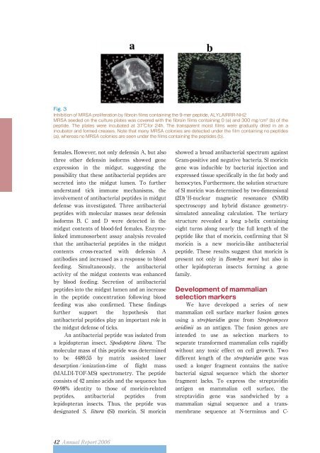
Fig. 3<br />
Inhibition of MRSA proliferation by fibroin films containing the 9-mer peptide, ALYLAIRRR-NH2<br />
MRSA seeded on the culture plates was covered with the fibroin films containing 0 (a) and 300 mg/cm 2 (b)ofthe<br />
peptide. The plates were incubated at 37for 24h. The transparent moist films were gradually dried in an a<br />
incubator and formed creases. Note that many MRSA colonies are detected under the film containing no peptides<br />
(a), whereas no MRSA colonies are seen under the films containing the peptides (b).<br />
females. However, not only defensin A, but also<br />
three other defensin isoforms showed gene<br />
expression in the midgut, suggesting the<br />
possibility that these antibacterial peptides are<br />
secreted into the midgut lumen. To further<br />
understand tick immune mechanisms, the<br />
involvement of antibacterial peptides in midgut<br />
defense was investigated. Three antibacterial<br />
peptides with molecular masses near defensin<br />
isoforms B, C and D were detected in the<br />
midgut contents of blood-fed females. Enzymelinked<br />
immunosorbent assay analysis revealed<br />
that the antibacterial peptides in the midgut<br />
contents cross-reacted with defensin A<br />
antibodies and increased as a response to blood<br />
feeding. Simultaneously, the antibacterial<br />
activity of the midgut contents was enhanced<br />
by blood feeding. Secretion of antibacterial<br />
peptides into the midgut lumen and an increase<br />
in the peptide concentration following blood<br />
feeding was also confirmed. These findings<br />
further support the hypothesis that<br />
antibacterial peptides play an important role in<br />
the midgut defense of ticks.<br />
An antibacterial peptide was isolated from<br />
a lepidopteran insect, The<br />
molecular mass of this peptide was determined<br />
to be 4489.55 by matrix assisted laser<br />
desorption/ ionization-time of flight mass<br />
(MALDI-TOF-MS) spectrometry. The peptide<br />
consists of 42 amino acids and the sequence has<br />
69-98% identity to those of moricin-related<br />
peptides, antibacterial peptides from<br />
lepidopteran insects. Thus, the peptide was<br />
designated (Sl) moricin. Sl moricin<br />
showed a broad antibacterial spectrum against<br />
Gram-positive and negative bacteria. Sl moricin<br />
gene was inducible by bacterial injection and<br />
expressed tissue specifically in the fat body and<br />
hemocytes. Furthermore, the solution structure<br />
of Sl moricin was determined by two-dimensional<br />
(2D) 1 H-nuclear magnetic resonance (NMR)<br />
spectroscopy and hybrid distance geometrysimulated<br />
annealing calculation. The tertiary<br />
structure revealed a long a-helix containing<br />
eight turns along nearly the full length of the<br />
peptide like that of moricin, confirming that Sl<br />
moricin is a new moricin-like antibacterial<br />
peptide. These results suggest that moricin is<br />
present not only in but also in<br />
other lepidopteran insects forming a gene<br />
family.<br />
Development of mammalian<br />
selection markers<br />
We have developed a series of new<br />
mammalian cell surface marker fusion genes<br />
using a gene from <br />
as an antigen. The fusion genes are<br />
intended to use as selection markers to<br />
separate transformed mammalian cells rapidly<br />
without any toxic effect on cell growth. Two<br />
different length of the gene was<br />
used: a longer fragment contains the native<br />
bacterial signal sequence which the shorter<br />
fragment lacks. To express the streptavidin<br />
antigen on mammalian cell surface, the<br />
streptavidin gene was sandwiched by a<br />
mammalian signal sequence and a transmembrane<br />
sequence at N-terminus and C-