CMDITR Review of Undergraduate Research - Pluto - University of ...
CMDITR Review of Undergraduate Research - Pluto - University of ...
CMDITR Review of Undergraduate Research - Pluto - University of ...

You also want an ePaper? Increase the reach of your titles
YUMPU automatically turns print PDFs into web optimized ePapers that Google loves.
were calcined at 700 o C in air for 3h. On the other<br />
hand, no further heat treatment was required for<br />
PVDF fibers as the evaporation <strong>of</strong> solvent<br />
occurred readily during electrospinning.<br />
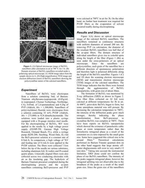
Figure 1. (A) Optical microscope image <strong>of</strong> BaTiO 3<br />
nan<strong>of</strong>ibers after calcination in air at 700 o C for 3 h. (B)<br />
Domain structure <strong>of</strong> BaTiO 3 nan<strong>of</strong>ibers revealed under a<br />
polarizing optical microscope. (C) SEM image taken from the<br />
sample shown in A. (D) High-magnification TEM image and<br />
electron diffraction (inset) <strong>of</strong> BaTiO 3 nan<strong>of</strong>ibers showing the<br />
polycrystalline nature <strong>of</strong> the calcined nan<strong>of</strong>ibers.<br />
Experiment<br />
Nan<strong>of</strong>ibers <strong>of</strong> BaTiO 3 were electrospun<br />
from a solution containing 5mL <strong>of</strong> Barium-<br />
Titanium ethylhexano-isopropoxide (0.05g/mL<br />
in isopropanol, Chemat Technology, Northridge,<br />
CA), 0.05mL <strong>of</strong> 2,4-pentanedione and 0.20g <strong>of</strong><br />
PVP (Aldrich, Mw ≈ 1,300,000). Nan<strong>of</strong>ibers <strong>of</strong><br />
poly(vinylidene) fluoride were electrospun from<br />
a solution containing 35 wt% PVDF (Aldrich,<br />
Mw ≈ 275,000) in N,N-dimethylacetamide. The<br />
solutions were loaded into a plastic syringe<br />
equipped with a 26-gauge stainless steel needle.<br />
For the electrospinning <strong>of</strong> BaTiO 3 , 9kV were<br />
applied to the needle by a high voltage power<br />
supply (ES30P-5W, Gamma High Voltage<br />
<strong>Research</strong>, Ormand Beach, FL), while a syringe<br />
pump (KDS-200, Stoelting, Wood Dale, IL) fed<br />
the BaTi-precursor solution at a constant rate <strong>of</strong><br />
0.5mL/h. In comparison, a high voltage <strong>of</strong> 7kV<br />
and feeding rate <strong>of</strong> 0.1mL/h were applied to the<br />
PVDF solution. The fibers were collected 7.5cm<br />
below the tip <strong>of</strong> the needle on various substrates,<br />
including aluminum foil, Si wafers and Pt-coated<br />
quartz plates. Uniaxially aligned fibers were also<br />
collected across two conducting electrodes with<br />
air as the insulating gap. The hydrolysis <strong>of</strong><br />
Barium-Titanium precursor completed during the<br />
electrospinning process and the as-spun<br />
nan<strong>of</strong>ibers containing BaTi-precursor and PVP<br />
Results and Discussion<br />
Figure 1(A) shows an optical microscope<br />
image <strong>of</strong> the calcined BaTiO 3 nan<strong>of</strong>ibers. The<br />
nan<strong>of</strong>ibers remained as continuous structures<br />
with uniform diameters <strong>of</strong> around 200 nm. By<br />
removing PVP via calcination, the diameter <strong>of</strong><br />
the resultant BaTiO 3 nan<strong>of</strong>ibers was half that <strong>of</strong><br />
the as-spun fibers. The domain structure <strong>of</strong><br />
individual nan<strong>of</strong>ibers gives rise to colorful spots<br />
along the length <strong>of</strong> the fiber (Figure 1 (B)) as<br />
seen under the cross-polarizers <strong>of</strong> an optical<br />
microscope. Since the nan<strong>of</strong>ibers are<br />
polycrystalline, each grain <strong>of</strong> the nan<strong>of</strong>iber has a<br />
different direction <strong>of</strong> spontaneous polarization<br />
and therefore light is polarized differently along<br />
the length <strong>of</strong> the BaTiO 3 nan<strong>of</strong>iber. Figures 1 (C)<br />
and 1D show the scanning electron microscope<br />
(SEM) and transmission electron microscope<br />
(TEM) images <strong>of</strong> the calcined nan<strong>of</strong>ibers. The<br />
TEM image indicates that the fibers were formed<br />
through the agglomeration <strong>of</strong> BaTiO 3<br />
nanoparticles, with grain size <strong>of</strong> about 30nm.<br />
The formation <strong>of</strong> BaTiO 3 was monitored by<br />
X-ray diffraction (XRD) as shown in Figure 2.<br />
Free fiber films collected on Al foil were<br />
calcined at different temperatures for 3h in air.<br />
At 600 o C, perovskite BaTiO 3 began to form, but<br />
some amorphous material was still present. By<br />
increasing the calcination temperature to 700 o C,<br />
the intensity <strong>of</strong> the diffraction peaks grew much<br />
stronger, thereby indicating the phase<br />
transformation from BaTi-precursor to<br />
crystalline BaTiO 3 was complete at 700 o C. There<br />
have been reports on BaTiO 3 nanoparticles<br />
which showed the existence <strong>of</strong> paraelectric cubic<br />
phase at room temperature rather than the<br />
ferroelectric tetragonal phase as a result <strong>of</strong> the<br />
constraint on c/a ratio imposed by the small grain<br />
size. Some have suggested this critical size to be<br />
around 30nm. 18 Theoretical calculations<br />
performed on Barium Titanate quantum dots on<br />
the other hand suggests that large atomic <strong>of</strong>fcenter<br />
displacements still exist in very small (