Proceedings of the meeting - Department of Physics - University of ...
Proceedings of the meeting - Department of Physics - University of ...
Proceedings of the meeting - Department of Physics - University of ...

Create successful ePaper yourself
Turn your PDF publications into a flip-book with our unique Google optimized e-Paper software.
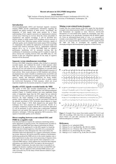
Recent advances in EEG/fMRI integrationStefan Debener 1,21 MRC Institute <strong>of</strong> Hearing Research, Royal South Hants Hospital, Southampton SO14 OYG, UK2 Clinical Neurosciences, School <strong>of</strong> Medicine, <strong>University</strong> <strong>of</strong> Southampton, Southampton, UKI2IntroductionElectroencephalography (EEG) and functional magnetic resonanceimaging (fMRI) provide complimentary information regarding <strong>the</strong>temporal and spatial resolution <strong>of</strong> brain activity. Accordingly, <strong>the</strong>integration <strong>of</strong> both signals holds great promise for a betterunderstanding <strong>of</strong> how cognitive processes are implemented in distinctcortical areas. Following an overview about <strong>the</strong> virtues and pitfalls <strong>of</strong>simultaneous and separate recordings, it will be described howartifacts unique to inside scanner EEG recordings can be removed. Itwill be shown that, following scanner artifact correction, signals <strong>of</strong>sufficient quality can be obtained from inside scanner EEG recordings(1). Importantly, this enables <strong>the</strong> application <strong>of</strong> advanced single-trialoriented EEG analysis techniques such as independent componentanalysis (ICA, e.g., 2). A recent EEG/fMRI study on cognitiveperformance monitoring will be presented showing that <strong>the</strong>combination <strong>of</strong> simultaneous recordings and ICA enables to study <strong>the</strong>direct trial-by-trial coupling between EEG and fMRI data (3). Thebenefits <strong>of</strong> this new integration approach for <strong>the</strong> study <strong>of</strong> cognitivefunction will be discussed.Mining event-related brain dynamicsUsually only <strong>the</strong> evoked fraction <strong>of</strong> <strong>the</strong> EEG signal, <strong>the</strong> time-domainaveraged event-related potential (ERP), is considered and all trial-bytrialfluctuations are regarded as noise. However, trial-by-trialfluctuations <strong>of</strong> event-related EEG signals are meaningful, since <strong>the</strong>yhave been shown to be specifically correlated with <strong>the</strong> BOLD signal(4). From an information-based point <strong>of</strong> view, it is reasonable toconsider event-related EEG signals within a multi-dimensional signalspace (5). It is proposed that <strong>the</strong> consideration <strong>of</strong> EEG signals beyond<strong>the</strong> ERP can help to investigate <strong>the</strong> coupling betweenelectrophysiological and hemodynamic properties <strong>of</strong> brain activity.Separate versus simultaneous recordingsPrevious EEG/fMRI integration attempts <strong>of</strong>ten focused on separatelyrecorded signals, and combined <strong>the</strong> results <strong>of</strong> both measures ra<strong>the</strong>rthan <strong>the</strong> signals directly. However, separate EEG/fMRI recordingprotocols are not well suited for <strong>the</strong> investigation <strong>of</strong> a possible directcoupling between hemodynamic and electrophysiological measures <strong>of</strong>brain activity. More recent advances in EEG hardware and s<strong>of</strong>twaredevelopments have made it feasible to simultaneously record EEG andfMRI data. Only simultaneous recordings guarantee that <strong>the</strong> identicalsensory, mental, and behavioral conditions are being studied.Moreover, simultaneous recording protocols allow to jointly exploit<strong>the</strong> trial-by-trial fluctuations <strong>of</strong> both measures, which can revealimportant insights into <strong>the</strong> dynamic properties <strong>of</strong> cognitive function.Quality <strong>of</strong> EEG signals recorded inside <strong>the</strong> MRIThe quality <strong>of</strong> EEG data recorded simultaneously with fMRI ismassively compromised by gradient artefacts and ballistocardiogramartefacts (Figure 1). Gradient artefacts can cause amplitudes beingorders <strong>of</strong> magnitudes larger than <strong>the</strong> EEG signal, but are relativelyinvariant over time, and <strong>the</strong>refore can be subtracted out (Figure 1 B).However, <strong>the</strong> major challenge is <strong>the</strong> ballistocardiogram (BCG)artefact, which originates from <strong>the</strong> pulsatile movement <strong>of</strong> blood and<strong>the</strong> pulsatile movement <strong>of</strong> EEG electrodes placed adjacent to largerblood vessels (Figure 1 B-D). Both channel-wise and spatial filtertechniques have been proposed for BCG-correction. Results <strong>of</strong> asystematic comparison <strong>of</strong> three different BCG-correction approaches(1) will be presented. It will be shown that a multivariate combinedchannel-wise and spatial filter approach based on ICA is superior toalternative techniques. Following appropriate BCG-correction, EEGsignals <strong>of</strong> reasonable quality can be obtained.Direct coupling between event-related EEG andfMRI in a cognitive taskGoal-directed behavior requires <strong>the</strong> continuous monitoring and dynamicadjustment <strong>of</strong> ongoing actions. We simultaneously recorded 32channel EEG and fMRI data from subjects performing an Eriksenflanker task (3). By applying ICA to <strong>the</strong> EEG signals, we found <strong>the</strong>single-trial error-related negativity <strong>of</strong> <strong>the</strong> EEG to be systematicallyrelated to behavior in <strong>the</strong> subsequent trial. This result reflects <strong>the</strong>immediate behavioral adjustment <strong>of</strong> a cognitive performance monitoringsystem. Moreover, <strong>the</strong> trial-by-trial EEG measure <strong>of</strong> performancemonitoring predicted <strong>the</strong> fMRI activity in <strong>the</strong> rostral cingulate zone, abrain region known to play a key role in processing <strong>of</strong> response errors.Importantly, this result was more specific when compared to a conventionalfMRI analysis, which supports <strong>the</strong> conclusion that temporalinformation, provided by <strong>the</strong> EEG, can improve <strong>the</strong> processing <strong>of</strong>fMRI signals. Fur<strong>the</strong>rmore, <strong>the</strong> study shows that it is possible to investigate<strong>the</strong> dynamic coupling <strong>of</strong> non-invasively recorded EEG andfMRI data. This new approach, EEG-informed fMRI analysis, providesa powerful way for <strong>the</strong> study <strong>of</strong> brain function related to cognitiveprocessing.Figure 1: Example 10-sec traces from inside scanner EEG (A-C) andEKG (D) recordings. EEG data from six representative channels areshown before (A) and after correction for MR gradient (B) andballistocardiogram (C) artefacts.References(1) Debener S et al. submitted (2006)(2) Debener S et al. Cog Brain Res. 22, 309-321 (2005)(3) Debener S et al. J Neurosci. 25, 11730-11737 (2005)(4) Debener S et al. Neuroimage 31, S31 (2006)(5) Makeig S et al. Trends in Cog Sci 8, 204-210 (2004)