Proceedings of the meeting - Department of Physics - University of ...
Proceedings of the meeting - Department of Physics - University of ...
Proceedings of the meeting - Department of Physics - University of ...

Create successful ePaper yourself
Turn your PDF publications into a flip-book with our unique Google optimized e-Paper software.
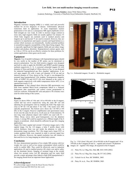
Low field, low cost multi-nuclear imaging research systemsEugeny Krjukov, James Wild, Martyn PaleyAcademic Radiology, <strong>University</strong> <strong>of</strong> Sheffield, Royal Hallamshire Hospital, Sheffield, UKP13IntroductionMagnetic resonance imaging (MRI) is a widely used and powerfulmethod for in-vivo diagnosis <strong>of</strong> disease. Unfortunately physicalrestrictions limit its application. At room temperature nuclearpolarisation is low and as a consequence MR signal amplitudes at lowfield strength are very weak. In order to increase image contrast tonoise ratio, high magnetic fields are usually applied. For instance 3Twhole body magnets are routinely used in medical practice andmagnets up to and beyond 7T are used in research. However, <strong>the</strong>y areexpensive to manufacture and require cryostats and liquid helium as acoolant which increases cost dramatically. Ano<strong>the</strong>r potential problemis nonuniform magnetic susceptibility <strong>of</strong> <strong>the</strong> object being imaged. Thisis especially significant for high magnetic fields and can reduce imagecontrast and produce distortion. Hyperpolarized contrast agents suchas 3-He or 129-Xe can be an effective solution for <strong>the</strong>se problems forcertain applications where gases can be used.EquipmentMagnets: Use <strong>of</strong> parallel techniques with hyperpolarised gases shouldbe very useful as <strong>the</strong> number <strong>of</strong> RF pulses can be minimised ropreserve polarisation. Two low field resistive magnets were designedand built in order to apply <strong>the</strong> MAMBA and SPIRIT parallel imagingmethods respectively (1, 2). A corrected solenoidal magnet (S) withdiameter <strong>of</strong> 20cm and length <strong>of</strong> 100cm shown in Fig. 1a was designedfor industrial hyperpolarised gas flow chamber’ applications. A sixcoil open magnet (H) with a main coil diameter <strong>of</strong> 60 cm and anintercoil distance <strong>of</strong> 15cm, shown in Fig. 1b and 1c was designed forneonatal lung hyperpolarised 3-He imaging. Homogeneous magneticfields <strong>of</strong> 0.008T (S) and 0.015T (H) were obtained at <strong>the</strong> centre <strong>of</strong>both magnets respectively with stable temperatures below 60 o C (1) asreported previously (3, 4).Spectrometer: A four channel direct detection MR spectrometer wasbuilt from standard Mini-Circuit components linked to a NationalInstruments data acquisition and control system programmed inLabView s<strong>of</strong>tware. Solenoidal RF coils and passive TR switches wereused for initial testing <strong>of</strong> <strong>the</strong> system.ResultsFigure 2a shows FIDs <strong>of</strong> 3-He and 1-H at 450 kHz in <strong>the</strong> H magnet,central and top curves respectively using <strong>the</strong> same RF coil andadjusting for gyromagnetic ratio by ramping <strong>the</strong> field. Flip angle wasclose to 90 o for 1-H and about 2 o for 3-He. 3-He nuclei were 30%optically prepolarized. The bottom curve shows <strong>the</strong> 1-H FID at 350kHz in <strong>the</strong> S magnet. Receiver amplification and spectrometer noiselevels were <strong>the</strong> same for all curves. In all cases an 18 mm diameter, 70mm long water phantom doped with CuSO4 was used with T1 ~100ms. Decay curves show good homogeneity over <strong>the</strong> phantomvolume. A strong FID could be acquired over a 300mm length <strong>of</strong> <strong>the</strong> Smagnet, as predicted, which should be sufficient for gas flow imagingapplications.Figure 2b-d show sagittal and coronal 1-H proton images and a 3-Hesagittal image. For <strong>the</strong> 1-H images, a letter F pr<strong>of</strong>ile object withsection thickness 2mm was put inside <strong>the</strong> phantom in order todemonstrate image resolution. The 3-He image used a larger FOV asweaker gradient fields were required in order to prevent signal lossdue to diffusion. However, <strong>the</strong> spatial resolution was still similar tothat used for in vivo lung imaging on whole body systems.ConclusionWe have demonstrated operation <strong>of</strong> two simple MR systems with lowweight, low cost as well as low energy consumption which could beuseful in mobile MRI. They could be effectively used in industrialapplications (S) and in medical practice (H) where cost and/or safetyare <strong>the</strong> dominant factors. Imaging <strong>of</strong> neonatal lungs at <strong>the</strong> cotsidewhere low acoustic noise, low RF absorption and low static fieldprovide a major advantage when linked to high SNR hyperpolarised 3-He imaging should form an important future application.AcknowledgementWe acknowledge support from <strong>the</strong> Royal Society Paul InstrumentFund and DH-NEAT for this workFig. 1 a – Solenoidal magnet; 1b and 1c – Helmholtz magnet.FID (V)1050-5-101aHe 3 450kHzH 1 450kHzH 1 350kHz0 5 10 15 20 25time (ms)2cFig. 2 a – Fid’s from 3-He and 1-H at 450 kHz in <strong>the</strong> H magnet and 1-H at350 kHz in <strong>the</strong> S magnet;2b and 2c – sagittal and coronal 1-H phantomimages; 2d – sagittal 3-He image, all acquired in <strong>the</strong> H magnet.References2a1b1c(1) Paley M et al. Mag. Res. Med. 48, 1043-1050 (2002)(2) Paley M et al. Mag. Res. Imag. 24, 557-562 (2006)(3) Fichele S et al. Proc. BC ISMRM., 2005.(4) Paley M et al., Proc. BC ISMRM, 2005.2b2d