Proceedings of the meeting - Department of Physics - University of ...
Proceedings of the meeting - Department of Physics - University of ...
Proceedings of the meeting - Department of Physics - University of ...

You also want an ePaper? Increase the reach of your titles
YUMPU automatically turns print PDFs into web optimized ePapers that Google loves.
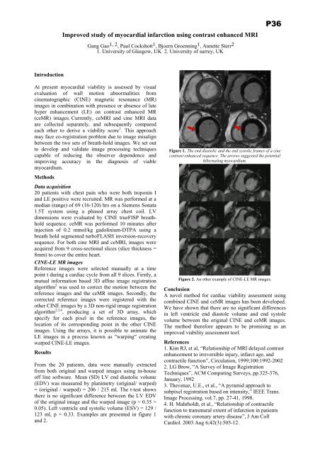
Improved study <strong>of</strong> myocardial infarction using contrast enhanced MRIGang Gao 1, 2 , Paul Cockshott 1 , Bjoern Groenning 1 , Annette Sterr 21. <strong>University</strong> <strong>of</strong> Glasgow, UK 2. <strong>University</strong> <strong>of</strong> surrey, UKP36IntroductionAt present myocardial viability is assessed by visualevaluation <strong>of</strong> wall motion abnormalities fromcinematographic (CINE) magnetic resonance (MR)images in combination with presence or absence <strong>of</strong> latehyper enhancement (LE) on contrast enhanced MR(ceMR) images. Currently, ceMRI and cine MRI dataare collected separately, and subsequently comparedeach o<strong>the</strong>r to derive a viability score 1 . This approachmay face co-registration problem due to image misalignbetween <strong>the</strong> two sets <strong>of</strong> breath-hold images. We set outto develop and validate image processing techniquescapable <strong>of</strong> reducing <strong>the</strong> observer dependence andimproving accuracy in <strong>the</strong> diagnosis <strong>of</strong> viablemyocardium.MethodsData acquisition20 patients with chest pain who were both troponin Iand LE positive were recruited. MR was performed at amedian (range) <strong>of</strong> 69 (16-120) hrs on a Siemens Sonata1.5T system using a phased array chest coil. LVdimensions were evaluated by CINE trueFISP breathholdsequence. ceMR was performed 10 minutes afterinjection <strong>of</strong> 0.2 mmol/kg gadolinium-DTPA using abreath hold segmented turboFLASH inversion-recoverysequence. For both cine MRI and ceMRI, images wereacquired from 9 cross-sectional slices (slice thickness =8mm) to cover <strong>the</strong> entire heart.CINE-LE MR imagesReference images were selected manually at a timepoint t during a cardiac cycle from all 9 slices. Firstly, amutual information based 3D affine image registrationalgorithm 2 was used to correct <strong>the</strong> motion between <strong>the</strong>reference images and <strong>the</strong> ceMR images. Secondly, <strong>the</strong>corrected reference images were registered with <strong>the</strong>o<strong>the</strong>r CINE images by a 3D non-rigid image registrationalgorithm 2,3,4 , producing a set <strong>of</strong> 3D array, whichspecify for each pixel in <strong>the</strong> reference images, <strong>the</strong>location <strong>of</strong> its corresponding point in <strong>the</strong> o<strong>the</strong>r CINEimages. Using <strong>the</strong> arrays, it is possible to animate <strong>the</strong>LE images in a process known as "warping" creatingwarped CINE-LE images.ResultsFrom <strong>the</strong> 20 patients, data were manually extractedfrom both original and warped images using in-house<strong>of</strong>f line s<strong>of</strong>tware. Mean (SD) LV end diastolic volume(EDV) was measured by planimetry (original/ warped)= (original / warped) = 206 / 215 ml. The t-test shows<strong>the</strong>re is no significant difference between <strong>the</strong> LV EDV<strong>of</strong> <strong>the</strong> original image and <strong>the</strong> warped image (p = 0.35 >0.05). Left ventricle end systolic volume (ESV) = 129 /123 ml, p = 0.33. Examples are presented in figure 1and 2.Figure 1. The end diastolic and <strong>the</strong> end systolic frames <strong>of</strong> a cinecontrast enhanced sequence. The arrows suggested <strong>the</strong> potentialhibernating myocardium.Figure 2. An o<strong>the</strong>r example <strong>of</strong> CINE-LE MR images.ConclusionA novel method for cardiac viability assessment usingcombined CINE and ceMR images has been developed.We have shown that <strong>the</strong>re are no significant differencesin left ventricle end diastole volume and end systolevolume between <strong>the</strong> original CINE and ceMR images.The method <strong>the</strong>refore appears to be promising as animproved viability assessment tool.References1. Kim RJ, et al, “Relationship <strong>of</strong> MRI delayed contrastenhancement to irreversible injury, infarct age, andcontractile function”, Circulation, 1999;100:1992-20022. LG Brow, “A Survey <strong>of</strong> Image RegistrationTechniques”, ACM Computing Surveys, pp.325-376,January, 19923. Thevenaz, U.E., et al., “A pyramid approach tosubpixel registration based on intensity,” IEEE Trans.Image Processing, vol.7, pp. 27-41, 1998.4. H. Mahrholdt, et al., “Relationship <strong>of</strong> contractilefunction to transmural extent <strong>of</strong> infarction in patientswith chronic coronary artery disease”, J Am CollCardiol. 2003 Aug 6;42(3):505-12.