Proceedings of the meeting - Department of Physics - University of ...
Proceedings of the meeting - Department of Physics - University of ...
Proceedings of the meeting - Department of Physics - University of ...

You also want an ePaper? Increase the reach of your titles
YUMPU automatically turns print PDFs into web optimized ePapers that Google loves.
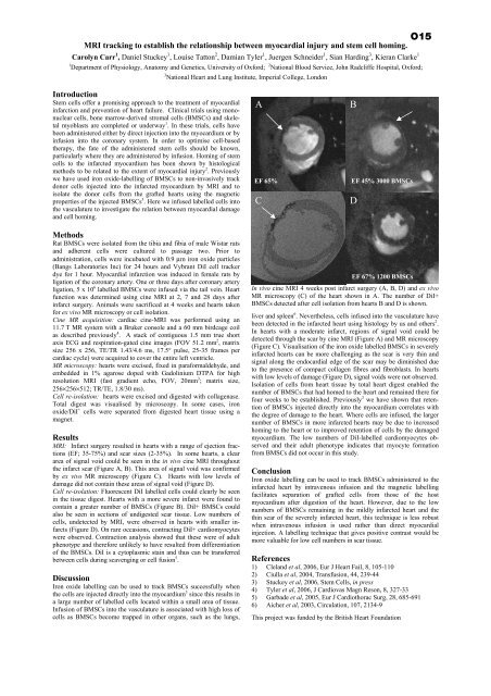
O15MRI tracking to establish <strong>the</strong> relationship between myocardial injury and stem cell homing.Carolyn Carr 1 , Daniel Stuckey 1 , Louise Tatton 2 , Damian Tyler 1 , Juergen Schneider 1 , Sian Harding 3 , Kieran Clarke 11 <strong>Department</strong> <strong>of</strong> Physiology, Anatomy and Genetics, <strong>University</strong> <strong>of</strong> Oxford; 2 National Blood Service, John Radcliffe Hospital, Oxford;3 National Heart and Lung Institute, Imperial College, LondonIntroductionStem cells <strong>of</strong>fer a promising approach to <strong>the</strong> treatment <strong>of</strong> myocardialinfarction and prevention <strong>of</strong> heart failure. Clinical trials using mononuclearcells, bone marrow-derived stromal cells (BMSCs) and skeletalmyoblasts are completed or underway 1 . In <strong>the</strong>se trials, cells havebeen administered ei<strong>the</strong>r by direct injection into <strong>the</strong> myocardium or byinfusion into <strong>the</strong> coronary system. In order to optimise cell-based<strong>the</strong>rapy, <strong>the</strong> fate <strong>of</strong> <strong>the</strong> administered stem cells should be known,particularly where <strong>the</strong>y are administered by infusion. Homing <strong>of</strong> stemcells to <strong>the</strong> infarcted myocardium has been shown by histologicalmethods to be related to <strong>the</strong> extent <strong>of</strong> myocardial injury 2 . Previouslywe have used iron oxide-labelling <strong>of</strong> BMSCs to non-invasively trackdonor cells injected into <strong>the</strong> infarcted myocardium by MRI and toisolate <strong>the</strong> donor cells from <strong>the</strong> grafted hearts using <strong>the</strong> magneticproperties <strong>of</strong> <strong>the</strong> injected BMSCs 3 . Here we infused labelled cells into<strong>the</strong> vasculature to investigate <strong>the</strong> relation between myocardial damageand cell homing.AEF 65%CBEF 45% 3000 BMSCsDMethodsRat BMSCs were isolated from <strong>the</strong> tibia and fibia <strong>of</strong> male Wistar ratsand adherent cells were cultured to passage two. Prior toadministration, cells were incubated with 0.9 µm iron oxide particles(Bangs Laboratories Inc) for 24 hours and Vybrant DiI cell trackerdye for 1 hour. Myocardial infarction was induced in female rats byligation <strong>of</strong> <strong>the</strong> coronary artery. One or three days after coronary arteryligation, 5 x 10 6 labelled BMSCs were infused via <strong>the</strong> tail vein. Heartfunction was determined using cine MRI at 2, 7 and 28 days afterinfarct surgery. Animals were sacrificed at 4 weeks and hearts takenfor ex vivo MR microscopy or cell isolation.Cine MR acquisition: cardiac cine-MRI was performed using an11.7 T MR system with a Bruker console and a 60 mm birdcage coilas described previously 4 . A stack <strong>of</strong> contiguous 1.5 mm true shortaxis ECG and respiration-gated cine images (FOV 51.2 mm 2 , matrixsize 256 x 256, TE/TR 1.43/4.6 ms, 17.5° pulse, 25-35 frames percardiac cycle) were acquired to cover <strong>the</strong> entire left ventricle.MR microscopy: hearts were excised, fixed in paraformaldehyde, andembedded in 1% agarose doped with Gadolinium DTPA for highresolution MRI (fast gradient echo, FOV, 20mm 3 ; matrix size,256×256×512; TR/TE, 1.8/30 ms).Cell re-isolation: hearts were excised and digested with collagenase.Total digest was visualised by microscopy. In some cases, ironoxide/DiI + cells were separated from digested heart tissue using amagnet.ResultsMRI: Infarct surgery resulted in hearts with a range <strong>of</strong> ejection fractions(EF; 35-75%) and scar sizes (2-35%). In some hearts, a cleararea <strong>of</strong> signal void could be seen in <strong>the</strong> in vivo cine MRI throughout<strong>the</strong> infarct scar (Figure A, B). This area <strong>of</strong> signal void was confirmedby ex vivo MR microscopy (Figure C). Hearts with low levels <strong>of</strong>damage did not contain <strong>the</strong>se areas <strong>of</strong> signal void (Figure D).Cell re-isolation: Fluorescent DiI labelled cells could clearly be seenin <strong>the</strong> tissue digest. Hearts with a more severe infarct were found tocontain a greater number <strong>of</strong> BMSCs (Figure B). DiI+ BMSCs couldalso be seen in sections <strong>of</strong> undigested scar tissue. Low numbers <strong>of</strong>cells, undetected by MRI, were observed in hearts with smaller infarcts(Figure D). On rare occasions, contracting DiI+ cardiomyocyteswere observed. Contraction analysis showed that <strong>the</strong>se were <strong>of</strong> adultphenotype and <strong>the</strong>refore unlikely to have resulted from differentiation<strong>of</strong> <strong>the</strong> BMSCs. DiI is a cytoplasmic stain and thus can be transferredbetween cells during scavenging or cell fusion 5 .DiscussionIron oxide labelling can be used to track BMSCs successfully when<strong>the</strong> cells are injected directly into <strong>the</strong> myocardium 3 since this results ina large number <strong>of</strong> labelled cells located within a small area <strong>of</strong> tissue.Infusion <strong>of</strong> BMSCs into <strong>the</strong> vasculature is associated with high loss <strong>of</strong>cells as BMSCs become trapped in o<strong>the</strong>r organs, such as <strong>the</strong> lungs,EF 67% 1200 BMSCsIn vivo cine MRI 4 weeks post infarct surgery (A, B, D) and ex vivoMR microscopy (C) <strong>of</strong> <strong>the</strong> heart shown in A. The number <strong>of</strong> DiI+BMSCs detected after cell isolation from hearts B and D is shown.liver and spleen 6 . Never<strong>the</strong>less, cells infused into <strong>the</strong> vasculature havebeen detected in <strong>the</strong> infarcted heart using histology by us and o<strong>the</strong>rs 2 .In hearts with a moderate infarct, regions <strong>of</strong> signal void could bedetected through <strong>the</strong> scar by cine MRI (Figure A) and MR microscopy(Figure C). Visualisation <strong>of</strong> <strong>the</strong> iron oxide labelled BMSCs in severelyinfarcted hearts can be more challenging as <strong>the</strong> scar is very thin andsignal along <strong>the</strong> endocardial edge <strong>of</strong> <strong>the</strong> scar may be diminished dueto <strong>the</strong> presence <strong>of</strong> compact collagen fibres and fibroblasts. In heartswith low levels <strong>of</strong> damage (Figure D), signal voids were not observed.Isolation <strong>of</strong> cells from heart tissue by total heart digest enabled <strong>the</strong>number <strong>of</strong> BMSCs that had homed to <strong>the</strong> heart and remained <strong>the</strong>re forfour weeks to be established. Previously 3 we have shown that retention<strong>of</strong> BMSCs injected directly into <strong>the</strong> myocardium correlates with<strong>the</strong> degree <strong>of</strong> damage to <strong>the</strong> heart. Where cells are infused, <strong>the</strong> largernumber <strong>of</strong> BMSCs in more infarcted hearts may be due to increasedhoming to <strong>the</strong> heart or to improved retention <strong>of</strong> cells by <strong>the</strong> damagedmyocardium. The low numbers <strong>of</strong> DiI-labelled cardiomyocytes observedand <strong>the</strong>ir adult phenotype indicates that myocyte formationfrom BMSCs did not occur in this study.ConclusionIron oxide labelling can be used to track BMSCs administered to <strong>the</strong>infarcted heart by intravenous infusion and <strong>the</strong> magnetic labellingfacilitates separation <strong>of</strong> grafted cells from those <strong>of</strong> <strong>the</strong> hostmyocardium after digestion <strong>of</strong> <strong>the</strong> heart. However, due to <strong>the</strong> lownumbers <strong>of</strong> BMSCs remaining in <strong>the</strong> mildly infarcted heart and <strong>the</strong>thin scar <strong>of</strong> <strong>the</strong> severely infarcted heart, this technique is less robustwhen intravenous infusion is used ra<strong>the</strong>r than direct myocardialinjection. A labelling technique that gives positive contrast would bemore valuable for low cell numbers in scar tissue.References1) Cleland et al, 2006, Eur J Heart Fail, 8, 105-1102) Ciulla et al, 2004, Transfusion, 44, 239-443) Stuckey et al, 2006, Stem Cells, in press4) Tyler et al, 2006, J Cardiovas Magn Reson, 8, 327-335) Garbade et al, 2005, Eur J Cardiothorac Surg, 28, 685-6916) Aicher et al, 2003, Circulation, 107, 2134-9This project was funded by <strong>the</strong> British Heart Foundation